. 2014 Aug;233(4):368-79.
doi: 10.1002/path.4363. Epub 2014 Jun 2.

MicroRNA-519a is a novel oncomir conferring tamoxifen resistance by targeting a network of tumour-suppressor genes in ER+ breast cancer

Affiliations
Free PMC article

MicroRNA-519a is a novel oncomir conferring tamoxifen resistance by targeting a network of tumour-suppressor genes in ER+ breast cancer

Aoife Ward et al. J Pathol. 2014 Aug.
Free PMC article

Abstract

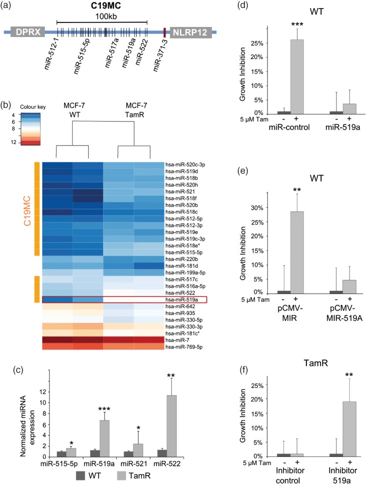
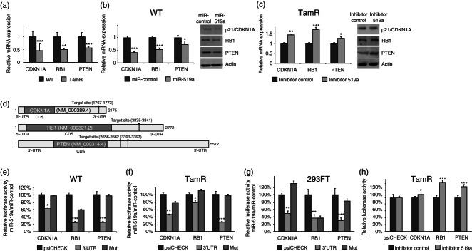
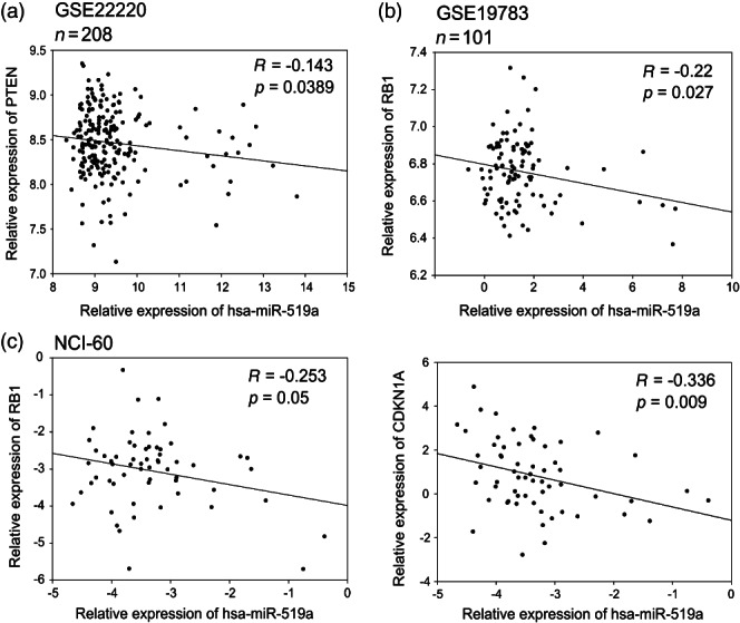
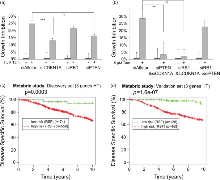
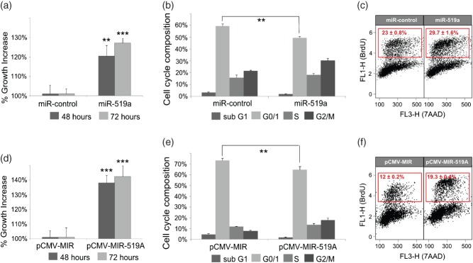
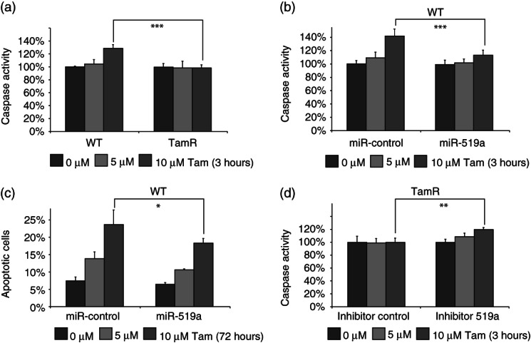
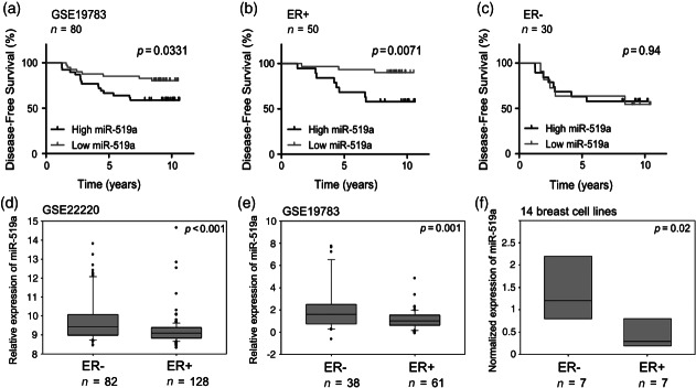
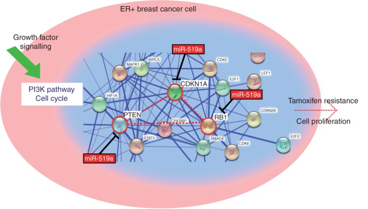
Tamoxifen is an endocrine therapy which is administered to up to 70% of all breast cancer patients with oestrogen receptor alpha (ERα) expression. Despite the initial response, most patients eventually acquire resistance to the drug. MicroRNAs (miRNAs) are a class of small non-coding RNAs which have the ability to post-transcriptionally regulate genes. Although the role of a few miRNAs has been described in tamoxifen resistance at the single gene/target level, little is known about how concerted actions of miRNAs targeting biological networks contribute to resistance. Here we identified the miRNA cluster, C19MC, which harbours around 50 mature miRNAs, to be up-regulated in resistant cells, with miRNA-519a being the most highly up-regulated. We could demonstrate that miRNA-519a regulates tamoxifen resistance using gain- and loss-of-function testing. By combining functional enrichment analysis and prediction algorithms, we identified three central tumour-suppressor genes (TSGs) in PI3K signalling and the cell cycle network as direct target genes of miR-519a. Combined expression of these target genes correlated with disease-specific survival in a cohort of tamoxifen-treated patients. We identified miRNA-519a as a novel oncomir in ER+ breast cancer cells as it increased cell viability and cell cycle progression as well as resistance to tamoxifen-induced apoptosis. Finally, we could show that elevated miRNA-519a levels were inversely correlated with the target genes' expression and that higher expression of this miRNA correlated with poorer survival in ER+ breast cancer patients. Hence we have identified miRNA-519a as a novel oncomir, co-regulating a network of TSGs in breast cancer and conferring resistance to tamoxifen. Using inhibitors of such miRNAs may serve as a novel therapeutic approach to combat resistance to therapy as well as proliferation and evasion of apoptosis in breast cancer.

Keywords: C19MC cluster; breast cancer; cell cycle network; microRNAs; tamoxifen resistance.


Figures

Figure 1
Figure 1
C19MC is up-regulated in TamR cells and miRNA-519a regulates tamoxifen resistance. (a) The C19MC locus comprises approximately 50 mature miRNAs and spans 100 kb on chromosome 19q13.42 bordered by genes DPRX distal and NLRP12, as well as the unrelated miR-371-3 cluster, proximal. (b) Heat map showing 18 members of the miRNA cluster C19MC, depicted with an orange bar, which are up-regulated in MCF-7 TamR cells. miR-519a is boxed in red. (c) qRT-PCR validation of the up-regulation of selected C19MC members in TamR cells compared with WT cells, normalized to RNU44 and RNU48. (d) Overexpressing miRNA-519a in WT cells using mimics or an overexpressing vector (e) confers resistance to tamoxifen after 72 h, while (f) inhibition of miRNA-519a in TamR cells sensitizes cells to tamoxifen treatment. All results are shown as an average of three biological and three technical replicates. One asterisk (*) denotes a p value of less than 0.05, two (**) denote p < 0.01, and three (***) denote p < 0.001, determined by a two-sided t-test, hereafter.
Figure 2
Figure 2
Validation of CDKN1A, RB1, and PTEN as direct targets of miRNA-519a. (a) qRT-PCR reveals that predicted targets of miRNA-519a are down-regulated in TamR cells. (b) Predicted targets are down-regulated 48 h after transfection with mimic in WT cells at RNA as well as protein levels and (c) up-regulated after 48 h transfection with inhibitor in TamR cells. (d) mRNA structure of target genes showing the predicted target sites of miRNA-519a in the respective 3′UTRs. (e) Luciferase assays show that the activity of each 3′UTR is reduced when WT (e), TamR (f), and HEK 293FT (g) cells are transfected with miRNA-519a compared with control. The signal is rescued after mutating the binding site for miRNA-519a. (h) Luciferase signal when TamR cells are transfected with miR-519a inhibitor. Luciferase signal was normalized to empty vector. All results are shown as an average of three biological and three technical replicates, with significance levels explained in Figure 1.
Figure 3
Figure 3
Correlation analysis of miRNA-519a and its target genes in patient datasets and NCI-60 cell line panel presented as scatter plots. miRNA-519a expression is inversely correlated with expression of target genes in two patient datasets consisting of 208 patients (a) and 101 patients (b), as well as in the NCI-60 cell line panel (http://dtp.nci.nih.gov/index.html; experiment ID: 372685) (c).
Figure 4
Figure 4
Target genes of miRNA-519a co-operatively regulate tamoxifen resistance. Inhibiting individual (a) and combinations (b) of the TSGs using siRNAs in WT cells promotes resistance to tamoxifen after 72 h. Results are shown as an average of three biological and three technical replicates, with significance levels explained in Figure 1. (c) Random survival forests (RSF) analysis of 632 (d) and 586 breast cancer patients who had received hormone therapy (HT) reveals a significant correlation between survival and expression of target genes.
Figure 5
Figure 5
miRNA-519a effects on viability and cell cycle. (a) Viability of MCF-7 WT cells was measured 48 and 72 h after transfection with miR-control or miRNA-519a. 7-AAD (b) and BrdU (c) staining was measured 72 h after transfection with miR-control or miRNA-519a to assay cell cycle phases. The S-phase population is gated in the red box for BrdU-positive cells. (d–f) Cell viability and cell cycle assays similar to a–c except that empty vector, pCMV-MIR or pCMV–MIR-519a overexpressing miRNA-519a were transfected as indicated. The results are presented as an average of two biological and three technical replicates, with significance levels explained in Figure 1.
Figure 6
Figure 6
miRNA-519a reduces tamoxifen-induced apoptosis. (a) Caspase activity is elevated only in WT cells after 3 h treatment with 10 µm tamoxifen. (b) Caspase activity is increased only in miR-control transfected cells and is abrogated by transfection with a miRNA-519a mimic. (c) PI staining reveals that more apoptotic cells are present in miR-control than in miRNA-519a mimic-transfected cells after 72 h treatment with tamoxifen. (d) Caspase activity is increased only in inhibitor 519a-transfected TamR cells. The results are presented as an average of two biological and three technical replicates each, with significance levels explained in Figure 1.
Figure 7
Figure 7
Increased expression of miRNA-519a is significantly correlated with poorer disease-free survival in ER+ breast cancer patients and poor clinicopathological features in breast cancer. Patients were separated according to miRNA-519a expression (median cut-off) in a cohort of breast cancer patients and miRNA-519a expression was significantly correlated with survival (a). When patients were further sub-classified according to ER status, miRNA-519a only correlated with survival in ER+ patients (b), but not in ER− patients (c). miRNA-519a was found to be significantly more highly expressed in ER− patients in two patient datasets (GEO Accession GSE19783 and GSE22220 26) (d, e) and in 14 breast cell lines, analysed by qRT-PCR (f).
Figure 8
Figure 8
miR-519a targets central nodes (PTEN, CDKN1A, and RB1) from PI3K and cell cycle network leading to increased proliferation and tamoxifen resistance. A snapshot from STRING (http://string-db.org) analysis depicts how the three validated direct target genes of miR-519a form a central connection in the network, highlighted in red.

Similar articles

Cited by

References

    1. Vargo-Gogola T, Rosen JM. Modelling breast cancer: one size does not fit all. Nature Rev Cancer. 2007;7:659–672. - PubMed
    1. Jordan VC, Murphy CS. Endocrine pharmacology of antiestrogens as antitumor agents. Endocr Rev. 1990;11:578–610. - PubMed
    1. Katzenellenbogen BS, Miller MA, Mullick A, et al. Antiestrogen action in breast cancer cells: modulation of proliferation and protein synthesis, and interaction with estrogen receptors and additional antiestrogen binding sites. Breast Cancer Res Treat. 1985;5:231–243. - PubMed
    1. Ring A, Dowsett M. Mechanisms of tamoxifen resistance. Endocr Relat Cancer. 2004;11:643–658. - PubMed
    1. Shiau AK, Barstad D, Loria PM, et al. The structural basis of estrogen receptor/coactivator recognition and the antagonism of this interaction by tamoxifen. Cell. 1998;95:927–937. - PubMed

Publication types

MeSH terms