. 2014 Aug;98(15):6689-700.
doi: 10.1007/s00253-014-5730-7. Epub 2014 Apr 22.

Generation of food-grade recombinant Lactobacillus casei delivering Myxococcus xanthus prolyl endopeptidase

Affiliations

Generation of food-grade recombinant Lactobacillus casei delivering Myxococcus xanthus prolyl endopeptidase

Patricia Alvarez-Sieiro et al. Appl Microbiol Biotechnol. 2014 Aug.

Abstract

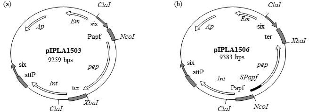
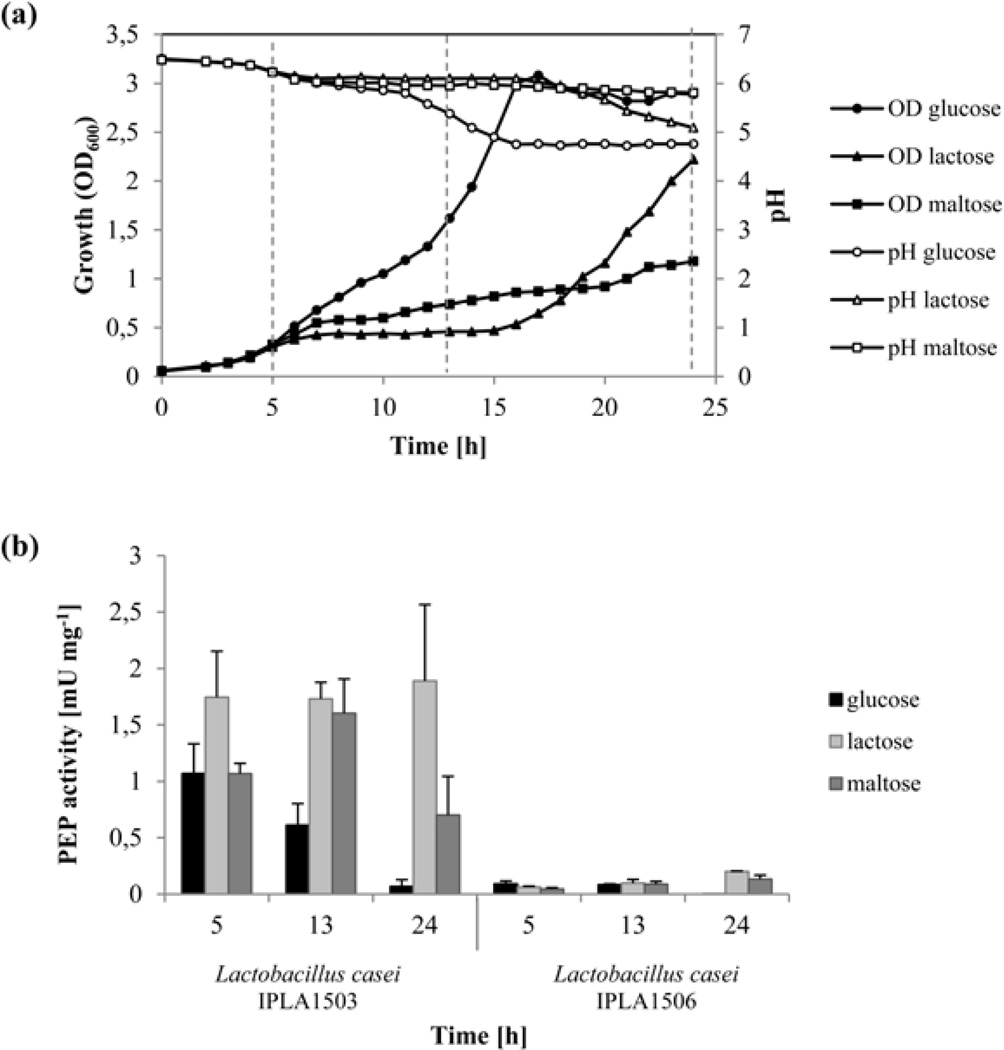
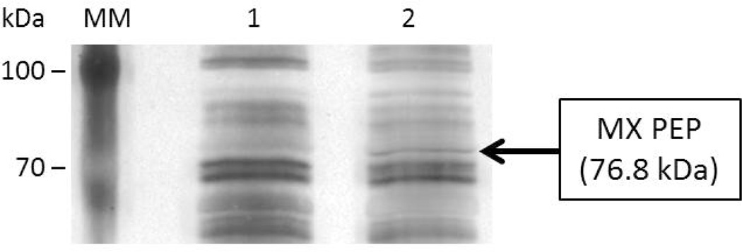
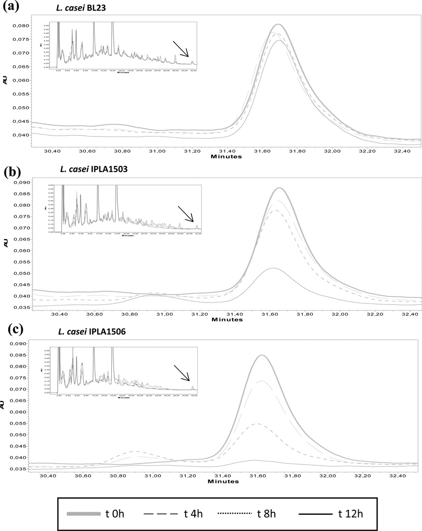
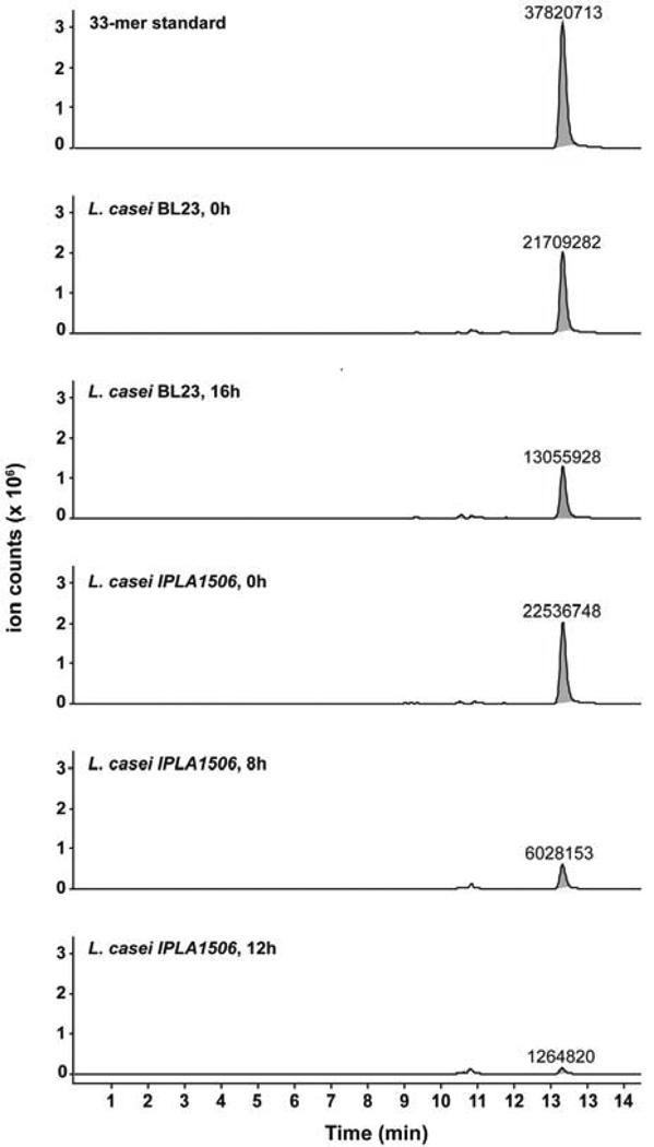
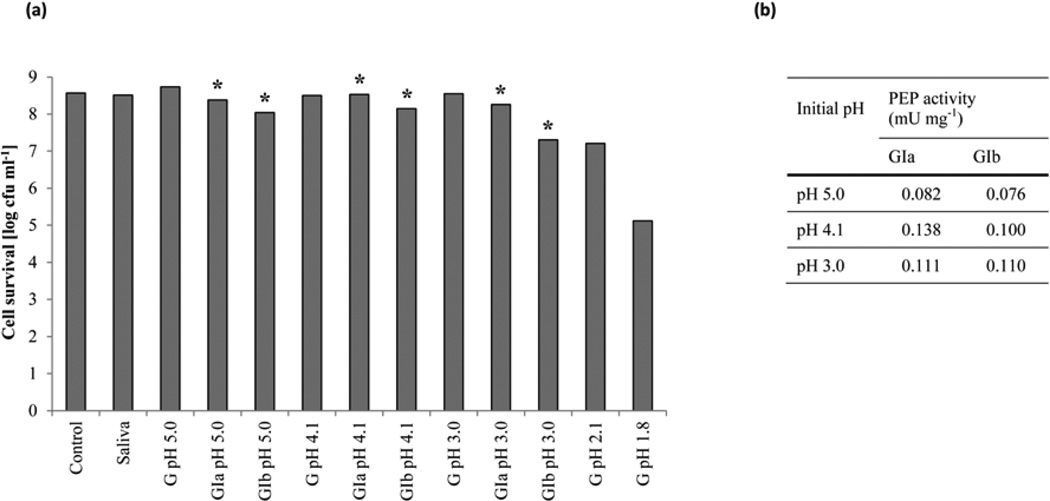
Prolyl endopeptidases (PEP) (EC 3.4.21.26), a family of serine proteases with the ability to hydrolyze the peptide bond on the carboxyl side of an internal proline residue, are able to degrade immunotoxic peptides responsible for celiac disease (CD), such as a 33-residue gluten peptide (33-mer). Oral administration of PEP has been suggested as a potential therapeutic approach for CD, although delivery of the enzyme to the small intestine requires intrinsic gastric stability or advanced formulation technologies. We have engineered two food-grade Lactobacillus casei strains to deliver PEP in an in vitro model of small intestine environment. One strain secretes PEP into the extracellular medium, whereas the other retains PEP in the intracellular environment. The strain that secretes PEP into the extracellular medium is the most effective to degrade the 33-mer and is resistant to simulated gastrointestinal stress. Our results suggest that in the future, after more studies and clinical trials, an engineered food-grade Lactobacillus strain may be useful as a vector for in situ production of PEP in the upper small intestine of CD patients.


Figures

Fig. 1
Fig. 1. Physical map of plasmids generated in this work
The physical map and relevant features of plasmids pIPLA1503 (a) and pIPLA1506 (b) are shown. Indicated are the ampicillin (Ap) and erythromycin (Em) resistance genes; the integration region of bacteriophage A2 (int-attP); the β -recombinase binding sites (six); the promoter, terminator and signal peptide of the aggregation-promoting factor gene of Lactobacillus crispatus (Papf, ter and SPapf) and the prolyl endopeptidase gene of Myxococcus xanthus (pep). Relevant restriction sites are also shown.
Fig. 2
Fig. 2. Effect of different carbon sources on the growth and PEP production ofL. casei IPLA1503 and L. casei IPLA1506
L. casei IPLA1506 was grown at 37°C in CFB media with different carbon sources: glucose, lactose and maltose. Panel (a) shows measurements of OD (600 nm) and pH values at a time intervals of one hour under each carbon source. Discontinuous lines show the points were the PEP activity was measured. Panel (b) shows the PEP production by L. casei IPLA1503 (left) and L. casei IPLA1506 (right) at three different points of the growth curve (5, 13 and 24 h) in three different carbon sources (glucose, lactose and maltose). The enzyme activities assays were performed in triplicate.
Fig. 3
Fig. 3. Time course of the PEP production of L. casei IPLA1506 at different pH
The L. casei IPLA1506 strain was grown through fermentations at free pH and diverse controlled pH values: pH 5.0, pH 6.0 and pH 7.0. The PEP activity was determined in the supernatant of cell culture.
Fig. 4
Fig. 4. SDS-PAGE analysis of the proteins secreted by L. casei BL23 and L. casei IPLA1506
The proteins of the supernatant of L. casei BL23 and L. casei IPLA1506 cultures were precipitated and analysed by SDS-PAGE. Lane 1: L. casei BL23. Lane 2: L. casei IPLA1506.
Fig. 5
Fig. 5. Hydrolysis of the immunotoxic 33-mer peptide
The strains L. casei BL23 (a), L. casei IPLA1503 (b) and L. casei IPLA1506 (c) were incubated with the 33-mer (25.5 µM) during 12 hours. Every four hours aliquots were removed and the substrate concentration was monitored by RP-HPLC analysis. Chromatograms show the hydrolysis time course of 33-mer peptide by the strains. The complete chromatogram appears in a box with the substrate arrowed.
Fig. 6
Fig. 6. LC-MS analysis of 33-mer proteolysis products
The strains L. casei BL23 and L. casei IPLA1506 were incubated with the 33-mer peptide (25.5 µM) for 16 hours at 37°C. Aliquots were taken at the times shown and the analyzed by LC-MS. After 16 hours of incubation with the IPLA1506 strain, the 33-mer is below the instrument limit of quantification. The 33-mer peak is highlighted in gray, and labels represent absolute ion counts.
Fig. 7
Fig. 7. Cell survival (a) and prolyl endopeptidase production (b) after gastric and gastrointestinal stresses of L. casei IPLA1506
The L. casei strain IPLA1506 was resuspended in milk and submitted to simulated gastrointestinal conditions to determine the cell survival. To simulate the gastric stress (G) samples were incubated with lysozyme and pepsin in a decreasing pH values. To simulate the gastrointestinal stress, samples taken from pH 5.0, pH 4.1 and pH 3.0 where further incubated with bile salts and pancreatic enzymes during 20 (GIa) and 120 minutes (Gib). Samples from Gia and Gib (*) were used as preinoculum in a new basal medium. After 4 hours of grow PEP activity was assayed in the supernatant of cell culture.

References

    1. Alvarez MA, Herrero M, Suárez JE. The site-specific recombination system of the Lactobacillus species bacteriophage A2 integrates in gram-positive and gram-negative bacteria. Virology. 1998;250(1):185–193. doi: http://dx.doi.org/10.1006/viro.1998.9353. - DOI - PubMed
    1. Axelsson L. Lactic acid bacteria: Classification and physiology. In: Salminen SaVW A, editor. Lactic acid bacteria Microbiological and functional aspects. Second edition edn. 1998. pp. 1–66.
    1. Bethune MT, Borda JT, Ribka E, Liu MX, Phillippi-Falkenstein K, Jandacek RJ, Doxiadis GG, Gray GM, Khosla C, Sestak K. A non-human primate model for gluten sensitivity. PLoS One. 2008;3(2):e1614. - PMC - PubMed
    1. Bethune MT, Khosla C. Oral enzyme therapy for celiac sprue. Methods Enzymol. 2012;502:241–271. - PMC - PubMed
    1. Carr FJ, Chill D, Maida N. The lactic acid bacteria: A literature survey. critical reviews in microbiology. 2002;28(4):281–370. - PubMed

Publication types

MeSH terms