. 2014 Jul;34(13):2409-17.
doi: 10.1128/MCB.00104-14. Epub 2014 Apr 21.

The canonical wnt signal restricts the glycogen synthase kinase 3/fbw7-dependent ubiquitination and degradation of eya1 phosphatase

Affiliations

The canonical wnt signal restricts the glycogen synthase kinase 3/fbw7-dependent ubiquitination and degradation of eya1 phosphatase

Ye Sun et al. Mol Cell Biol. 2014 Jul.

Abstract

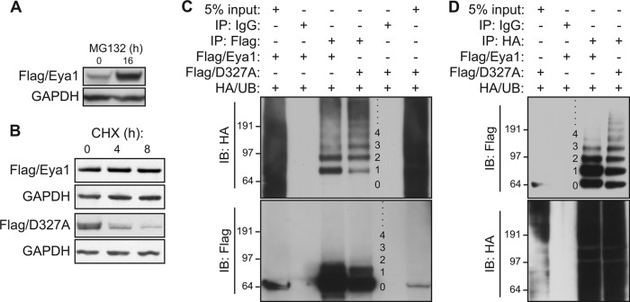
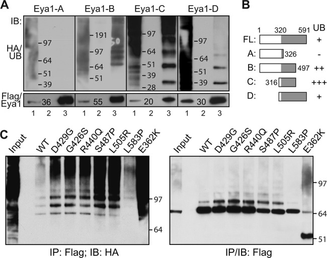
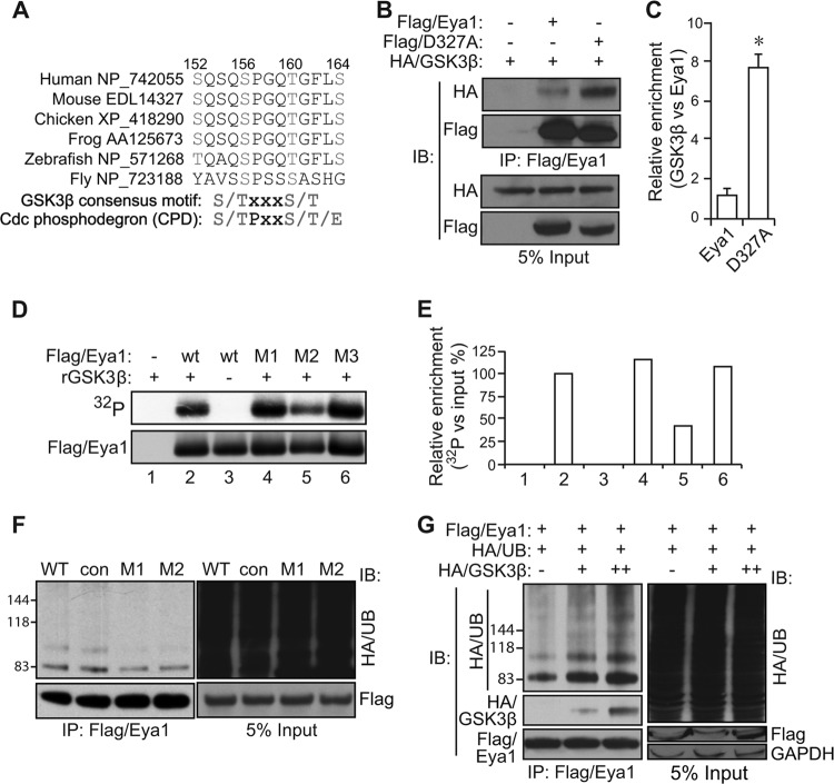
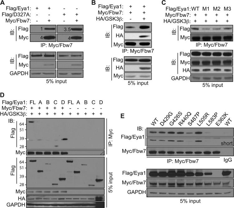
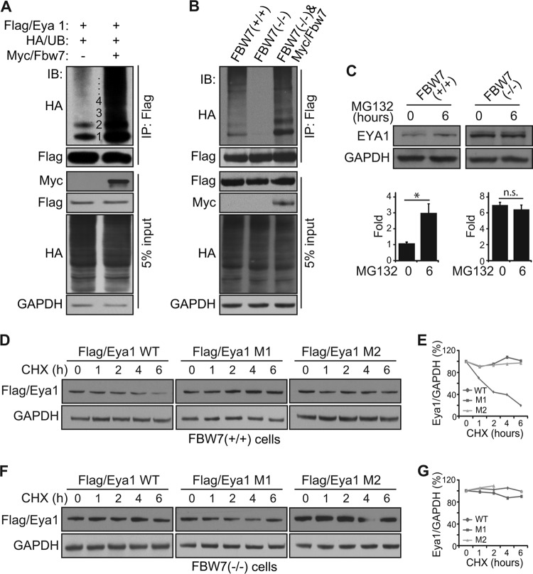
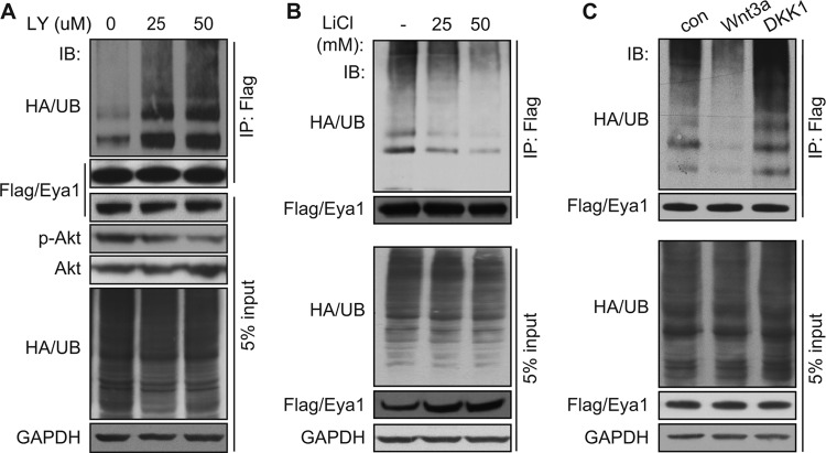
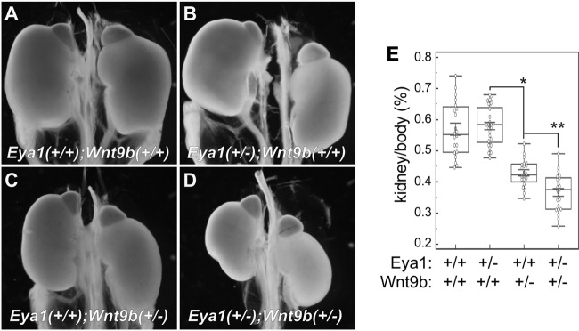
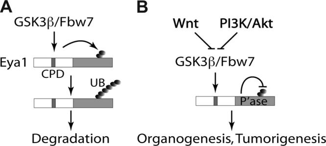
Haploinsufficiency of Eya1 causes the branchio-oto-renal (BOR) syndrome, and abnormally high levels of Eya1 are linked to breast cancer progression and poor prognosis. Therefore, regulation of Eya1 activity is key to its tissue-specific functions and oncogenic activities. Here, we show that Eya1 is posttranslationally modified by ubiquitin and that its ubiquitination level is self-limited to prevent premature degradation. Eya1 has an evolutionarily conserved CDC4 phosphodegron (CPD) signal, a target site of glycogen synthase kinase 3 (GSK3) kinase and Fbw7 ubiquitin ligase, which is required for Eya1 ubiquitination. Genetic deletion of Fbw7 and pharmacological inhibition of GSK3 significantly decrease Eya1 ubiquitination. Conversely, activation of the phosphatidylinositol 3-kinase (PI3K)/Akt and the canonical Wnt signal suppresses Eya1 ubiquitination. Compound Eya1(+/-); Wnt9b(+/-) mutants exhibit an increased penetrance of renal defect, indicating that they function in the same genetic pathway in vivo. Together, these findings reveal that the canonical Wnt and PI3K/Akt signal pathways restrain the GSK3/Fbw7-dependent Eya1 ubiquitination, and they further suggest that dysregulation of this novel axis contributes to tumorigenesis.


Figures

FIG 1
FIG 1
Eya1 is posttranslationally modified by ubiquitin. (A and B) A proteasome inhibitor, MG132, increases the Eya1 protein level. HEK293 cells that were stably transfected with a Flag-Eya1 expression vector were treated with MG132 (12.5 μM). The Flag/Eya1 protein level in total cell lysates was determined using a Flag-specific antibody after 16 h of MG132 treatment (A) or during a time course (B). GAPDH was used as a protein loading control. DMSO mock treatment was considered the starting point (0 h). (C and D) Eya1 is ubiquitinated in HEK293 cells. Flag/Eya1, Flag/D327A, and HA-tagged ubiquitin (HA/UB) were transiently coexpressed in HEK293 cells. An immunoprecipitation/immunoblotting (IP/IB) assay was done using the indicated antibodies or control IgG at 48 h after transfection. Numbers on the side of each panel indicate the ubiquitin chain length.
FIG 2
FIG 2
The conserved Eya tyrosine phosphatase domain is ubiquitinated. (A and B) The C terminus of Eya1 is ubiquitinated. Flag-tagged Eya1 truncated fragments (as indicated) and HA/UB were coexpressed in HEK293 cells, and total cell lysates were subjected to an IP/IB assay at 48 h after transfection. Results are summarized in panel B. Lane 1, total cell lysate; lane 2, IP with control IgG; lane 3, IP with a Flag-specific antibody. (C) Ubiquitination patterns of several Flag-tagged Eya1 variants that were found in human branchio-oto-renal (BOR) syndrome. WT, wild type.
FIG 3
FIG 3
GSK3β phosphorylates and enhances the ubiquitination level of Eya1. (A) Sequence alignment of Eya1 proteins reveals a conserved GSK3β consensus motif and the Cdc4 (also known as Fbw7) phosphodegron (CPD) signal sequence. (B and C) GSK3β, wild-type Eya1, and a phosphatase mutant D327A variant were transiently expressed in HEK293 cells, and total cell lysates were used for a co-IP/IB assay. Quantitative results are shown in panel C. *, P < 0.05 (n = 3). (D and E) An autoradiograph of radiolabeled Flag/Eya1 (top panel) and immunoblot (bottom panel) following an in vitro kinase reaction using recombinant GSK3β protein, immunopurified Flag/Eya1, and radiolabeled [γ-32P]ATP is shown in panel D. The relative density of 32P labeling was normalized against the input Flag/Eya1 level and presented in panel E. (F) Mutation of the GSK3β consensus motif/CPD signal sequence reduces the Eya1 ubiquitination level. Flag/Eya1 variants and HA/UB were transiently expressed in HEK293 cells, and the Flag/Eya1 ubiquitination level was determined using the indicated antibodies in an IP/IB assay. (G) GSK3β was coexpressed with Flag/Eya1 and HA/UB to assess its impact on Eya1 ubiquitination using an IP/IB assay. WT, wild type; con, control (SSSEIAS [aa 48 to 54] to AAAEIAS); M1, S152A/S156A; M2, S156A/T160A; M3, T160A/S164A.
FIG 4
FIG 4
Eya1 interacts with Fbw7 ubiquitin ligase. (A and B) Wild-type (Flag/Eya1) and phosphatase mutant (Flag/D327A) Eya1 were coexpressed with Myc/Fbw7 in HEK293 cells with (B) or without (A) HA/GSK3β. Interaction between Eya1 and Fbw7 was examined by the IP/IB assay using the indicated antibodies. The numbers in panel A indicate relative levels. (C) Combinations of Flag/Eya1 variants, Myc/Fbw7, and HA/GSK3β were coexpressed and examined using the indicated antibodies in a co-IP/IB assay. M1, S152A/S156A; M2, S156A/T160A; M3, T160A/S164A. (D and E) Interactions between Fbw7 and Eya1 fragments (D) as well as BOR mutant variants (E) as assessed by an IP/IB assay. Asterisk, nonspecific signal.
FIG 5
FIG 5
Fbw7 regulates endogenous the Eya1 ubiquitination level and half-life. (A and B) Flag/Eya1, HA/UB, and Myc/Fbw7 were coexpressed in HEK293 cells (A) or FBW7 knockout HCT116 cells (B), and the Flag/Eya1 ubiquitination level was analyzed using the indicated antibodies in an IP/IB assay. (C) Endogenous EYA1 protein levels in wild-type and FBW7−/− HCT116 cells. (D to G) Flag/Eya1 and variants were expressed in wild-type (D and E) and FBW7−/− (F and G) HCT116 cells. Cells were treated with cycloheximide (CHX) (20 μg/ml), and the Flag/Eya1 protein level was chased in a period of 6 h and quantified using ImageJ based on signal density and a protein loading control (GAPDH).
FIG 6
FIG 6
The canonical Wnt signal pathway regulates the Eya1 ubiquitination level. (A) Flag/Eya1 ubiquitination levels in transiently transfected cells with an increasing amount of LY294002, a pharmacological inhibitor of the PI3K/Akt signal pathway. (B and C) Activation of the canonical Wnt signal pathway by LiCl (B) and Wnt3a (C) reduces Eya1 ubiquitination. Dkk1 (C), an antagonist of canonical Wnt signal, enhanced Eya1 ubiquitination. Flag/Eya1 and HA/UB-expressing cells were treated with LY294002 (A), LiCl (B), or Wnt or Dkk1 conditioned medium (C), followed by IP/IB analysis using the indicated antibodies.
FIG 7
FIG 7
Wnt9b and Eya1 synergistically regulate kidney growth. (A to D) Wnt9b and Eya1 synergistically regulate kidney growth. Representative kidney images from newborn pups with different combinations of genotypes are shown. (E) Quantitative analysis of kidney/body weight ratio. *, P < 0.005; **, P < 0.001.
FIG 8
FIG 8
A proposed model of Eya1 ubiquitination and regulation. (A) A conserved N-terminal CPD signal facilitates Eya1 ubiquitination at the C-terminal tyrosine phosphatase domain. This is in part mediated by GSK3β kinase and Fbw7 ubiquitin ligase. (B) The canonical Wnt signal, PI3K/Akt signal, and Eya1 intrinsic tyrosine phosphatase activity restrict Eya1 ubiquitination.

References

    1. Bonini NM, Leiserson WM, Benzer S. 1993. The eyes absent gene: genetic control of cell survival and differentiation in the developing Drosophila eye. Cell 72:379–395. 10.1016/0092-8674(93)90115-7 - DOI - PubMed
    1. Bonini NM, Bui QT, Gray-Board GL, Warrick JM. 1997. The Drosophila eyes absent gene directs ectopic eye formation in a pathway conserved between flies and vertebrates. Development 124:4819–4826 - PubMed
    1. Pignoni F, Hu B, Zavitz KH, Xiao J, Garrity PA, Zipursky SL. 1997. The eye-specification proteins So and Eya form a complex and regulate multiple steps in Drosophila eye development. Cell 91:881–891. 10.1016/S0092-8674(00)80480-8 - DOI - PubMed
    1. Xu PX, Adams J, Peters H, Brown MC, Heaney S, Maas R. 1999. Eya1-deficient mice lack ears and kidneys and show abnormal apoptosis of organ primordia. Nat. Genet. 23:113–117. 10.1038/12722 - DOI - PubMed
    1. Li X, Oghi KA, Zhang J, Krones A, Bush KT, Glass CK, Nigam SK, Aggarwal AK, Maas R, Rose DW, Rosenfeld MG. 2003. Eya protein phosphatase activity regulates Six1-Dach-Eya transcriptional effects in mammalian organogenesis. Nature 426:247–254. 10.1038/nature02083 - DOI - PubMed

Publication types

MeSH terms

LinkOut - more resources