Review
. 2015 Jan;73(1):376-89.
doi: 10.1002/mrm.25263. Epub 2014 Apr 21.

Biodegradable, polymer encapsulated, metal oxide particles for MRI-based cell tracking

Affiliations
Review

Biodegradable, polymer encapsulated, metal oxide particles for MRI-based cell tracking

Erik M Shapiro. Magn Reson Med. 2015 Jan.

Abstract

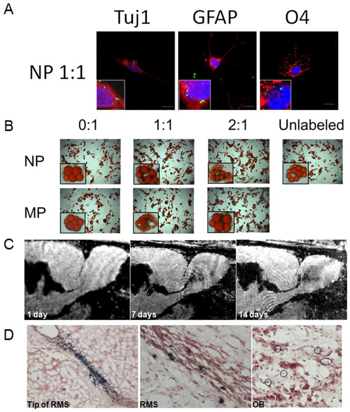
Metallic particles have shaped the use of magnetic resonance imaging (MRI) for molecular and cellular imaging. Although these particles have generally been developed for extracellular residence, either as blood pool contrast agents or targeted contrast agents, the coopted use of these particles for intracellular labeling has grown over the last 20 years. Coincident with this growth has been the development of metal oxide particles specifically intended for intracellular residence, and innovations in the nature of the metallic core. One promising nanoparticle construct for MRI-based cell tracking is polymer encapsulated metal oxide nanoparticles. Rather than a polymer coated metal oxide nanocrystal of the core: shell type, polymer encapsulated metal oxide nanoparticles cluster many nanocrystals within a polymer matrix. This nanoparticle composite more efficiently packages inorganic nanocrystals, affording the ability to label cells with more inorganic material. Further, for magnetic nanocrystals, the clustering of multiple magnetic nanocrystals within a single nanoparticle enhances r2 and r2* relaxivity. Methods for fabricating polymer encapsulated metal oxide nanoparticles are facile, yielding both varied compositions and synthetic approaches. This review presents a brief history into the use of metal oxide particles for MRI-based cell tracking and details the development and use of biodegradable, polymer encapsulated, metal oxide nanoparticles and microparticles for MRI-based cell tracking.

Keywords: MRI; particles; stem cells; iron oxide; polymer.


Figures

Figure 1
Figure 1
Schematic illustration of fabrication scheme for PLGA encapsulated iron oxide nanocrystals. Iron oxide nanocrystals (black circles), along with PLGA, are dissolved in dichloromethane. This oil phase is added to the aqueous phase, comprised of 0–5% PVA. For nanoparticles, an oil-in-water emulsion is formed by tip sonication; microparticles are formed by homogenization. Mechanical stirring for three hours allows dichloromethane to evaporate, generating hardened PLGA nanoparticles encapsulating iron oxide nanocrystals.
Figure 2
Figure 2
Left 2: SEM and Right 2: TEM micrographs of interferon-loaded magnetic (a) PLGA microspheres and (b) PLA microspheres. Adapted with permission from: Zhou S, Sun J, Sun L, Dai Y, Liu L, Li X, Wang J, Weng J, Jia W, Zhang Z. Preparation and characterization of interferon-loaded magnetic biodegradable microspheres. J Biomed Mater Res B Appl Biomater 2008;87(1):189–196.
Figure 3
Figure 3
Scanning electron microscopic micrographs of A) PLGA encapsulated iron oxide nanoparticles and B) microparticles. Insets show transmission electron microscopic images showing inner distribution of iron oxide nanocrystals. Adapted from: Nkansah MK, Thakral D, Shapiro EM. Magnetic poly(lactide-co-glycolide) and cellulose particles for MRI-based cell tracking. Magn Reson Med 2011;65(6):1776–1785.
Figure 4
Figure 4
Scanning electron micrographs of magnetic PLGA particles made with 2 concentrations of magnetite — first column is 0:1 magnetite/PLGA, second column is 1:1 magnetite/PLGA and third column is 2:1 magnetite/PLGA. Top row is nanoparticles and bottom row is microparticles. Note the regular, spherical appearance of all fabricated particles. Scale bar is 1 μm for NPs and 10 μm for MPs. Adapted from: Granot D, Nkansah MK, Bennewitz MF, Tang KS, Markakis EA, Shapiro EM. Clinically viable magnetic poly(lactide-co-glycolide) particles for MRI-based cell tracking. Magn Reson Med 2014;71(3):1238–1250.
Figure 5
Figure 5
Subcellular tracking of nanovaccine carriers by confocal imaging and TEM. (A) Confocal laser scanning microscopy of PLGA harboring iron oxide nanocrystals and fluorescently labeled antigen. Nanoparticles traffic to endosomes/lysosomes as evidenced by positive costaining of EEA1 and LAMP1, stain for the early endosomal marker and the lysosomal marker, respectively, with fluorescence from the nanoparticles. (B) TEM of nanoparticles in subcellular organelles. Part of the nanoparticles was found in the endocytic vesicles (1). A proportion of the SPIO was already released from nanoparticles and associated with endosomal or lysosomal membranes (2). Some nanoparticles also appeared to be localized close to, or within, the vesicle membrane at the endosomal/cytosolic interface (3); other nanoparticles were localized within the cytoplasm (4). Adapted with permission from: Cruz LJ, Tacken PJ, Bonetto F, Buschow SI, Croes HJ, Wijers M, de Vries IJ, Figdor CG. Multimodal imaging of nanovaccine carriers targeted to human dendritic cells. Mol Pharm 2011;8(2):520–531.
Figure 6
Figure 6
A) Confocal fluorescence microscopy analysis of neural stem cell differentiation following labeling by PLGA encapsulated iron oxide nanoparticles. Scale bar is 10 microns. Insets show digital expansions of cells highlighting intracellular presence of particles. Column headers indicate particle type and antigen stained for. B) Light microscopy of mesenchymal stem cell differentiation down adipogenic lineage following labeling with PLGA encapsulated iron oxide nanoparticles (NP) or microparticles (MP). Row and column headers indicate particle formulation. Panels d and l) are differentiation of unlabeled cells. Insets are optical zoomed images of individual adipocytes containing particles, or no particles for unlabeled cells. C) In vivo MRI of endogenous neuroblast migration. MRI montage of same animal at level of SVZ – RMS – OB injected with PLGA encapsulated iron oxide microparticles. Days of MRI are given in panels. D) Prussian blue staining for iron in SVZ, RMS, and OB showing presence of iron specifically and throughout the migratory pathway. Adapted from: Granot D, Nkansah MK, Bennewitz MF, Tang KS, Markakis EA, Shapiro EM. Clinically viable magnetic poly(lactide-co-glycolide) particles for MRI-based cell tracking. Magn Reson Med 2014;71(3):1238–1250.
Figure 7
Figure 7
Dissolution profile of (a) magnetic PLGA nano- and microparticles and (b) magnetic cellulose and cellulose acetate nanoparticles, in citrate buffer (pH 5.5) over 100 days. Adapted from: Nkansah MK, Thakral D, Shapiro EM. Magnetic poly(lactide-co-glycolide) and cellulose particles for MRI-based cell tracking. Magn Reson Med 2011;65(6):1776–1785.
Figure 8
Figure 8
(a) T2* MR images of rat livers and (b) Prussian blue staining for PLGA encapsulated iron oxide nanoparticles in the liver before treatment and at the indicated times after intravascular injection. Adapted with permission from: Lee PW, Hsu SH, Wang JJ, Tsai JS, Lin KJ, Wey SP, Chen FR, Lai CH, Yen TC, Sung HW. The characteristics, biodistribution, magnetic resonance imaging and biodegradability of superparamagnetic core-shell nanoparticles. Biomaterials 2010;31(6):1316–1324.
Figure 9
Figure 9
A) In vivo biodegradation over 12 weeks following intravenous injection of Feridex, inert MPIOs and PLGA encapsulated iron oxide nanoparticles. Data are mean +/− SEM. MRI of mice livers are at TE = 6 ms for mice injected as indicated, both at 1 day and 12 weeks following injection. B) Superposition of in vivo nanoparticle degradation time line over in vitro dissolution and in vitro degradation time lines. Adapted from: Nkansah MK, Thakral D, Shapiro EM. Magnetic poly(lactide-co-glycolide) and cellulose particles for MRI-based cell tracking. Magn Reson Med 2011;65(6):1776–1785. and Granot D, Nkansah MK, Bennewitz MF, Tang KS, Markakis EA, Shapiro EM. Clinically viable magnetic poly(lactide-co-glycolide) particles for MRI-based cell tracking. Magn Reson Med 2014;71(3):1238–1250.
Figure 10
Figure 10
Scanning electron microscopic micrographs of cellulose encapsulate iron oxide nanoparticles. Inset shows transmission electron microscopic images showing inner distribution of iron oxide nanocrystals. Adapted from: Nkansah MK, Thakral D, Shapiro EM. Magnetic poly(lactide-co-glycolide) and cellulose particles for MRI-based cell tracking. Magn Reson Med 2011;65(6):1776–1785.
Figure 11
Figure 11
A) Transmission electron microscopy of manganese oxide nanocrystals, B) scanning electron microscopy of PLGA encapsulated manganese oxide nanoparticles, C) Transmission electron microscopy of PLGA encapsulated manganese oxide nanoparticles; inset shows expansion of a single nanoparticle, D) fluorescence and optical microscopy of cell labeled with fluorescently labeled PLGA encapsulated manganese oxide nanoparticles. Adapted with permission from: Bennewitz MF, Lobo TL, Nkansah MK, Ulas G, Brudvig GW, Shapiro EM. Biocompatible and pH-Sensitive PLGA Encapsulated MnO Nanocrystals for Molecular and Cellular MRI. ACS Nano 2011;5(5):3438–3446.

References

    1. Bloch F, Hansen WW, Packard M. The Nuclear Induction Experiment. Physical Review. 1946;70(7–8):474–485.
    1. Bloembergen N, Purcell EM, Pound RV. Relaxation Effects in Nuclear Magnetic Resonance Absorption. Physical Review. 1948;73(7):679–712.
    1. Lauterbur PC. Image Formation by Induced Local Interactions - Examples Employing Nuclear Magnetic-Resonance. Nature. 1973;242(5394):190–191. - PubMed
    1. Lauterbur PC. Progress in n.m.r zeugmatography imaging. Philos Trans R Soc Lond B Biol Sci. 1980;289(1037):483–487. - PubMed
    1. Ohgushi M, Nagayama K, Wada A. Dextran-magnetite: a new relaxation reagent and its application to T2 measurements in gel systems. J Magn Reson. 1978;29:599–601.

Publication types