. 2014 Apr;6(4):264-80.
doi: 10.18632/aging.100651.

Prelamin A accumulation and stress conditions induce impaired Oct-1 activity and autophagy in prematurely aged human mesenchymal stem cell

Affiliations

Prelamin A accumulation and stress conditions induce impaired Oct-1 activity and autophagy in prematurely aged human mesenchymal stem cell

Arantza Infante et al. Aging (Albany NY). 2014 Apr.

Abstract

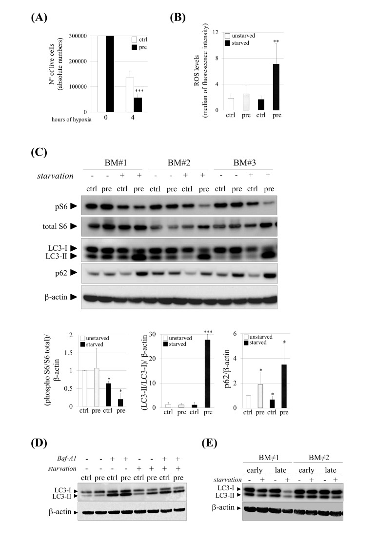
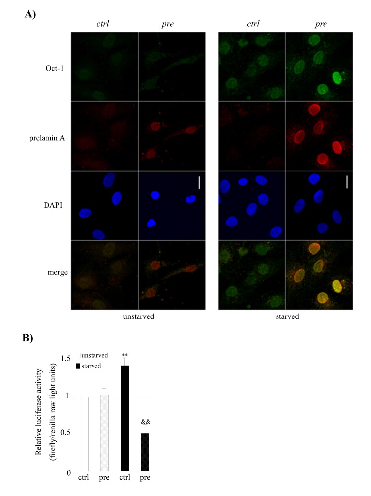
Aging, a time-dependent functional decline of biological processes, is the primary risk factor in developing diseases such as cancer, cardiovascular or degenerative diseases. There is a real need to understand the human aging process in order to increase the length of disease-free life, also known as "health span". Accumulation of progerin and prelamin A are the hallmark of a group of premature aging diseases but have also been found during normal cellular aging strongly suggesting similar mechanisms between healthy aging and LMNA-linked progeroid syndromes. How this toxic accumulation contributes to aging (physiological or pathological) remains unclear. Since affected tissues in age-associated disorders and in pathological aging are mainly of mesenchymal origin we propose a model of human aging based on mesenchymal stem cells (hMSCs) which accumulate prelamin A. We demonstrate that prelamin A-accumulating hMSCs have a premature aging phenotype which affects their functional competence in vivo. The combination of prelamin A accumulation and stress conditions enhance the aging phenotype by dysregulating the activity of the octamer binding protein Oct-1This experimental model has been fundamental to identify a new role for Oct-1 in hMSCs aging.


Conflict of interest statement

No conflict of interest could be disclosed for any author.

Figures

Figure 1
Figure 1
Prelamin A accumulation induces shorter telomeres and increased DNA damage signalling in hMSCs. (A) Telomere length and (B) the percentage of short telomeres were analyzed by HT-Q-FISH. At least 17,000 nuclei were analyzed per sample. Bars are average +/- standard deviation of 3 independent donors. (C) Representative immunofluorescence micrograph and (D) quantification of γ-H2AX foci. Blue: DAPI, red: γ-H2AX. Scale bar: 10 μm. At least 100 nuclei were analyzed per sample. χ2 was used for statistical significance. *** p<0.001 when compared versus ctrl-hMSCs. &&& p< 0.001 when compared versus ctrl-hMSCs starved. (pre): prelamin A-accumulating hMSCs, (ctrl): control-hMSCs.
Figure 2
Figure 2
Increased susceptibility and impaired autophagy under stress conditions in pre-hMSCs. (A) Number of live cells after submitting hMSCs to hypoxia. (B) Reactive Oxygen Species (ROS) measurement of hMSCs cultured under basal (unstarved) or starvation conditions (starved). (C) Western blot of indicated proteins in ctrl and pre-hMSCs cultured under basal or serum starvation conditions. β-actin was used as loading control. hMSCs from three independent bone marrow donors (BM) where analyzed together. Densitometry for each immunoblot is provided. (D) Determination of autophagic flux of hMSCs treated with Bafilomycin (Baf-A1) 0.1 μM during 9 hours. LC3-I and LC3-II expression levels are shown and β-actin was used as loading control. (E) Western blot of LC3-I and LC3-II expression levels in replicative senescent hMSCs (Early: early-passage hMSCs; Late: late-passage hMSCs), Bars are average +/- standard deviation of 3 independent donors. Differences marked with asterisks are significantly different from control unstarved (panel A and D) or control starved cells (panel B) *** p<0.001, ** p<0.01, *p<0.05. (pre):prelamin A-accumulating hMSCs, (ctrl):control-hMSCs.
Figure 3
Figure 3
Dys-functional activity of hMSCs which accumulate prelamin A in vivo. (A) Perfusion rate in mice after transplanting hMSCs. ** p<0.01, * p<0.05. (B) Laser-Doppler quantification of hind limb blood flow over time after hMSCs transplantation (Post-isch = blood flow measured immediately after ischemia). ** p<0.01 and * p<0.05 when compared versus vehicle, & p<0.05 when compared versus control hMSCs. (C) Representative photographs of animal hind limbs (left) and PET images (right) 14 days after ischemia and hMSCs transplantation. Bars are average +/- standard deviation (panel A) or standard error of the mean (panel B). (pre): prelamin A-accumulating hMSCs, (ctrl): control-hMSCs.
Figure 4
Figure 4
hMSCs show an altered transcriptomic profile and phenotype of senescence under prelamin A accumulation and serum starvation conditions. (A) Q-RT-PCR validation for a subset of genes grouped in oxidation-reduction and response to oxidative stress categories and (B) for genes known to be up-regulated in senescent hMSCs. The gene expression ratio for pre-hMSCs versus control-hMSCs under starved conditions is shown in A and in the case of B it has been included too the comparison of pre-hMSCs versus ctrl-hMSCs ratio under unstarved conditions. For gene expression normalization GAPDH was used. Bars are mean +/- standard error mean of 3 independent donors. ** p< 0.01, * p<0.05. (C) Senescence-associated morphological changes in pre-hMSCs under serum starvation conditions. Bright field images (scale bar: 100 μm) and confocal immunofluorescence images (scale bar: 20 μm) are shown. Red: beta-tubulin, blue: DAPI. (D) SA β-gal quantification in hMSCs under basal (unstarved) or serum starvation conditions (starved). Bars are average +/- standard deviation. ** p< 0.01, * p<0.05. (E) DiRE analysis of genes found to be dys-regulated in pre-hMSCs. The graph shows the top 10 candidate transcription factors ranked by importance. (pre): prelamin A-accumulating hMSCs, (ctrl): control-hMSCs.
Figure 5
Figure 5
Prelamin A accumulation and serum starvation conditions induce the overexpression of Oct-1 and its impaired activity in hMSCs. (A) Representative confocal immunofluorescence staining showing the expression of Oct-1 and prelamin A in hMSCs under basal (unstarved) or serum starvation conditions (starved). Scale bar: 20 μm. (B) Luciferase reporter assays of Oct-1 transcription factor activity in hMSCs cultured under under basal (unstarved) or serum starvation conditions (starved). Bars are average +/- standard deviation of 3 independent donors. Differences that are significant are marked as follows: ** p<0.01 when compared to control-hMSCs, && p<0.01 when compared to control-hMSCs starved.
Figure 6
Figure 6
Induction of senescence and autophagy in Oct-1 silenced hMSCs. (A) Western blot showing decreased Oct-1 expression after Oct-1 shRNA plasmids transduction in hMSCs, NT: Non Targeting shRNA. Numbers show densitometric quantification for Oct-1 expression. (B) Cell survival after Oct-1 silencing in hMSCs cultured under basal (unstarved) or serum starvation conditions (starved). (C) Representative SA β-gal staining (left) and quantification (right) in hMSCs transduced with NT or Oct-1 shRNA. Scale bar: 100 μm. (D) Western blot of indicated proteins from hMSCs silenced for Oct-1 or not (NT). β-actin was used as loading control. Densitometry for each immunoblot is provided. For these experiments hMSCs from two independent donors were used(BM≠1 and BM≠2). Bars are mean +/- standard deviation

References

    1. Scaffidi P, Misteli T. Lamin A-dependent nuclear defects in human aging. Science. 2006;312:1059–1063. - PMC - PubMed
    1. Ragnauth CD, Warren DT, Liu Y, McNair R, Tajsic T, Figg N, Shroff R, Skepper J, Shanahan CM. Prelamin A acts to accelerate smooth muscle cell senescence and is a novel biomarker of human vascular aging. Circulation. 2010;121:2200–2210. - PubMed
    1. De Sandre-Giovannoli A, Bernard R, Cau P, Navarro C, Amiel J, Boccaccio I, Lyonnet S, Stewart CL, Munnich A, Le Merrer M, Lévy N. Lamin a truncation in Hutchinson-Gilford progeria. Science. 2003;300:2055. - PubMed
    1. Capanni C, Mattioli E, Columbaro M, Lucarelli E, Parnaik VK, Novelli G, Wehnert M, Cenni V, Maraldi NM, Squarzoni S, Lattanzi G. Altered pre-lamin A processing is a common mechanism leading to lipodystrophy. Hum Mol Genet. 2005;14:1489–1502. - PubMed
    1. Shackleton S, Smallwood DT, Clayton P, Wilson LC, Agarwal AK, Garg A, Trembath RC. Compound heterozygous ZMPSTE24 mutations reduce prelamin A processing and result in a severe progeroid phenotype. J Med Genet. 2005;42:e36. - PMC - PubMed

Publication types

MeSH terms