. 2014 Jun 6;289(23):16148-63.
doi: 10.1074/jbc.M114.549980. Epub 2014 Apr 21.

Rabies virus envelope glycoprotein targets lentiviral vectors to the axonal retrograde pathway in motor neurons

Affiliations

Rabies virus envelope glycoprotein targets lentiviral vectors to the axonal retrograde pathway in motor neurons

James N Hislop et al. J Biol Chem. .

Abstract

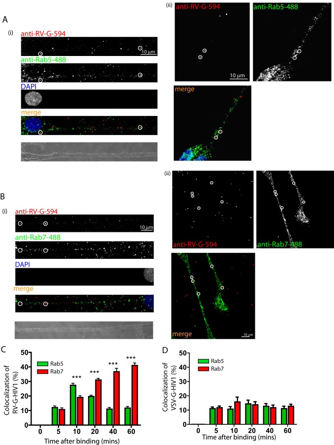
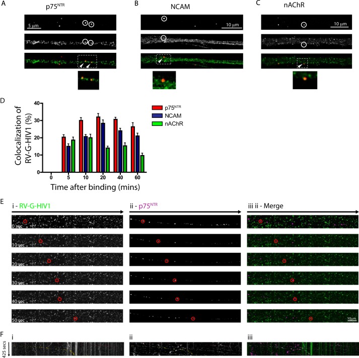
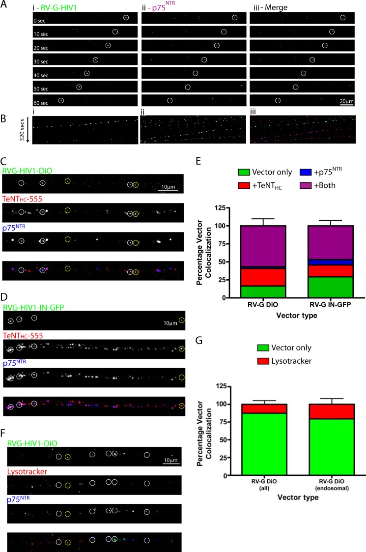
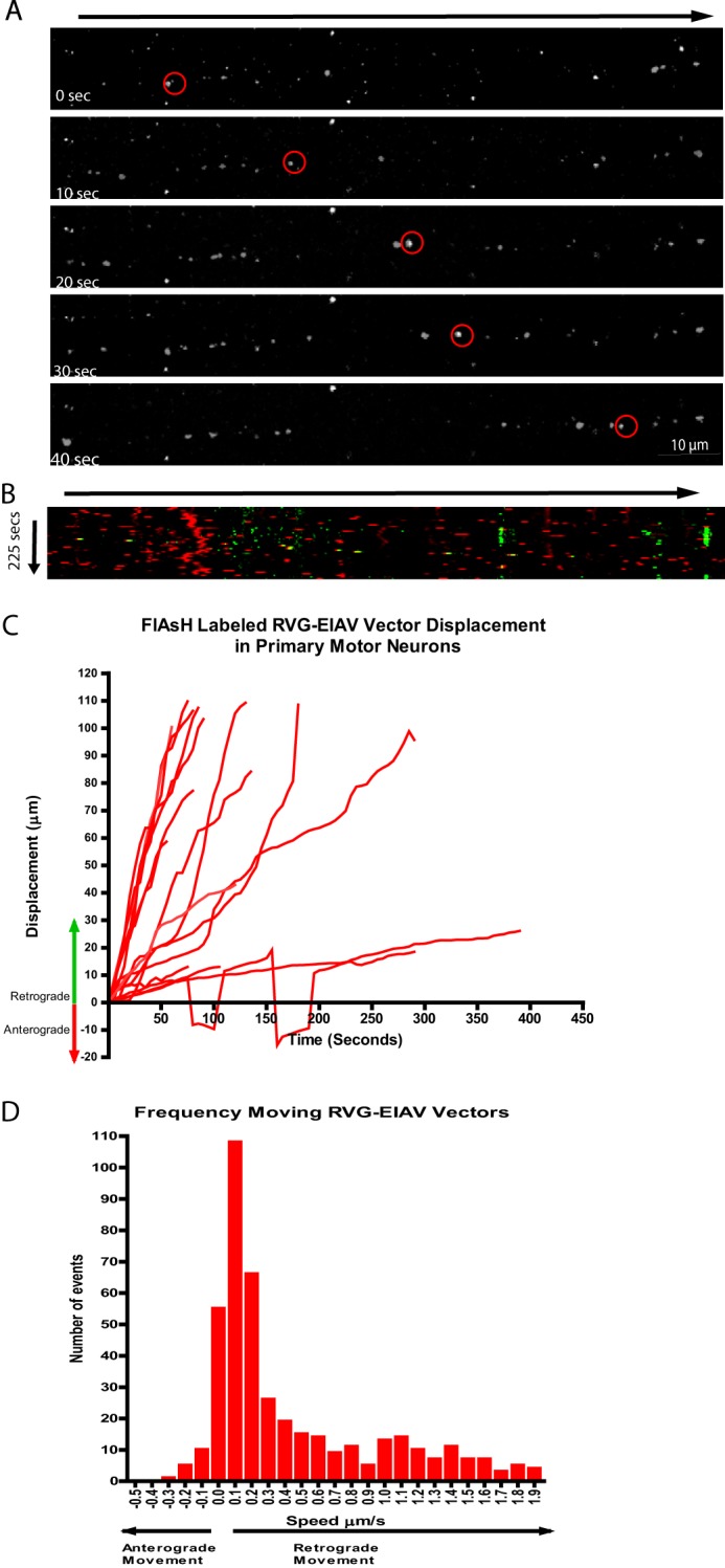
Rabies pseudotyped lentiviral vectors have great potential in gene therapy, not least because of their ability to transduce neurons following their distal axonal application. However, very little is known about the molecular processes that underlie their retrograde transport and cell transduction. Using multiple labeling techniques and confocal microscopy, we demonstrated that pseudotyping with rabies virus envelope glycoprotein (RV-G) enabled the axonal retrograde transport of two distinct subtypes of lentiviral vector in motor neuron cultures. Analysis of this process revealed that these vectors trafficked through Rab5-positive endosomes and accumulated within a non-acidic Rab7 compartment. RV-G pseudotyped vectors were co-transported with both the tetanus neurotoxin-binding fragment and the membrane proteins thought to mediate rabies virus endocytosis (neural cell adhesion molecule, nicotinic acetylcholine receptor, and p75 neurotrophin receptor), thus demonstrating that pseudotyping with RV-G targets lentiviral vectors for transport along the same pathway exploited by several toxins and viruses. Using motor neurons cultured in compartmentalized chambers, we demonstrated that axonal retrograde transport of these vectors was rapid and efficient; however, it was not able to transduce the targeted neurons efficiently, suggesting that impairment in processes occurring after arrival of the viral vector in the soma is responsible for the low transduction efficiency seen in vivo, which suggests a novel area for improvement of gene therapy vectors.

Keywords: Axon; Endosome; Glycoprotein; Lentivirus; Rab Proteins; Trafficking.


Figures

FIGURE 1.
FIGURE 1.
Insertion of tetracysteine tag in the matrix protein allows normal processing of Gag and production of high titer lentiviral vectors. Diagrammatic representation of HIV-1 Gag (A) and EIAV Gag (B) and the positions of insertion of the tetracysteine tag. The tags were inserted in regions of variability as deduced from multiple sequence alignment of all primate lentivirus gag. CA, capsid; CA-N, capsid N terminal; NC, nucleocapsid; NC-N, nucleocapsid N terminal. C and E, vector-associated Gag was detected by Western blot using anti-HIV-1 Gag and -EIAV Gag antibodies showing the effect of the tetracysteine tag on the efficiency of Gag processing by proteases in HIV-1 (C) and EIAV (E). D and F, vector samples were used to transduce 293T cells. The transduced cells were harvested and fixed 72 h after the addition of vectors, and transduction levels were measured by FACS to generate biological titers of the different HIV-1 (D) and EIAV (F) tagged constructs. TU, transducing units.
FIGURE 2.
FIGURE 2.
Trafficking of RV-G pseudotyped lentiviral vectors in NSC-34 cells. Differentiated NSC-34 cells were incubated with RV-G pseudotyped HIV-1 particles labeled with Vybrant DiD for 10 min (m.o.i. 10) prior to imaging. A, representative time series from supplemental Movie 1 showing retrograde trafficking particles (red and yellow circles; cell body is on the left). Asterisks indicate static particles. B, the same time series represented as a kymograph. C, single vector analysis showing the displacement from the origin of each particle analyzed (red, retrograde trafficking particles (38 tracks); blue, anterograde trafficking particles (five tracks)). D, speed distribution analysis of individual particle movements (speed between frames) (red, retrograde tracks (1386 individual events); blue, anterograde tracks (141 individual events)).
FIGURE 3.
FIGURE 3.
Trafficking of RV-G pseudotyped lentiviral vectors in primary motor neurons. A and B, primary motor neurons (DIV 3) were incubated with FlAsH-labeled RV-G pseudotyped HIV-1 vectors (green) for 30 min on ice (panels i–iii) before placing at 37 °C for 45 min (panels iv–vi), fixing under non-permeabilizing conditions, and labeling with anti-RV-G (red). Surface vectors are labeled with both markers and appear yellow in the merged images (panels iii and vi), whereas internal vectors are inaccessible to anti RV-G and appear green. These data were quantified and expressed as a percentage of total vectors counted (B; n = 3, 30 cells, 550 particles). Error bars represent S.D. C–F, motor neurons (DIV 3) were incubated with RV-G pseudotyped HIV-1 vectors (Vybrant DiO- or DiD-labeled) for 30 min (m.o.i. 10) before confocal imaging. C, representative image series from supplemental Movie 3. Asterisks indicate static particles. D, kymograph of the same image series. Individual track analysis (E) and speed distribution analysis (F) of moving particles (>20 μm in one direction, 20 tracks, 458 individual events) are shown.
FIGURE 4.
FIGURE 4.
RV-G pseudotyped HIV-1 vectors enter a sequential Rab5 and Rab7 endocytic pathway. Primary motor neurons (DIV3) were incubated with RV-G pseudotyped HIV-1 vectors (A and B) for 10 min and then incubated at 37 °C for the indicated times before fixing and staining with anti-RV-G (A and B, red) and either anti-Rab5 (A, green) or anti-Rab7 (B, green). A and B show representative examples of colocalization (circles) after 10 (A, panels i and ii) or 40 min (B, panels i and ii). Vector colocalization was then quantified and expressed as a percentage of total vectors (C; n = 3, 15–20 vectors per time point; ***, p > 0.001, two-way analysis of variance). Similar experiments were performed with VSV-G pseudotyped HIV-1 vectors, and the data were quantified using anti-VSV-G (D). Error bars represent S.D.
FIGURE 5.
FIGURE 5.
Endocytosed RV-G pseudotyped lentiviral vectors colocalize with the cognate receptors for rabies virus and cotraffic within the same compartment. A–D, primary motor neurons (DIV3) were incubated with RV-G pseudotyped HIV-1 vectors (A–C) for 10 min and then incubated at 37 °C for the indicated times before fixing and staining. Colocalization with the indicated receptors was quantified and then expressed as a percentage of total vectors detected (D; n = 3, 15–20 particles per time point). Error bars represent S.D. Shown are representative images of anti-RV-G (A–C, red) and either anti-p75NTR (A, green), anti-NCAM (B, green), or anti-nAChR (C, green). The segment denoted by a white rectangle is enlarged below to show colocalization in the axon. E and F, motor neurons (DIV 3) were incubated with RV-G pseudotyped HIV-1 vectors (Vybrant DiO-labeled) and anti-p75NTR-Alexa Fluor 647 for 30 min (m.o.i. 10) before time lapse confocal imaging. E, representative image series of trafficking vector particles from supplemental Movie 5 showing RV-G pseudotyped HIV-1 (panel i; green in merged image in panel iii) and p75NTR (panel ii; magenta in merged image in panel iii). F, kymographs of the same image series.
FIGURE 6.
FIGURE 6.
Retrograde trafficking of RV-G pseudotyped lentiviral vectors in compartmented primary motor neuron cultures. A and B, primary motor neurons plated in microfluidic chambers (DIV 7) were incubated with RV-G pseudotyped HIV-1 vectors (Vybrant DiO-labeled) and anti-p75NTR-Alexa Fluor 647 for 60 min in the axonal chamber before time lapse confocal imaging of a region of axon >200 μm or more from the axonal compartment. A, representative image series of trafficking vector particles from supplemental Movie 6 showing RV-G pseudotyped HIV-1 (panel i; green in merged image in panel iii) and p75NTR (panel ii; magenta in merged image in panel iii). B, kymographs of the same image series. C–E, motor neurons (DIV 7) grown in microfluidic chambers were incubated with either DiO-labeled RV-G pseudotyped HIV-1 (C) or RV-G-HIV1-IN-eGFP (D) with TeNT-Alexa Fluor 555 and anti-p75NTR-Alexa Fluor 647 for 2 h at 37 °C before fixing and imaging by confocal microscopy. Shown are representative maximum projections of Z-stack images (C and D) where white circles show examples of colocalization of vectors with both markers and yellow circles show examples of colocalization with just TeNT Hc-Alexa Fluor 555. E shows quantification of colocalization (n = 3, 172 RV-G pseudotyped DiO-labeled vector particles, 101 RV-G pseudotyped IN-eGFP vector particles). Error bars represent S.D. F and G, motor neurons (DIV 7) were grown in microfluidic chambers and incubated with LysoTracker DND (10 nm) for 2 h followed by DiO-labeled RV-G pseudotyped HIV-1 and anti-p75NTR-Alexa Fluor 647 for 2 h at 37 °C before fixing and imaging by confocal microscopy. Shown is a representative maximum projection of Z-stack images (F) where white circles show examples of vectors with p75NTR alone and yellow circles show examples of colocalization with LysoTracker. G shows quantification of colocalization of all vectors or of vectors within endosomes that are either p75NTR- and/or LysoTracker-positive (n = 3, 127 particles or 78 particles, respectively). Error bars represent S.D.
FIGURE 7.
FIGURE 7.
Pseudotyping with RV-G targets EIAV vectors for retrograde endosomal transport in primary motor neuron cultures. A and B, primary motor neurons were incubated with FlAsH-labeled RV-G pseudotyped EIAV vectors for 30 min with Alexa Fluor 647-labeled anti-p75NTR antibody before confocal imaging. A, representative time series from supplemental Movie 7 showing retrograde trafficking of vector particle (red circles; cell body is on the right). B, kymographs of the same image series (yellow spots). C, single vector track analysis showing the displacement of each moving particle (16 tracks). D, speed distribution analysis of individual particle movements (441 events).

References

    1. Wong L.-F., Goodhead L., Prat C., Mitrophanous K. A., Kingsman S. M., Mazarakis N. D. (2006) Lentivirus-mediated gene transfer to the central nervous system: therapeutic and research applications. Hum. Gene Ther. 17, 1–9 - PubMed
    1. Sakuma T., Barry M. A., Ikeda Y. (2012) Lentiviral vectors: basic to translational. Biochem. J. 443, 603–618 - PubMed
    1. Cronin J., Zhang X.-Y., Reiser J. (2005) Altering the tropism of lentiviral vectors through pseudotyping. Curr. Gene Ther. 5, 387–398 - PMC - PubMed
    1. Mazarakis N. D., Azzouz M., Rohll J. B., Ellard F. M., Wilkes F. J., Olsen A. L., Carter E. E., Barber R. D., Baban D. F., Kingsman S. M., Kingsman A. J., O'Malley K., Mitrophanous K. A. (2001) Rabies virus glycoprotein pseudotyping of lentiviral vectors enables retrograde axonal transport and access to the nervous system after peripheral delivery. Hum. Mol. Genet. 10, 2109–2121 - PubMed
    1. Azzouz M., Le T., Ralph G. S., Walmsley L., Monani U. R., Lee D. C., Wilkes F., Mitrophanous K. A., Kingsman S. M., Burghes A. H., Mazarakis N. D. (2004) Lentivector-mediated SMN replacement in a mouse model of spinal muscular atrophy. J. Clin. Investig. 114, 1726–1731 - PMC - PubMed

Publication types

Substances

LinkOut - more resources