. 2014 Jul;23(7):954-61.
doi: 10.1002/pro.2478. Epub 2014 May 6.

Crystal structure of the Campylobacter jejuni CmeC outer membrane channel

Affiliations

Crystal structure of the Campylobacter jejuni CmeC outer membrane channel

Chih-Chia Su et al. Protein Sci. 2014 Jul.

Abstract

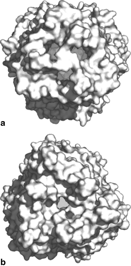
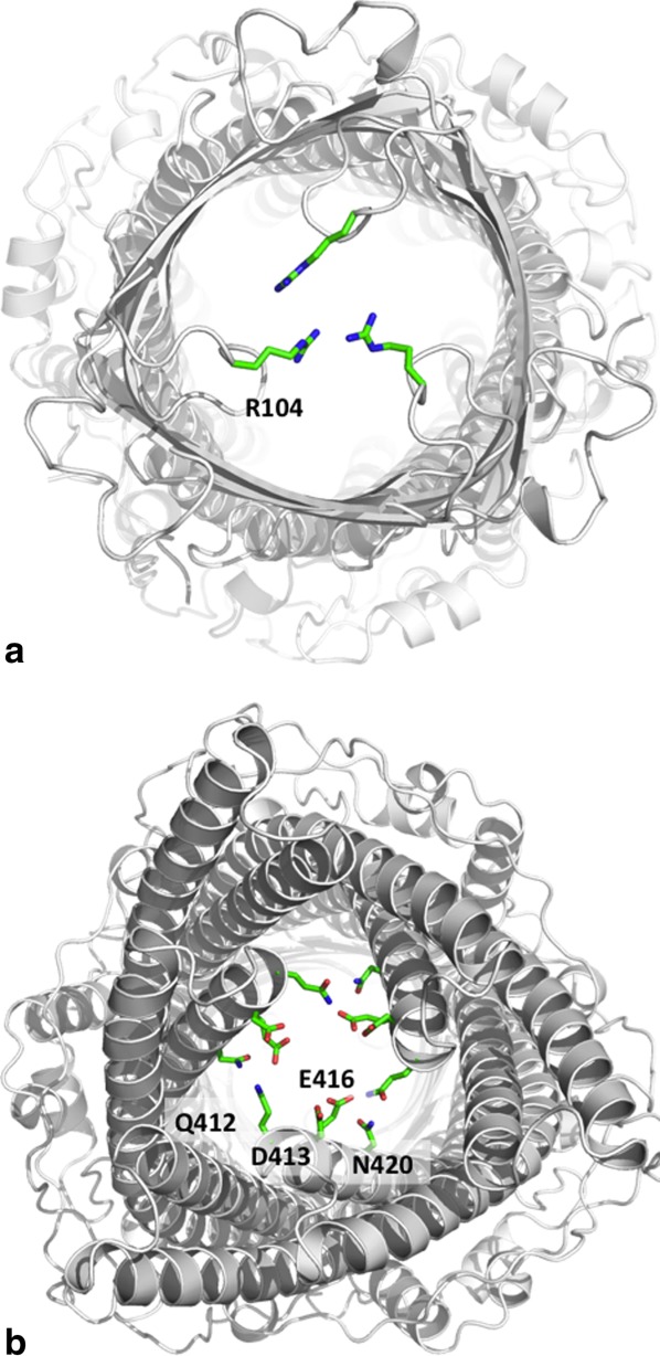
As one of the world's most prevalent enteric pathogens, Campylobacter jejuni is a major causative agent of human enterocolitis and is responsible for more than 400 million cases of diarrhea each year. The impact of this pathogen on children is of particular significance. Campylobacter has developed resistance to many antimicrobial agents via multidrug efflux machinery. The CmeABC tripartite multidrug efflux pump, belonging to the resistance-nodulation-cell division (RND) superfamily, plays a major role in drug resistant phenotypes of C. jejuni. This efflux complex spans the entire cell envelop of C. jejuni and mediates resistance to various antibiotics and toxic compounds. We here report the crystal structure of C. jejuni CmeC, the outer membrane component of the CmeABC tripartite multidrug efflux system. The structure reveals a possible mechanism for substrate export.

Keywords: Campylobacter jejuni; efflux channel; membrane protein; multidrug resistance; resistance-nodulation-cell division.


Figures

Figure 1
Figure 1
Stereo view of the experimental electron density map of the CmeC channel at a resolution of 2.37 Å. (a) Anomalous maps of the 15 selenium sites (contoured at 4 σ). The selenium sites corresponding to the five methionines from each protomer of the CmeC trimer are in orange. The Cα traces of CmeC are in green, magenta, and white, respectively. (b) Representative section of the electron density at the interface between H8 and H9 of the periplasmic domain of CmeC. The electron density (colored white) is contoured at the 1.2 σ level and superimposed with the final refined model (green, carbon; red, oxygen; blue, nitrogen). An interactive view is available in the electronic version of the article.
Figure 2
Figure 2
Structure of the C. jejuni CmeC channel protein. (a) Ribbon diagram of a protomer of CmeC viewed in the membrane plane. The molecule is colored using a rainbow gradient from the N-terminus (blue) to the C-terminus (red). (b) Ribbon diagram of the CmeC trimer viewed in the membrane plane. Each subunit of CmeC is labeled with a different color. The CmeC protomer is acylated (in sticks) through the first N-terminal cysteine residue to anchor onto the outer membrane.
Figure 3
Figure 3
Secondary structural topology of the CmeC monomer. The topology was constructed based on the crystal structure of CmeC. The α-helices and β-strands are colored orange and green, respectively.
Figure 4
Figure 4
Electrostatic surface potentials of CmeC. Surface representations of the (a) outside and (b) inside the CmeC channel colored by charge (red; negative −15 kT/e, blue; positive +15 kT/e).
Figure 5
Figure 5
Surface representations of the trimeric CmeC channel. The views from both the (a) extracellular and (b) periplasmic sides suggest that the CmeC channel is in its closed form.
Figure 6
Figure 6
The interior of the CmeC channel. (a) Extracellular view of the CmeC trimer. The three R104 residues of the trimer are found to interact and block the channel. (b) Periplasmic view of the CmeC trimer. The charged and polar residues Q412, D413, E416, and N420 are found to interact with each other and block this end of the channel.

References

    1. Ruiz-Palacios GM. The health burden of Campylobacter infection and the impact of antimicrobial resistance: Playing chicken. Clin Infect Dis. 2007;44:701–703. - PubMed
    1. van Doorn PA, Ruts L, Jacobs BC. Clinical features, pathogenesis, and treatment of Guillain-Barre syndrome. Lancet Neurol. 2008;7:939–950. - PubMed
    1. Engberg J, Aarestrup FM, Taylor DE, Gerner-Smidt P, Nachamkin I. Quinolone and macrolide resistance in Campylobacter jejuni and C. coli: Resistance mechanisms and trends in human isolates. Emerg Infect Dis. 2001;7:24–34. - PMC - PubMed
    1. Gibreel A, Taylor DE. Macrolide resistance in Campylobacter jejuni and Campylobacter coli. J Antimicrob Chemother. 2006;58:243–255. - PubMed
    1. Luangtongkum T, Jeon B, Han J, Plummer P, Logue CM, Zhang Q. Antibiotic resistance in Campylobacter: Emergence, transmission and persistence. Future Microbiol. 2009;4:189–200. - PMC - PubMed

Publication types

MeSH terms

Associated data

LinkOut - more resources