. 2014 May 6;111(18):E1833-42.
doi: 10.1073/pnas.1320122111. Epub 2014 Apr 21.

Myosin-10 produces its power-stroke in two phases and moves processively along a single actin filament under low load

Affiliations

Myosin-10 produces its power-stroke in two phases and moves processively along a single actin filament under low load

Yasuharu Takagi et al. Proc Natl Acad Sci U S A. .

Abstract

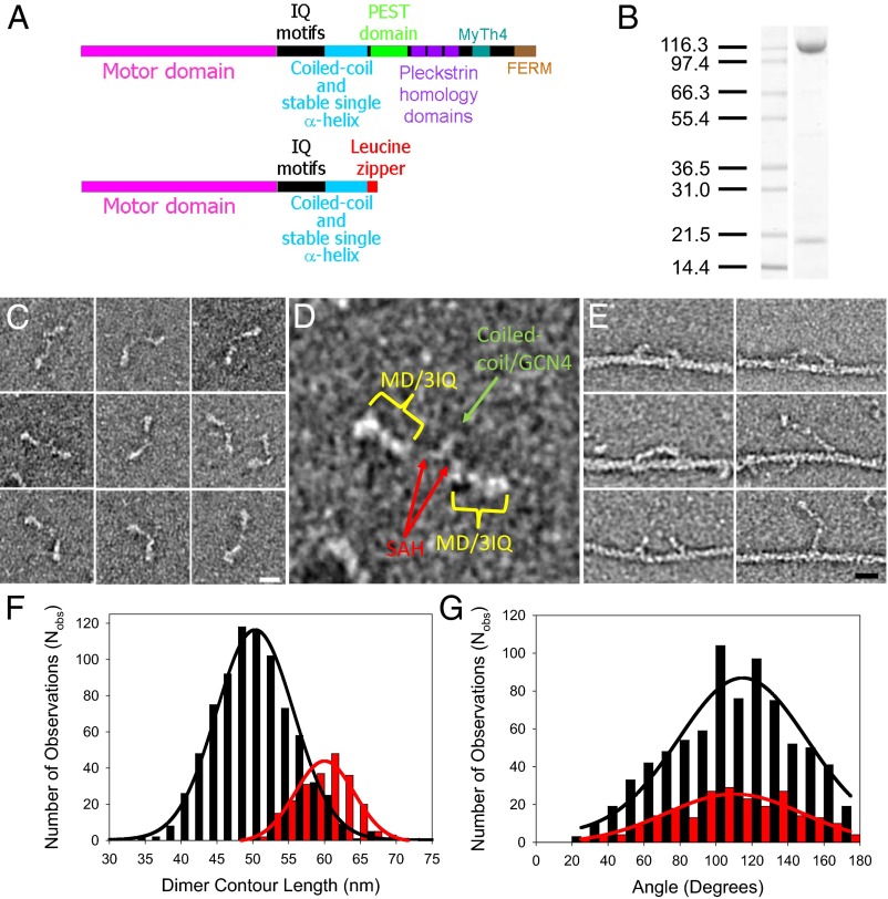
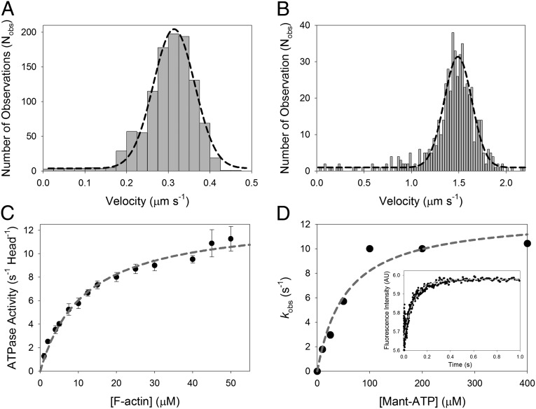
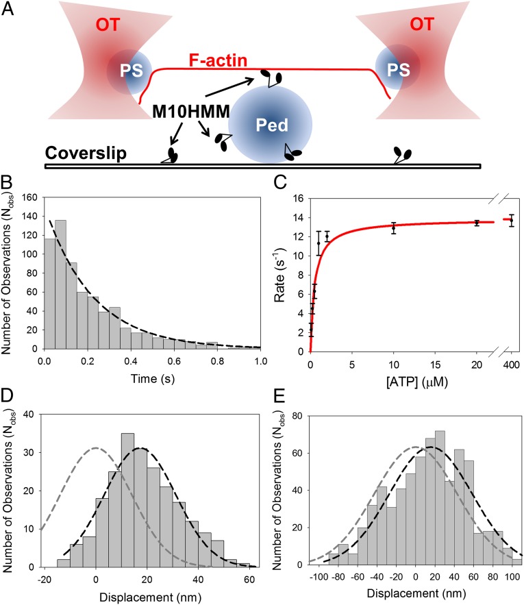
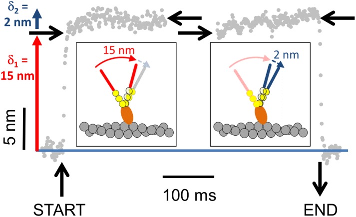
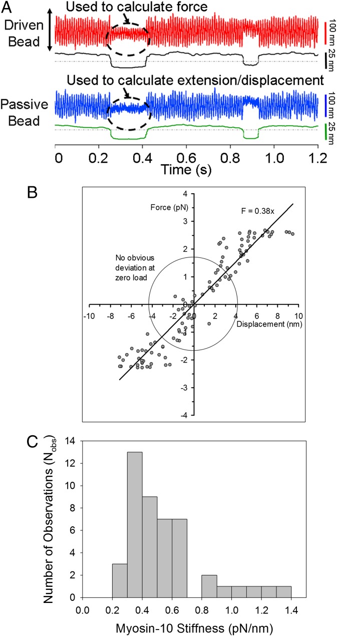
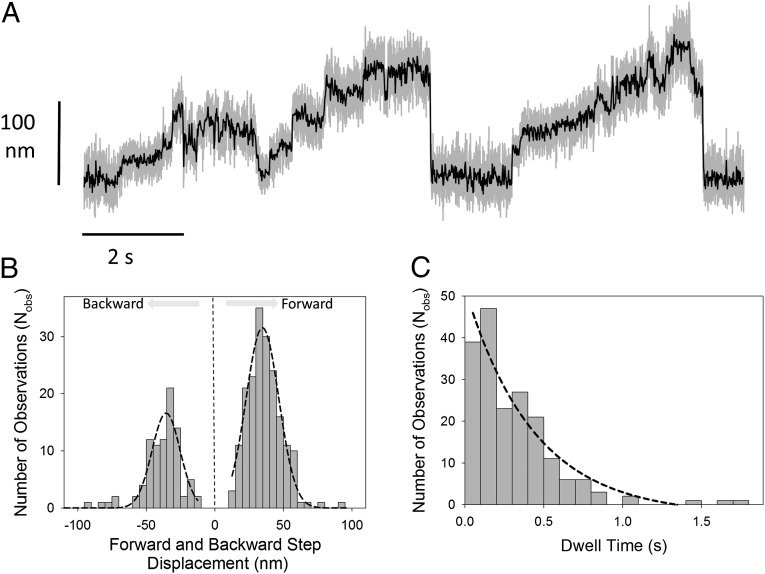
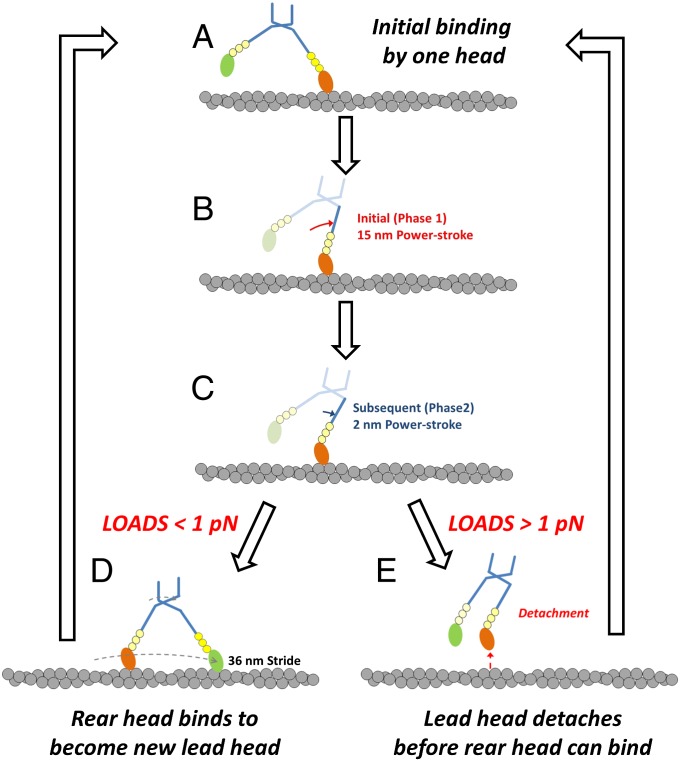
Myosin-10 is an actin-based molecular motor that participates in essential intracellular processes such as filopodia formation/extension, phagocytosis, cell migration, and mitotic spindle maintenance. To study this motor protein's mechano-chemical properties, we used a recombinant, truncated form of myosin-10 consisting of the first 936 amino acids, followed by a GCN4 leucine zipper motif, to force dimerization. Negative-stain electron microscopy reveals that the majority of molecules are dimeric with a head-to-head contour distance of ∼50 nm. In vitro motility assays show that myosin-10 moves actin filaments smoothly with a velocity of ∼310 nm/s. Steady-state and transient kinetic analysis of the ATPase cycle shows that the ADP release rate (∼13 s(-1)) is similar to the maximum ATPase activity (∼12-14 s(-1)) and therefore contributes to rate limitation of the enzymatic cycle. Single molecule optical tweezers experiments show that under intermediate load (∼0.5 pN), myosin-10 interacts intermittently with actin and produces a power stroke of ∼17 nm, composed of an initial 15-nm and subsequent 2-nm movement. At low optical trap loads, we observed staircase-like processive movements of myosin-10 interacting with the actin filament, consisting of up to six ∼35-nm steps per binding interaction. We discuss the implications of this load-dependent processivity of myosin-10 as a filopodial transport motor.

Keywords: actomyosin; myosin X; myosin-5a; optical trapping; stable single alpha-helix.


Conflict of interest statement

The authors declare no conflict of interest.

Figures

Fig. 1.
Fig. 1.
Myosin-10 heavy meromyosin-like (M10HMM) construct design. (A) Illustration of the full-length myosin-10 (Upper) and the M10HMM construct (Lower) structural organization. (B) Image of a typical SDS-polyacrylamide gel electrophoretogram (4–20%) of M10HMM after elution from an anti-FLAG resin. Approximately 0.4–0.8 mg of protein was purified per preparation. (C) Collage of M10HMM dimers negatively stained with 1% uranyl acetate (Each window = 92 × 92 nm). From electron micrographs it was determined that 87.2% of heavy chains were dimerized (Nobservations = 1,089). (Scale bar for all panels, 20 nm.) (D) An example M10HMM image from a panel from C with identification of regions, including motor domain (MD)/3 IQ motifs, the proximal coiled-coil region, and regions that most likely represents the stable SAH domains. Window size = 92 × 92 nm. (E) Images of M10HMM bound to actin in the presence of 1 μM ATP. Both single-headed binding and double-headed binding are seen. Examples can be seen in which the two heads of M10HMM span the actin pseudorepeat. (Scale bar for all panels, 20 nm.) (F) Histogram showing the contour length of M10HMM dimers (black bins), measured from the tip of one motor domain to the tip of the other [black line = Gaussian fit; peak = 51 ± 5.5 nm (SD); Nobs = 795; R2 = 0.99]. As a comparison, the contour length of myosin-5a-HMM (M5aHMM) (red bins) are shown [red line = Gaussian fit; peak = 60 ± 3.8 nm (SD); Nobs= 221; R2 = 0.96]. (G) Histogram showing the distribution of head-head angular variation of M10HMM (black bins; Nobs = 782) and M5aHMM (red bins; Nobs = 230) dimers. Gaussian fits showed peaks at 115 ± 2.7° (SEM) for M10HMM (black line; R2 = 0.89) and 110 ± 3.1° (SEM) (red line; R2 = 0.86) for M5aHMM.
Fig. 2.
Fig. 2.
Ensemble mechanics and solution kinetics data of acto-M10HMM. (A) In vitro actin gliding assay using M10HMM construct at 23 °C. Average velocity = 310 ± 70 nm/s (SD) (Nfilaments tracked = 1,076). (B) In vitro actin gliding assay using M10HMM at 37 °C. Average velocity = 1,500 ± 120 nm/s (SD) (Nfilaments tracked = 541). (C) Plot of the actin-activated steady-state ATPase of M10HMM. In the presence of actin, the steady-state activity of M10HMM reached a maximal rate of 13.0 ± 0.47 s−1 (SD) (Vmax). Half-maximal activation was achieved at 12 ± 1.2 μM (SD) (KATPase) (Npreparations = 5). In the absence of actin, the basal steady-state activity of the construct was 0.04 ± 0.01 s−1 (SD). Data were collected at 25 °C. (D) To determine the ADP release rate from the acto-M10HMM complex, we used various mant-ATP concentrations to chase the dissociation of ADP from the M10HMM-ADP complex. A premixture of 1 μM M10HMM with 12 μM filamentous actin was incubated with 30 μM ADP. The acto-M10HMM-ADP complex was chased with different concentrations of mant-ATP. Fitting the kobs at different mant-ATP concentrations with a hyperbola was used to determine the maximal kobs, ∼12.7 s−1. (Inset) Transient record at 200 μM mant-ATP (postmixture concentration) with a kobs = 11.6 s−1. Data were collected at 25 °C.
Fig. 3.
Fig. 3.
Single molecule optical tweezers assay. (A) Illustration of the three-bead assay. Two 1-μm polystyrene (PS) beads, coated with NeutrAvidin, are trapped in independent optical tweezers (OT), and a single actin filament (F-actin), containing 10% biotinylated globular-actin, is attached to form a dumbbell structure using a biotin-NeutrAvidin linkage at either end. The dumbbell is then brought to close proximity to a 2.4-μm glass pedestal (Ped) attached to the coverslip, sparsely coated with M10HMM (∼0.005–0.02 μg/mL). The bead positions are measured with nanometer precision using split photodiodes. In some experiments the optical tweezer was oscillated back and forth; this increases the signal-to-noise ratio and also allows the acto-myosin stiffness to be measured (SI Text). (B) An example of attached lifetime histogram for (Nobs= 667) acto-M10HMM events (measured at 250 nM ATP). The detachment rate was determined by fitting a single-exponential to the histogram [4.5 ± 0.5 s−1 (SD); R2 = 0.97; Table S1]. (C) ATP dependence of detachment rate, determined from event lifetime histograms measured at different ATP concentrations, exhibited Michaelis–Menten type kinetics with Vmax = 14.0 ± 0.5 s−1 (SD); KM = 0.44 ± 0.08 μM (SD) (R2 = 0.97). (D) Displacement histogram for acto-M10HMM (measured at 250 nM ATP) in the absence of oscillations. Power stroke determined from the Gaussian fit (black dashed line) was ∼17 nm [17.2 ± 0.8 nm (SEM); R2 = 0.95] (Nmyosins = 6; Nobs = 220) shifted from 0 nm (dark gray dashed line). (E) Example displacement histogram for acto-M10HMM (at 250 nM ATP) in the presence of oscillations. Power stroke was determined as the shift in the Gaussian peak (black dashed line) from 0 nm (dark gray dashed line). In this example, the average power-stroke is ∼16 nm [16 ± 3.1 nm (SEM); R2 = 0.9] (Nmyosins = 8; Nobs = 667).
Fig. 4.
Fig. 4.
Ensemble average showing the time course of the M10HMM power stroke. Binding events were synchronized to their start-and end times (n = 220, from six myosin molecules) by changes in signal variance. The position values were then averaged so that random thermal noise cancelled out, whereas movement due to the M10HMM power stroke was reinforced. The power stroke of shows two phases. First, a 15-nm step (δ1) then a second 2-nm step (δ2). Vertical black lines show the start and end of the synchronized interaction. Horizontal black arrows show the initial and final positions of start and end times for the synchronized plots. The cartoon (Inset) illustrates how a two-step power stroke might occur (for simplicity, only one myosin-10 head/lever is shown).
Fig. 5.
Fig. 5.
Single molecule acto-M10HMM stiffness. (A) Raw data for stiffness measurement of M10HMM using the three-bead assay. In each trace (driven and passive bead traces), top signal shows the raw data (red and blue) and the bottom signal (black) is variance. (B) Example force-displacement diagram of acto-M10HMM. The slope determines the stiffness of a single M10HMM bound to F-actin, which is ∼0.38 pN/nm, for this example. Within the circle (force ∼ ±1.5 pN and displacement ∼ ±4 nm), there seems to be no obvious nonlinearity behavior. (C) Distribution of acto-M10HMM stiffnesses spans from 0.2 to 1.5 pN/nm, with a median ∼0.45 pN/nm (Nmolecules = 59).
Fig. 6.
Fig. 6.
Processive behavior at low optical tweezers stiffness. (A) Using a very compliant (i.e., low stiffness) optical trap (∼0.005–0.008 pN/nm), we could observe staircase-like movements generated by a single M10HMM molecule (here recorded at 100 nM ATP). Gray lines are raw data sampled at 20 kHz; black lines are following a fourth-order Butterworth, 200 Hz low-pass filter. (B) Individual step amplitudes, identified by a t test step-finder algorithm (SI Text) were histogrammed, and Gaussian fitting showed that the forward step size had an averaged ∼35 nm [34.7 ± 0.6 nm (SEM); R2 = 0.97; Nobs = 198] and the backward steps averaged ∼36 nm [36 ± 1.1 nm (SEM); R2 = 0.86; Nobs = 91]. (C) Exponential fitting to the forward stepping dwell time histogram gave a step rate of ∼2.3 s−1 [2.3 ± 0.5 s−1 (SEM); R2 = 0.93; Nobs = 198].
Fig. 7.
Fig. 7.
Proposed model of the M10HMM translocation along a single actin filament incorporating mechanical data from study. Initial binding of M10HMM to F-actin occurs via a leading head (A), is followed by an initial rapid phase of the power stroke giving 15 nm of movement (phase 1; B), and then a subsequent 2 nm (phase 2; C). Then, if the external load on the molecule is <1 pN, the trailing M10HMM head may swing forward and bind at the next available actin site (36 nm) in front and therefore becomes the new leading head (D), allowing processive stepping movement to proceed. However, if the load exceeds 1 pN, the leading head detaches before the trailing head has a chance to bind so that the M10HMM molecule then detaches from F-actin, which terminates the processive movement (E).

Similar articles

Cited by

References

    1. Odronitz F, Kollmar M. Drawing the tree of eukaryotic life based on the analysis of 2,269 manually annotated myosins from 328 species. Genome Biol. 2007;8(9):R196. - PMC - PubMed
    1. Richards TA, Cavalier-Smith T. Myosin domain evolution and the primary divergence of eukaryotes. Nature. 2005;436(7054):1113–1118. - PubMed
    1. Berg JS, Derfler BH, Pennisi CM, Corey DP, Cheney RE. Myosin-X, a novel myosin with pleckstrin homology domains, associates with regions of dynamic actin. J Cell Sci. 2000;113(Pt 19):3439–3451. - PubMed
    1. Berg JS, Cheney RE. Myosin-X is an unconventional myosin that undergoes intrafilopodial motility. Nat Cell Biol. 2002;4(3):246–250. - PubMed
    1. Zhu XJ, et al. Myosin X regulates netrin receptors and functions in axonal path-finding. Nat Cell Biol. 2007;9(2):184–192. - PubMed

Publication types

MeSH terms

LinkOut - more resources