. 2014 May 6;111(18):6750-5.
doi: 10.1073/pnas.1400136111. Epub 2014 Apr 21.

An outer membrane channel protein of Mycobacterium tuberculosis with exotoxin activity

Affiliations

An outer membrane channel protein of Mycobacterium tuberculosis with exotoxin activity

Olga Danilchanka et al. Proc Natl Acad Sci U S A. .

Abstract

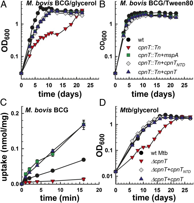
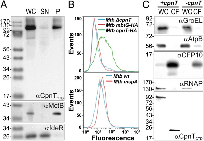
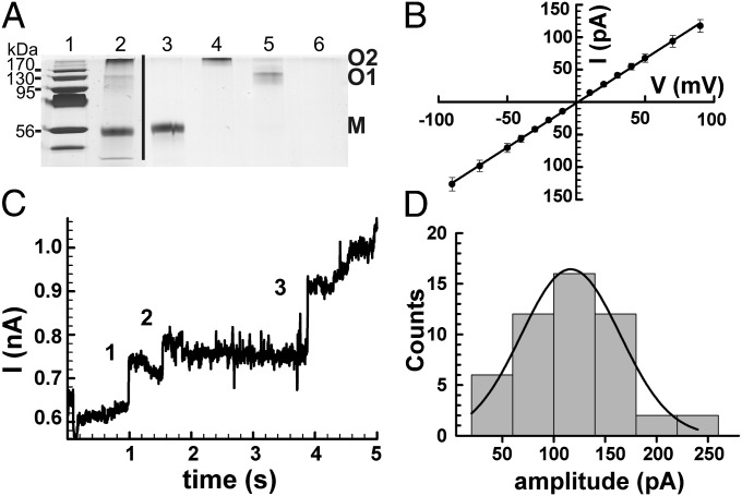
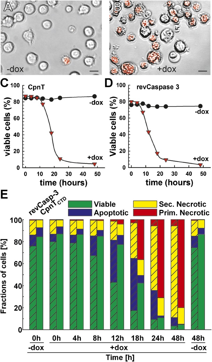
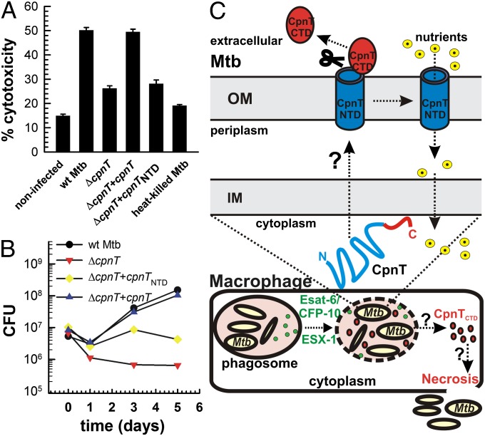
The ability to control the timing and mode of host cell death plays a pivotal role in microbial infections. Many bacteria use toxins to kill host cells and evade immune responses. Such toxins are unknown in Mycobacterium tuberculosis. Virulent M. tuberculosis strains induce necrotic cell death in macrophages by an obscure molecular mechanism. Here we show that the M. tuberculosis protein Rv3903c (channel protein with necrosis-inducing toxin, CpnT) consists of an N-terminal channel domain that is used for uptake of nutrients across the outer membrane and a secreted toxic C-terminal domain. Infection experiments revealed that CpnT is required for survival and cytotoxicity of M. tuberculosis in macrophages. Furthermore, we demonstrate that the C-terminal domain of CpnT causes necrotic cell death in eukaryotic cells. Thus, CpnT has a dual function in uptake of nutrients and induction of host cell death by M. tuberculosis.

Keywords: pore; secretion; transport.


Conflict of interest statement

The authors declare no conflict of interest.

Figures

Fig. 1.
Fig. 1.
CpnT is required for efficient growth on and uptake of glycerol by M. bovis bacillus Calmette–Guérin (BCG) and M. tuberculosis. Growth of WT M. bovis BCG (black circle), cpnT::Tn (red inverted triangle), cpnT::Tn complemented with mspA (green square), cpnTNTD (gray diamond), or cpnT (blue triangle) in minimal Hartmans-de Bont (HdB) medium supplemented with 0.1% glycerol (A) and 1% Tween-80 (B). Experiments were carried out at least three times. Representative growth curves are shown. (C) [14C]Glycerol uptake by M. bovis BCG. The uptake rate is expressed as nanomole of glycerol per milligram of cells. Uptake experiments were done in triplicate and mean values are shown with SDs. The P value determined by Student t test was less than 0.05 for WT versus the cpnT::Tn mutant for all time points. (D) Growth of WT M. tuberculosis (black circle), ∆cpnT (red inverted triangle), and ∆cpnT complemented with cpnTNTD (black diamond) or cpnT (blue triangle) in 7H9 medium with 0.2% glycerol. Experiments were carried out three times. A representative growth curve is shown.
Fig. 2.
Fig. 2.
Subcellular localization of CpnT in M. tuberculosis. (A) Immunoblot analysis of whole-cell lysates (WC), water-soluble supernatant (SN), and membrane-associated pellet (P) protein fractions obtained by ultracentrifugation of WT M. tuberculosis mc26206. IdeR and MctB were used as controls for soluble and membrane-associated proteins, respectively. CpnT was detected using an antibody recognizing the CTD. (B) Surface accessibility of CpnT. Cells of M. tuberculosis ΔcpnT and of M. tuberculosis overexpressing cpnTHA and mbtGHA were incubated with an anti-HA antibody followed by detection with anti-rabbit AlexaFluor 488-labeled antibody (Upper). WT M. tuberculosis and M. tuberculosis expressing the porin gene mspA of M. smegmatis were incubated with a monoclonal anti-MspA antibody (P2) and an anti-mouse FITC-labeled antibody (Lower). The fluorescence of surface-stained M. tuberculosis cells was measured by flow cytometry and is displayed as histograms (MFI, mean fluorescence intensity). (C) Secretion of the CTD of CpnT. Immunoblot analysis of whole cell lysates (WC) and culture filtrates (CF) of the M. tuberculosis ΔcpnT mutant grown in vitro carrying integrative vectors with (+cpnT) or without (-cpnT) the cpnT gene. The cytoplasmic proteins GroEL and RNA polymerase (RNAP), the inner membrane protein AtpB, and the secreted protein CFP-10 served as markers for the subcellular fractions.
Fig. 3.
Fig. 3.
The oligomeric NTD of CpnT forms water-filled membrane channels. (A) To preserve disulfide bridges, proteins were mixed with nonreducing loading buffer and samples were loaded without boiling. Lane 1, molecular weight marker; lane 2, CpnTNTD after anion exchange chromatography; lane 3, gel-purified monomer of CpnTNTD (M); lane 4, gel-purified oligomer O2 of CpnTNTD; lane 5, gel-purified oligomer O1 of CpnTNTD (O1); lane 6, “protein-free” control gel. The gel was stained with silver. (B) Average I/V characteristics of two CpnTNTD channels. The I/V curves were recorded for two individual channels reconstituted into lipid bilayers in two separate experiments. The single channel conductance was calculated from the slope of the fitted line and was determined as 1.36 ± 0.01 nS. (C) Current trace of the O2 oligomer of CpnTNTD recorded at +100 mV and filtered by a Gaussian low-pass filter with a bandwidth of 100 Hz. The protein concentration was ∼0.3 µg/mL. The current trace represents a 5-s recording of a continuous multichannel experiment. Events 1, 2, and 3 had current amplitudes (conductances) of 95 pA (1 nS), 65 pA (0.7 nS), and 152 pA (1.5 nS), respectively. (D) Distribution of the current amplitudes of 50 channels of the CpnTNTD O2 oligomer recorded from 25 experiments at +100 mV. The dotted line represents a Gaussian fit for the data with a mean of 116 ± 48 pA. The average single-channel conductance was 1.2 ± 0.5 nS.
Fig. 4.
Fig. 4.
The CTD of CpnT induces necrotic cell death in eukaryotic cells. The Jurkat T-cell line J644 containing an integrated Tet-regulated cpnTCTD expression cassette was uninduced (A) or induced with 100 ng/mL doxycycline (B) for 16 h, and subsequently stained for cell viability with 7-amino-actinomycin D (7AAD). Bright field images were merged with the fluorescence image visualizing stained, nonviable cells in red. Magnification, 40×. (Scale bar, 20 µm.) (C and D) Death kinetics of J644-derived cells after induction of expression of cpnTCTD (C) or a gene encoding activated caspase-3 (revCasp-3) (D) with doxycycline (1 µg/mL). Viable and dead cells were distinguished by flow cytometry according to their position in forward scatter and log side scatter. (E) Characterization of the cell death induced by CpnTCTD and activated caspase-3. Cell death kinetics of the J644-derived cell lines either uninduced or induced with 1 µg/mL doxycycline. Cells were grouped into viable, apoptotic, secondary, and primary necrotic using a six-parameter classification scheme (cell size, cell granularity, membrane integrity, phosphatidyl serine exposure, mitochondrial potential, and nuclear degeneration) (32). All bars of one column sum up to 100% with the cells gated from the living and dead cells at each time point.
Fig. 5.
Fig. 5.
CpnT is required for cytotoxicity and survival of M. tuberculosis in macrophages. (A) Differentiated THP-1 macrophages were infected with the indicated strains at an MOI of 20 for 2 d. Cytotoxicity was determined by flow cytometry after staining with the live/dead stain. (B) Differentiated THP-1 macrophages were infected with WT M. tuberculosis (black circle), ∆cpnT (red inverted triangle), and ∆cpnT complemented with cpnTNTD (yellow diamond) or cpnT (blue triangle) at an MOI of 10. Macrophages were lysed at the indicated time points, and the number of viable bacteria was counted as colony forming units (CFU) on agar plates. Bars represent mean ± SD. (C) Model of CpnT secretion and CpnT-induced cell death of macrophages. Secretion of CpnT by M. tuberculosis is mediated by unknown inner membrane (IM) and outer membrane (OM) components. We suggest that the C-terminal toxin is cleaved after integration of CpnT into the outer membrane. This leaves the N-terminal channel domain (CpnTNTD) in the outer membrane to enable uptake of nutrients. Esat-6 and CFP10 are secreted by ESX-1 and perforate the phagosomal membrane. Then, the secreted CTD of CpnT (CpnTCTD) probably gains access to the macrophage cytoplasm to induce necrosis by an unknown mechanism. These events enable M. tuberculosis to escape from the phagosome and eventually from the destroyed macrophage.

References

    1. Collier RJ. Understanding the mode of action of diphtheria toxin: A perspective on progress during the 20th century. Toxicon. 2001;39(11):1793–1803. - PubMed
    1. Gordon SV, Bottai D, Simeone R, Stinear TP, Brosch R. Pathogenicity in the tubercle bacillus: Molecular and evolutionary determinants. Bioessays. 2009;31(4):378–388. - PubMed
    1. Henkel JS, Baldwin MR, Barbieri JT. Toxins from bacteria. EXS. 2010;100:1–29. - PMC - PubMed
    1. Mukhopadhyay S, Nair S, Ghosh S. Pathogenesis in tuberculosis: Transcriptomic approaches to unraveling virulence mechanisms and finding new drug targets. FEMS Microbiol Rev. 2012;36(2):463–485. - PubMed
    1. World Health Organization 2013. Global Tuberculosis Control, WHO report 2012 (World Health Organization)

Publication types

MeSH terms

LinkOut - more resources