

Peripheral lymphadenopathy: approach and diagnostic tools
- PMID: 24753638
- PMCID: PMC3993046
Peripheral lymphadenopathy: approach and diagnostic tools
Abstract
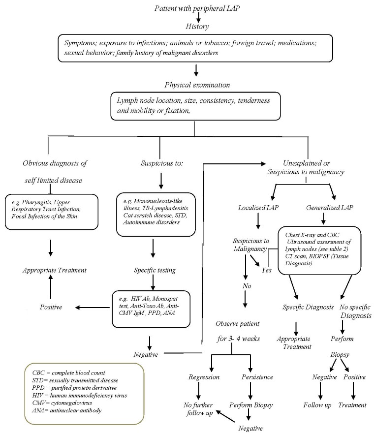
Peripheral lymph nodes, located deep in the subcutaneous tissue, clean antigens from the extracellular fluid. Generally, a normal sized lymph node is less than one cm in diameter. Peripheral lymphadenopathy (LAP) is frequently due to a local or systemic, benign, self-limited, infectious disease. However, it could be a manifestation of underlying malignancy. Seventy-five percent of all LAPs are localized, with more than 50% being seen in the head and neck area. LAP may be localized or generalized. Cervical lymph nodes are involved more often than the other lymphatic regions. Generally, it is due to infections, but most of the supraclavicular lymphadenopathies are associated with malignancy. Based on different geographical areas, the etiology is various. For example, in tropical areas, tuberculosis (TB) is a main benign cause of LAP in adults and children. Complete history taking and physical examination are mandatory for diagnosis; however, laboratory tests, imaging diagnostic methods, and tissue samplings are the next steps. Tissue diagnosis by fine needle aspiration biopsy or excisional biopsy is the gold standard evaluation for LAP. We concluded that in patients with peripheral LAP, the patient's age and environmental exposures along with a careful history taking and physical examination can help the physician to request step by step further work-up when required, including laboratory tests, imaging modalities, and tissue diagnosis, to reach an appropriate diagnosis.
Keywords: Benign; Diagnosis; Localization; Lymphadenopathy; Malignant.
Figures
References
-
- Ferrer R. Lymphadenopathy: differential diagnosis and evaluation. Am Fam Physician. 1998;58:1313–20. PubMed PMID: 9803196. - PubMed
-
- Slap GB, Brooks JS, Schwartz JS. When to perform biopsies of enlarged peripheral lymph nodes in young patients. JAMA. 1984;252:1321–6. doi:10.1001/jama.1984.03350100051031. PubMed PMID: 6471252. - PubMed
-
- Ochicha O, Edino ST, Mohammed AZ, Umar AB, Atanda AT. Pathology of peripheral lymph node biopsies in Kane, Northern Nigeria. Ann Afr Med. 2007;6:104–8. PubMed PMID: 18240497. - PubMed
-
- Okolo SN, Nwana EJ, Mohammed AZ. Histopathologic diagnoses of lymphadenopathy in children in Jos, Nigeria. Niger Postgrad Med J. 2003;10:165–7. PubMed PMID:14692059. - PubMed
Publication types
LinkOut - more resources
Full Text Sources
Research Materials