Susceptibility weighted magnetic resonance imaging of brain: A multifaceted powerful sequence that adds to understanding of acute stroke
- PMID: 24753661
- PMCID: PMC3992771
- DOI: 10.4103/0972-2327.128555
Susceptibility weighted magnetic resonance imaging of brain: A multifaceted powerful sequence that adds to understanding of acute stroke
Abstract
Context: To evaluate the additional information that susceptibility weighted sequences and datasets would provide in acute stroke.
Aims: The aim of this study were to assess the value addition of susceptibility weighted magnetic resonance imaging (SWI) of brain in patients with acute arterial infarct.
Materials and methods: All patients referred for a complete brain magnetic resonance imaging (MRI) between March 2010 and March 2011 at our institution had SWI as part of routine MRI (T1, T2, and diffusion imaging). Retrospective study of 62 consecutive patients with acute arterial infarct was evaluated for the presence of macroscopic hemorrhage, petechial micro-bleeds, dark middle cerebral artery (MCA) sign and prominent vessels in the vicinity of infarct.
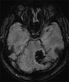
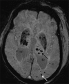
Results: SWI was found to detect hemorrhage not seen on other routine MRI sequences in 22 patients. Out of 62 patients, 17 (10 petechial) had hemorrhage less than 50% and 5 patients had greater than 50% area of hemorrhage. A "dark artery sign" due to thrombus within the artery was seen in 8 out of 62 patients. Prominent cortical and intraparenchymal veins were seen in 14 out of 62 patients.
Conclusions: SWI has been previously shown to be sensitive in detecting hemorrhage; however is not routinely used in stroke evaluation. Our study shows that SWI, by virtue of identifying unsuspected hemorrhage, central occluded vessel, and venous congestion is additive in value to the routine MR exam and should be part of a routine MR brain in patients suspected of having an acute infarct.
Keywords: Deoxyhaemoglobin; hemorrhage; infarct; ischemia; magnetic resonance imaging; susceptibility weighted imaging.
Conflict of interest statement
Figures


References
-
- Haacke EM, Xu Y, Cheng YC, Reichenbach JR. Susceptibility weighted imaging (SWI) Magn Reson Med. 2004;52:612–8. - PubMed
-
- Hermier M, Nighoghossian N. Contribution of susceptibility-weighted imaging to acute stroke assessment. Stroke. 2004;35:1989–94. - PubMed
-
- Sehgal V, Delproposto Z, Haacke EM, Tong KA, Wycliffe N, Kido DK, et al. Clinical applications of neuroimaging with susceptibility-weighted imaging. J Magn Reson Imaging. 2005;22:439–50. - PubMed
LinkOut - more resources
Full Text Sources
Other Literature Sources

