Acanthamoeba meningoencephalitis
- PMID: 24753675
- PMCID: PMC3992747
- DOI: 10.4103/0972-2327.128571
Acanthamoeba meningoencephalitis
Abstract
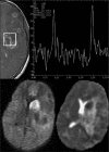
Report of a case of young immunocompetent male adult with autopsy proven acanthamoeba meningoencephalitis. The patient presented with a protracted febrile illness of 3 months duration with features of meningoencephalitis, this was followed by rapid deterioration while on anti tuberculous therapy and steroids and ended fatally. His magnetic resonance imaging showed features of hemorrhagic meningoencephalitis and magnetic resonance spectroscopy showed choline peak. Autopsy revealed necrotizing meningoencephalitis and intraocular colonization due to acanthamoeba.
Keywords: Acanthamoeba meningoencephalitis; immunocompetent; intraocular colonization; magnetic resonance imaging; magnetic resonance spectroscopy.
Conflict of interest statement
Figures


References
-
- Kidney DD, Kim SH. CNS infections with free-living amebas: Neuroimaging findings. AJR Am J Roentgenol. 1998;171:809–12. - PubMed
-
- Martínez AJ, Sotelo-Avila C, Garcia-Tamayo J, Morón JT, Willaert E, Stamm WP. Meningoencephalitis due to Acanthamoeba SP. Pathogenesis and clinico-pathological study. Acta Neuropathol. 1977;37:183–91. - PubMed
-
- Gordon SM, Steinberg JP, DuPuis MH, Kozarsky PE, Nickerson JF, Visvesvara GS. Culture isolation of Acanthamoeba species and leptomyxid amebas from patients with amebic meningoencephalitis, including two patients with AIDS. Clin Infect Dis. 1992;15:1024–30. - PubMed
-
- Ma P, Visvesvara GS, Martinez AJ, Theodore FH, Daggett PM, Sawyer TK. Naegleria and Acanthamoeba infections: Review. Rev Infect Dis. 1990;12:490–513. - PubMed
-
- Martínez AJ. Is Acanthamoeba encephalitis an opportunistic infection? Neurology. 1980;30:567–74. - PubMed
Publication types
LinkOut - more resources
Full Text Sources
Other Literature Sources

