Different effects of tetanic stimulation of facial nerve and ulnar nerve on transcranial electrical stimulation motor-evoked potentials
- PMID: 24753756
- PMCID: PMC3992401
Different effects of tetanic stimulation of facial nerve and ulnar nerve on transcranial electrical stimulation motor-evoked potentials
Abstract
Objective: Our objective was to examine whether prior tetanic stimulation of cranial nerves enhances the amplitudes of transcranial motor-evoked potentials (MEPs).
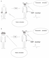
Methods: Thirty patients undergoing elective craniotomy under propofol-fentanyl anesthesia with partial neuromuscular blockade were enrolled. Both control and posttetanic MEPs (c-MEPs and p-MEPs) monitoring were performed with a train of five pulses delivered to C3 or C4. c-MEPs were recorded from target muscles and p-MEPs were obtained 1 s after tetanic stimulation to the ulnar nerves and facial nerves. The amplitudes of paired MEPs were compared with Wilcoxon's signed rank test.
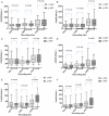
Results: When tetanic stimulation was separately applied to the facial nerves, amplitudes of p-MEPs from abductor pollicis brevis, orbicularis oculi or oris were similar with those of c-MEPs. When tetanic stimulations were separately applied to the ulnar nerves, the amplitudes of p-MEPs from the abductor pollicis brevis but not orbicularis oculi or oris were significantly enlarged compared with c-MEP.
Conclusions: We found that only prior tetanic stimulation of ulnar nerve but not facial nerve could enlarge the amplitudes of trancranial hand MEPs. Augmentation of MEP amplitude via prior tetanic stimulation of peripheral nerve seems to originate from the subcortical level but not motor cortex.
Keywords: Motor-evoked potentials; abductor pollicis brevis; facial nerve; orbicularis oculi; orbicularis oris; tetanic stimulation; ulnar nerve.
Figures


Similar articles
-
The application of tetanic stimulation of the unilateral tibial nerve before transcranial stimulation can augment the amplitudes of myogenic motor-evoked potentials from the muscles in the bilateral upper and lower limbs.Anesth Analg. 2008 Jul;107(1):215-20. doi: 10.1213/ane.0b013e318177082e. Anesth Analg. 2008. PMID: 18635490
-
The effects of the neuromuscular blockade levels on amplitudes of posttetanic motor-evoked potentials and movement in response to transcranial stimulation in patients receiving propofol and fentanyl anesthesia.Anesth Analg. 2008 Mar;106(3):930-4, table of contents. doi: 10.1213/ane.0b013e3181617508. Anesth Analg. 2008. PMID: 18292442 Clinical Trial.
-
Tetanic stimulation of the peripheral nerve before transcranial electrical stimulation can enlarge amplitudes of myogenic motor evoked potentials during general anesthesia with neuromuscular blockade.Anesthesiology. 2005 Apr;102(4):733-8. doi: 10.1097/00000542-200504000-00007. Anesthesiology. 2005. PMID: 15791101
-
Tetanic stimulation of the pudendal nerve prior to transcranial electrical stimulation augments the amplitude of motor evoked potentials during pediatric neurosurgery.J Neurosurg Pediatr. 2021 Apr 23;27(6):707-715. doi: 10.3171/2020.10.PEDS20674. Print 2021 Jun 1. J Neurosurg Pediatr. 2021. PMID: 33892470
-
Effects of intraoperative motor evoked potential amplification following tetanic stimulation of the pudendal nerve in pediatric craniotomy.J Neurosurg Pediatr. 2023 Feb 24;31(5):488-495. doi: 10.3171/2023.1.PEDS22505. Print 2023 May 1. J Neurosurg Pediatr. 2023. PMID: 36840735
Cited by
-
Variables associated with cortical motor mapping thresholds: A retrospective data review with a unique case of interlimb motor facilitation.Front Neurol. 2023 Apr 11;14:1150670. doi: 10.3389/fneur.2023.1150670. eCollection 2023. Front Neurol. 2023. PMID: 37114230 Free PMC article.
-
Tetanic stimulation of the peripheral nerve augments motor evoked potentials by re-exciting spinal anterior horn cells.J Clin Monit Comput. 2022 Feb;36(1):259-270. doi: 10.1007/s10877-020-00647-z. Epub 2021 Jan 9. J Clin Monit Comput. 2022. PMID: 33420971
-
Does Post-Tetanic Transcranial Stimulation Augment the Wave Amplitudes of Spinal Cord Evoked Potential (Tc-SCEP)?Global Spine J. 2025 Mar;15(2):1473-1478. doi: 10.1177/21925682241299713. Epub 2024 Nov 8. Global Spine J. 2025. PMID: 39512181 Free PMC article.
-
Utility of desflurane as an anesthetic in motor-evoked potentials in spine surgery and the facilitating effect in tetanic stimulation of bilateral median nerves.J Clin Monit Comput. 2024 Jun;38(3):663-670. doi: 10.1007/s10877-023-01096-0. Epub 2023 Nov 2. J Clin Monit Comput. 2024. PMID: 37917209
-
Motor-Evoked Potential Monitoring With Multi-train Electrical Stimulation During Thoracoabdominal Aortic Aneurysm Surgery: A Case Report.Cureus. 2024 Feb 8;16(2):e53872. doi: 10.7759/cureus.53872. eCollection 2024 Feb. Cureus. 2024. PMID: 38465173 Free PMC article.
References
-
- Kawaguchi M, Sakamoto T, Inoue S, Kakimoto M, Furuya H, Morimoto T, Sakaki T. Low dose propofol as a supplement to ketamine-based anesthesia during intraoperative monitoring of motor-evoked potentials. Spine (Phila Pa 1976) 2000;25:974–979. - PubMed
-
- Lotto ML, Banoub M, Schubert A. Effects of anesthetic agents and physiologic changes on intraoperative motor evoked potentials. J Neurosurg Anesthesiol. 2004;16:32–42. - PubMed
-
- Yamamoto Y, Kawaguchi M, Kakimoto M, Inoue S, Furuya H. The effects of dexmedetomidine on myogenic motor evoked potentials in rabbits. Anesth Analg. 2007;104:1488–1492. table of contents. - PubMed
-
- Yamamoto Y, Kawaguchi M, Kakimoto M, Takahashi M, Inoue S, Goto T, Furuya H. The effects of xenon on myogenic motor evoked potentials in rabbits: a comparison with propofol and isoflurane. Anesth Analg. 2006;102:1715–1721. - PubMed
-
- Kawaguchi M, Sakamoto T, Ohnishi H, Shimizu K, Karasawa J, Furuya H. Intraoperative myogenic motor evoked potentials induced by direct electrical stimulation of the exposed motor cortex under isoflurane and sevoflurane. Anesth Analg. 1996;82:593–599. - PubMed
LinkOut - more resources
Full Text Sources
Miscellaneous
