Review
. 2013;5(1):72-81.

The uterine myocyte as a target for prevention of preterm birth

Affiliations
Review

The uterine myocyte as a target for prevention of preterm birth

B F Mitchell et al. Facts Views Vis Obgyn. 2013.

Abstract

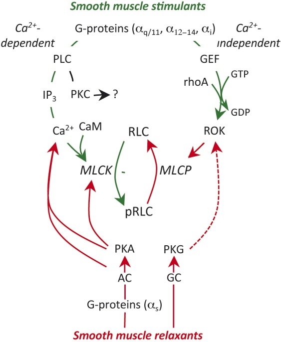
Preterm birth (PTB) remains the most common cause of neonatal morbidity and mortality as well as long-term disability. Current strategies to prevent or arrest spontaneous preterm labor (SPTL) have limited success. For almost three decades, there have been no novel pharmacological agents used clinically to address this important obstetrical complication. In this review, we focus on the uterine myocyte as a target for prevention of spontaneous PTB. After presenting an overview of intracellular signaling pathways that are important in regulation of smooth muscle contractility, we discuss previous and current pharmacological approaches to manage SPTL. We also present recent evidence from our own laboratories suggesting a potentially novel and uterine-specific approach to maintain or impose uterine relaxation. Finally, we briefly discuss extrinsic systems that might affect uterine activity and reinforce the concept that SPTL represents a syndrome that is the end result of a variety of pathophysiologic etiologies leading to PTB. We conclude by emphasizing the need for much more research to provide sufficient understanding of the mechanisms of SPTL and to make inroads towards reducing the incidence and adverse consequences of this common and serious syndrome.

Keywords: Prematurity; myosin regulatory light chain; preterm labour; rhoA-associated kinase; tocolysis; tocolysis contractility.


Figures

Fig. 1
Fig. 1
Regulation of smooth muscle contractility. Uterine stimulants generally stimulate specific G-protein coupled receptors (GPCR) on the myocyte membrane. This triggers two stimulatory pathways. The Gαq subunit activates phospholipase C (PLC) in the adjacent membrane and this results in hydrolysis of membrane phosphoinositides to from inositol trisphosphate (IP3) and diacyl glycerol, which can activate protein kinase C (PKC; see text). IP3 stimulates release of Ca2+ from the sarcoplasmic reticulum and the resulting decrease in cell membrane resting potential leads to massive influx of Ca2+ from the extracellular space through voltage–gated L-type Ca2+-channels. Ca2+ binds to calmodulin (CaM). The Ca2+-CaM complex interacts with and activates myosin regulatory light chain kinase (MLCK). MLCK mediates the phosphorylation of myosin regulatory lights chains (RLC) and initiates the contractile apparatus. The second stimulatory pathway is mediated through Gα12-14. This causes activation of a guanidine nucleotide exchange factor (GEF), which activates membrane rhoA-GDP by substituting GTP for the GDP. The rhoA-GTP activates rhoA-associated kinase (ROK). ROK phosphorylates myosin regulatory light chain phosphatase (MLCP), which inhibits its activity. Since MLCP is normally responsible for removing the phosphate from pRLC to cause uterine relaxation, enhanced ROK activity prolongs the effectiveness of pRLC and promotes enhanced contractile activity. The major inhibitory pathways might also be mediated by GPCR, linked through Gαs resulting in activation of adenylate cyclase (AC) or direct activation of guanylate cyclase (GC), which in turn activates protein kinase A (PKA) or PKG. These kinases inhibit contractility through a variety of mechanisms described in the text. The green lines indicate stimulatory activity and the red lines inhibitory. The broken line indicates an indirect effect.
Fig. 2
Fig. 2
Expression of mRNA for oxytocin receptor in myocytes derived from upper and lower uterine segments. PCR of extracts from primary cultures of uterine myocytes derived from paired samples of uterine muscle (n = 2 patients) obtained from the upper fundal portion of the uterus (U) or the lower uterine segment (L) along with molecular weight markers (M), blanks (Bl) and extracts from myometrial tissues (My). The cells were used at passage 3, 4 and 5 (P3-P5). The upper panels demonstrate expression of OTR (476 bp) and the bottom panels show expression of GAPDH (598 bp) as a control. OTR is expressed in upper and lower segment myocytes and does not change with passage number.
Fig. 3
Fig. 3
Intracellular concentrations of Ca2+ following stimulation of uterine myocytes with oxytocin. Primary cultures of fundal (upper panel) and lower (lower panel) segment uterine myocytes were plated in Greiner Bio-One black plates (5.0 × 104 cells/ml) and grown for 48 hours. Cells were then loaded with Fluo-4 dye and incubated at 37°C for 30 minutes. OT (0.1 µM, 1 µM, 10 µM) was added to the cells and fluorescence was measured. Data are presented as peak fluorescence over baseline fluorescence (F/F0; mean ± SEM; n = 4 patients).
Fig. 4
Fig. 4
Concentrations of pRLC in uterine myocytes from upper and lower uterine segments following stimulation by oxytocin. Primary cultures of myocytes from the uterine fundus and lower uterine segments (n = 5 patients) were treated with increasing concentrations of OT for 20 s and the response measured using a specific antibody for pRLC phosphorylated at S19 in an in-cell western assay. There were significant, concentration-dependent increases that were similar in cells from upper and lower segments. 15-minute pretreatment with the rho-kinase inhibitor (g-H; 1 µM) or the myosin regulatory light chain kinase inhibitor (ML7; 25 µM) caused approximately 20-30% suppression of basal pRLC but the response to OT was maintained.
Fig. 5
Fig. 5
Concentrations of ppRLC in uterine myocytes from upper and lower uterine segments following stimulation by oxytocin. Primary cultures of myocytes from the uterine fundus and lower uterine segments (n = 5 patients) were treated with increasing concentrations of OT for 20 s and the response measured using a specific antibody for ppRLC phosphorylated at T18 and S19 in an in-cell western assay. There were significant, concentration-dependent increases in ppRLC in cells from upper and lower segments. As for pRLC, 15-mimute pretreatment with the rho-kinase inhibitor (g-H; 1 µM) or the myosin regulatory light chain kinase inhibitor (ML7; 25 µM) caused approximately 20-30% suppression of basal ppRLC. However, in contrast to the results for pRLC, the ppRLC responses to OT were completely abolished in the presence of the kinase inhibitors.
Fig. 6
Fig. 6
Schematic diagram illustrating the unique pattern of phosphorylation of RLC in uterine myocytes. As in all smooth muscle, myosin regulatory light chain (RLC) is phosphorylated by myosin regulatory light chain kinase (MLCK) at S19. However, in uterine myocytes, pRLC is additionally phosphorylated at T18 to form ppRLC, a more potent activator of the myosin ATPase that supplies energy for the contraction. This diphosphorylation pathway might be a unique adaptive pathway for smooth muscle that contract in a phasic pattern. The green lines indicate stimulatory activity and the red lines inhibitory.

References

    1. Aguilar HN, Mitchell BF. Physiological Pathways and Molecular Mechanisms Regulating Uterine Contractility. Hum Reprod Update. 2010;16:725–744. - PubMed
    1. Aguilar HN, Tracey CN, Tsang SC, et al. Phos-Tag-Based Analysis of Myosin Regulatory Light Chain Phosphorylation in Human Uterine Myocytes. PLoS One. 6:e20903. - PMC - PubMed
    1. Aguilar HN, Tracey CN, Zielnik B, Mitchell BF. Quantification of Rapid Myosin Regulatory Light Chain Phosphorylation Using High-Throughput in-Cell Western Assays: Comparison to Western Immunoblots. PLoS One. 2010;5:e9965. - PMC - PubMed
    1. Aguilar HN, Tracey CN, Zielnik B, Mitchell BF. Rho-kinase mediates diphosphorylation of myosin regulatory light chain in cultured uterine but not vascular smooth muscle cells. http://onlinelibrary.wiley.com/doi/ 10.1111/j.1582-4934.2012.01625.x/abs... J Cell Mol Med. 2012 - PMC - PubMed
    1. Arthur P, Taggart MJ, Zielnik B, Mitchell BF. Relationship between Gene Expression and Function of Uterotonic Systems in the Rat During Gestation, Uterine Activation and Both Term and Preterm Labour. J Physiol. 2008;586:6063–6076. - PMC - PubMed

LinkOut - more resources