

Twin infant with lymphatic dysplasia diagnosed with Noonan syndrome by molecular genetic testing
- PMID: 24754368
- PMCID: PMC4086230
- DOI: 10.3109/15513815.2014.904026
Twin infant with lymphatic dysplasia diagnosed with Noonan syndrome by molecular genetic testing
Abstract
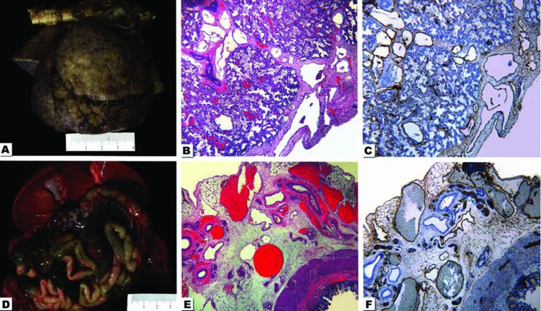
Noonan Syndrome is an autosomal dominant disorder characterized by short stature, congenital heart defects, developmental delay, dysmorphic facial features and occasional lymphatic dysplasias. The features of Noonan Syndrome change with age and have variable expression. The diagnosis has historically been based on clinical grounds. We describe a child that was born with congenital refractory chylothorax and subcutaneous edema suspected to be secondary to pulmonary lymphangiectasis. The infant died of respiratory failure and anasarca at 80 days. The autopsy confirmed lymphatic dysplasia in lungs and mesentery. The baby had no dysmorphic facial features and was diagnosed postmortem with Noonan syndrome by genomic DNA sequence analysis as he had a heterozygous mutation for G503R in the PTPN11 gene.
Keywords: Noonan syndrome; chylothorax; genomic DNA sequence; lymphatic dysplasia; pulmonary lymphangiectasis.
Figures
Similar articles
-
A PTPN11 mutation in a woman with Noonan syndrome and protein-losing enteropathy.BMC Gastroenterol. 2020 Feb 13;20(1):34. doi: 10.1186/s12876-020-01187-1. BMC Gastroenterol. 2020. PMID: 32054441 Free PMC article. Review.
-
Congenital Chylothorax as the Initial Presentation of PTPN11-Associated Noonan Syndrome.J Pediatr. 2017 Jun;185:248-248.e1. doi: 10.1016/j.jpeds.2017.02.042. Epub 2017 Mar 28. J Pediatr. 2017. PMID: 28363362 Free PMC article. No abstract available.
-
A mother and son with Noonan syndrome resulting from a PTPN11 mutation: first report of molecularly proven cases from Turkey.Turk J Pediatr. 2010 May-Jun;52(3):321-4. Turk J Pediatr. 2010. PMID: 20718194
-
Co-occurring SHOC2 and PTPN11 mutations in a patient with severe/complex Noonan syndrome-like phenotype.Am J Med Genet A. 2011 Jun;155A(6):1217-24. doi: 10.1002/ajmg.a.33987. Epub 2011 May 5. Am J Med Genet A. 2011. PMID: 21548061
-
Noonan syndrome, PTPN11 mutations, and brain tumors. A clinical report and review of the literature.Am J Med Genet A. 2017 Apr;173(4):1061-1065. doi: 10.1002/ajmg.a.38108. Am J Med Genet A. 2017. PMID: 28328117 Review.
Cited by
-
Severe generalized edema in a premature neonate: A case report and literature review.Clin Case Rep. 2024 Sep 3;12(9):e9341. doi: 10.1002/ccr3.9341. eCollection 2024 Sep. Clin Case Rep. 2024. PMID: 39229301 Free PMC article.
-
Mutational landscape, clonal evolution patterns, and role of RAS mutations in relapsed acute lymphoblastic leukemia.Proc Natl Acad Sci U S A. 2016 Oct 4;113(40):11306-11311. doi: 10.1073/pnas.1608420113. Epub 2016 Sep 21. Proc Natl Acad Sci U S A. 2016. PMID: 27655895 Free PMC article.
-
A PTPN11 mutation in a woman with Noonan syndrome and protein-losing enteropathy.BMC Gastroenterol. 2020 Feb 13;20(1):34. doi: 10.1186/s12876-020-01187-1. BMC Gastroenterol. 2020. PMID: 32054441 Free PMC article. Review.
-
Congenital Chylothorax as the Initial Presentation of PTPN11-Associated Noonan Syndrome.J Pediatr. 2017 Jun;185:248-248.e1. doi: 10.1016/j.jpeds.2017.02.042. Epub 2017 Mar 28. J Pediatr. 2017. PMID: 28363362 Free PMC article. No abstract available.
-
Case report: Noonan syndrome with protein-losing enteropathy.Front Genet. 2023 Sep 27;14:1237821. doi: 10.3389/fgene.2023.1237821. eCollection 2023. Front Genet. 2023. PMID: 37829277 Free PMC article.
References
-
- Noonan JA. Noonan syndrome a historical perspective. Heart Views. 2002;3:13–15.
-
- Kobylinski O. Ueber eine flughoutahnibiche ausbreitung. Am Hals Arch Anthoropol. 1883;14:342–348.
-
- Moore KL, Persaud TVN. The Developing Human: Clinically Oriented Embryology. 7th ed. Philadelphia, PA: Saunders; 2003.
Publication types
MeSH terms
Substances
Supplementary concepts
LinkOut - more resources
Full Text Sources
Other Literature Sources
Miscellaneous