. 2014 Jul;225(1):109-17.
doi: 10.1111/joa.12188. Epub 2014 Apr 23.

Adenosine A2B and A3 receptor location at the mouse neuromuscular junction

Affiliations

Adenosine A2B and A3 receptor location at the mouse neuromuscular junction

Neus Garcia et al. J Anat. 2014 Jul.

Abstract

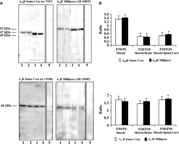
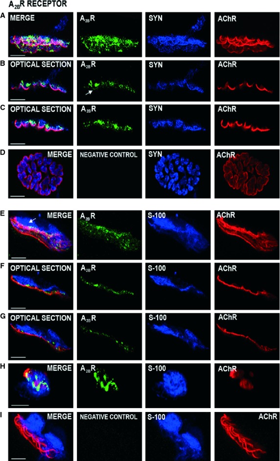
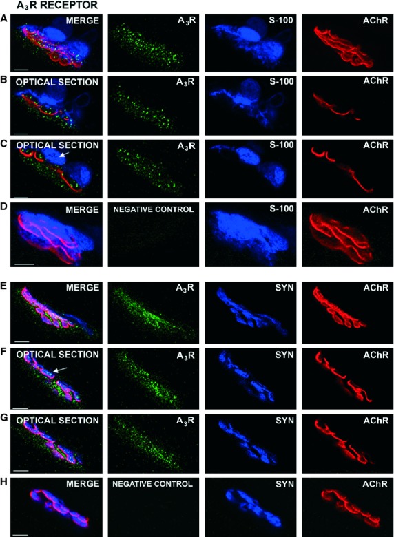
To date, four subtypes of adenosine receptors have been cloned (A(1)R, A(2A)R, A(2B)R, and A(3)R). In a previous study we used confocal immunocytochemistry to identify A(1)R and A(2A)R receptors at mouse neuromuscular junctions (NMJs). The data shows that these receptors are localized differently in the three cells (muscle, nerve and glia) that configure the NMJs. A(1)R localizes in the terminal teloglial Schwann cell and nerve terminal, whereas A(2A)R localizes in the postsynaptic muscle and in the axon and nerve terminal. Here, we use Western blotting to investigate the presence of A(2B)R and A(3)R receptors in striated muscle and immunohistochemistry to localize them in the three cells of the adult neuromuscular synapse. The data show that A(2B)R and A(3)R receptors are present in the nerve terminal and muscle cells at the NMJs. Neither A(2B)R nor A(3)R receptors are localized in the Schwann cells. Thus, the four subtypes of adenosine receptors are present in the motor endings. The presence of these receptors in the neuromuscular synapse allows the receptors to be involved in the modulation of transmitter release.

Keywords: adenosine receptors; cholinergic synapses; immunofluorescence; motor end-plate; motor nerve terminal.


Figures

Fig. 1
Fig. 1
Presence of A2BR and A3R in P6 and P30 LAL muscle. (A) Representative Western blotting analysis of A2BR (top) and A3R (bottom) (with antibodies from Millipore and Santa Cruz) protein presence in lysates of adult muscle (P30, line 1), neonatal muscle (P6, line 2), adult brain (line 3) and adult spinal cord (line 4). Samples are 100 μg of protein. Line 5 (for A2BR and A3R from Millipore) is the negative control, incubated with blocked peptide. Line 5 (for A3R from Santa Cruz) is the negative control incubated without primary antibody). As a positive control, we used adult brain and spinal cord. Actin immunoblots were used for protein loading controls (not shown). (B) Quantitative analysis of several A2BR (top) and A3R (bottom) Western blots normalized by actin staining. The data show that the presence of A2BR and A3R protein is more abundant in the adult muscle than in the newborn muscle when both antibodies are used. Also, A2BR is more abundant in brain and spinal cord than in muscle whereas A3R protein is slightly more abundant in muscle than in brain and spinal cord. Results are expressed as mean ± SEM of five independent experiments, *P < 0.05.
Fig. 2
Fig. 2
Localization of the A2BR receptors by immunohistochemistry at the neuromuscular junction. Immunofluorescence staining and confocal microscopy analysis. Triple labeling of A2BR protein (green fluorescence) with syntaxin or S-100 (blue fluorescence) and nAChR-alpha-bungarotoxin (red fluorescence). Images (A–C) were obtained using the rabbit polyclonal antibody against the adenosine A2BR (AB1589P, Millipore Corporation) and (E–H) images using the goat polyclonal antibody anti-A2BR-adenosine receptor, N-19 (sc-7506, Santa Cruz Biotechnology Inc.). A2BR immunoreactivity shows that the receptor is present in the nerve terminal. In (A), the A2BR label is observed in a wide area on the syntaxin positive nerve ending. In (B,C), the side view of the two individual optical sections of the synapse in (A) shows the A2BR spots colocalized with the blue syntaxin label in some synaptic gutters and in the postsynaptic membrane (arrow in B, green image). In addition, a fine line granular labeling occurs in the space of the nerve terminals between the blue Schwann cell and the red postsynaptic line (F and G are two individual optical sections of the synapse in E). The A2BR is not labeled in the area that corresponds to S-100 antibody (arrow in E). (H) shows a detail of A2BR positive synaptic boutons on a nerve terminal (in green) seen en face. (D,I) An example of negative control for A2BR proteins (see Results). Scale bar: 10 μm.
Fig. 3
Fig. 3
Localization of the A3R receptors by immunohistochemistry at the neuromuscular junction. In these adult muscles, A3R receptors are strongly immunolabeled in the neuromuscular junction. (A–D) images were obtained using the rabbit polyclonal antibody AB1590P (Millipore Corporation) and (E–H) images using the rabbit polyclonal antibody (H80) (sc-13938, Santa Cruz Biotechnology, Inc.). The merge images (from at least 10 confocal Z planes) of the synapses in (A,E) show an abundant granular A3R labeling in the synaptic area. The optical sections (from the synapse in A) in (B,C) show immunolabeled spots in the nerve terminal position (blue in the space between glial cells and red in the space between muscle cells). Syntaxin-labeled terminal axons are also positive to the A3R (arrow in F is an individual optical section of the synapse in E). Fine granular marks are observed in the muscle cell below the postsynaptic gutters (for instance in F,G). The A3R label does not colocalize with S-100 (arrow in C). (D,H) Examples of negative control for A3R proteins (see Results). Scale bar: 10 μm.

References

    1. Adamek S, Shakirzyanova AV, Malomouzh AI, et al. Interaction of glutamate- and adenosine-induced decrease of acetylcholine quantal release at frog neuromuscular junction. Physiol Res. 2010;59:803–810. - PubMed
    1. Baxter RL, Vega-Riveroll LJ, Deuchars J, et al. A2A adenosine receptors are located on presynaptic motor nerve terminals in the mouse. Synapse. 2005;57:229–234. - PubMed
    1. Bibel M, Barde YA. Neurotrophins: key regulators of cell fate and cell shape in the vertebrate nervous system. Genes Dev. 2000;14:2919–2937. - PubMed
    1. Carreira MC, Camiña JP, Díaz-Rodríguez E, et al. Adenosine does not bind to the growth hormone secretagogue receptor type-1a (GHS-R1a) J Endocrinol. 2006;191:147–157. - PubMed
    1. Christofi FL, Zhang H, Yu JG, et al. Differential gene expression of adenosine A1, A2a, A2b, and A3 receptors in the human enteric nervous system. J Comp Neurol. 2001;439:46–64. - PubMed

Publication types

Substances

LinkOut - more resources