

All-atomic molecular dynamic studies of human CDK8: insight into the A-loop, point mutations and binding with its partner CycC
- PMID: 24754906
- PMCID: PMC4122639
- DOI: 10.1016/j.compbiolchem.2014.03.003
All-atomic molecular dynamic studies of human CDK8: insight into the A-loop, point mutations and binding with its partner CycC
Abstract
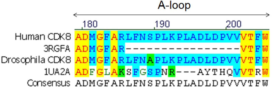
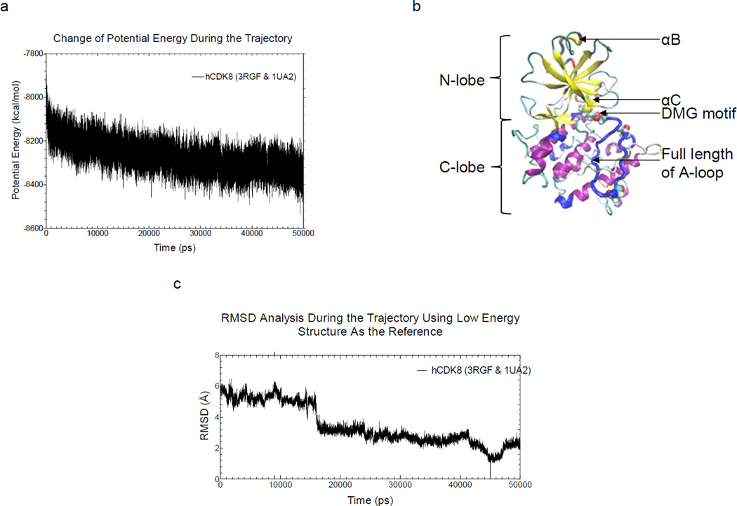
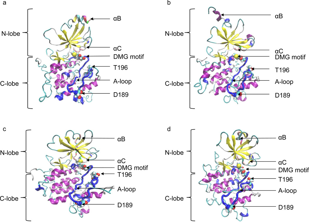
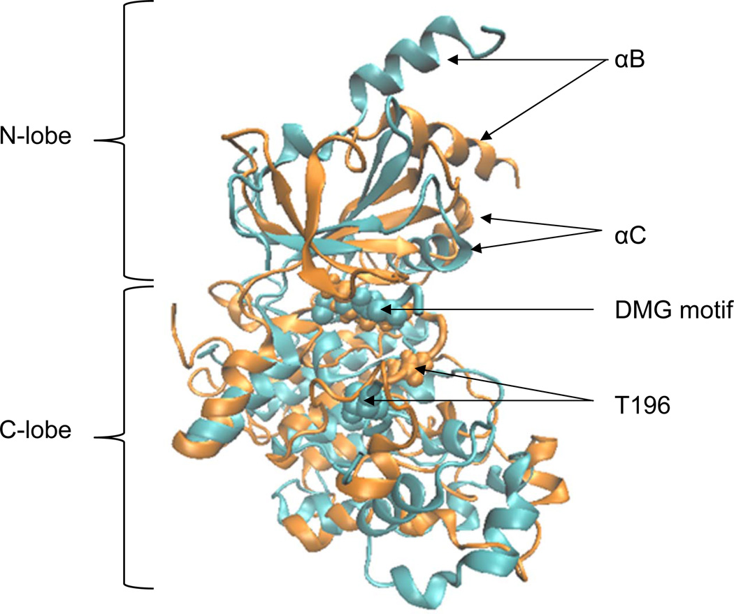
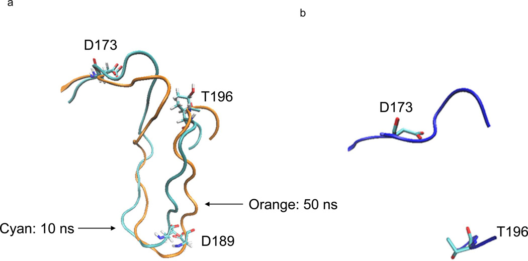
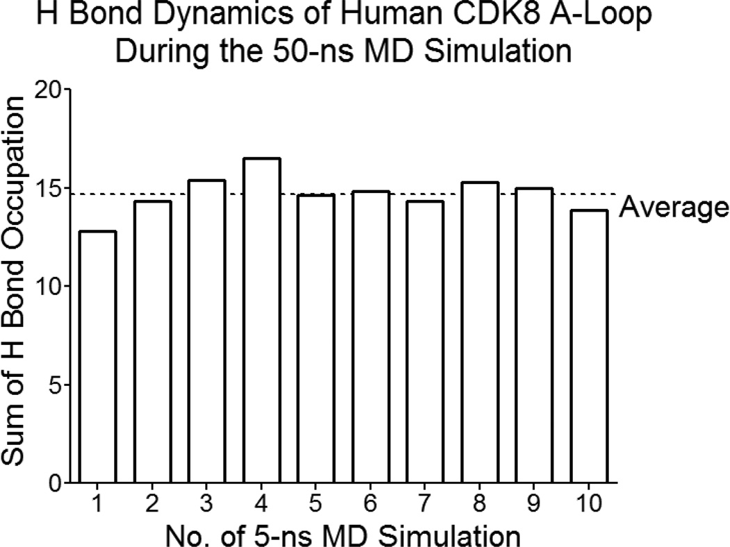
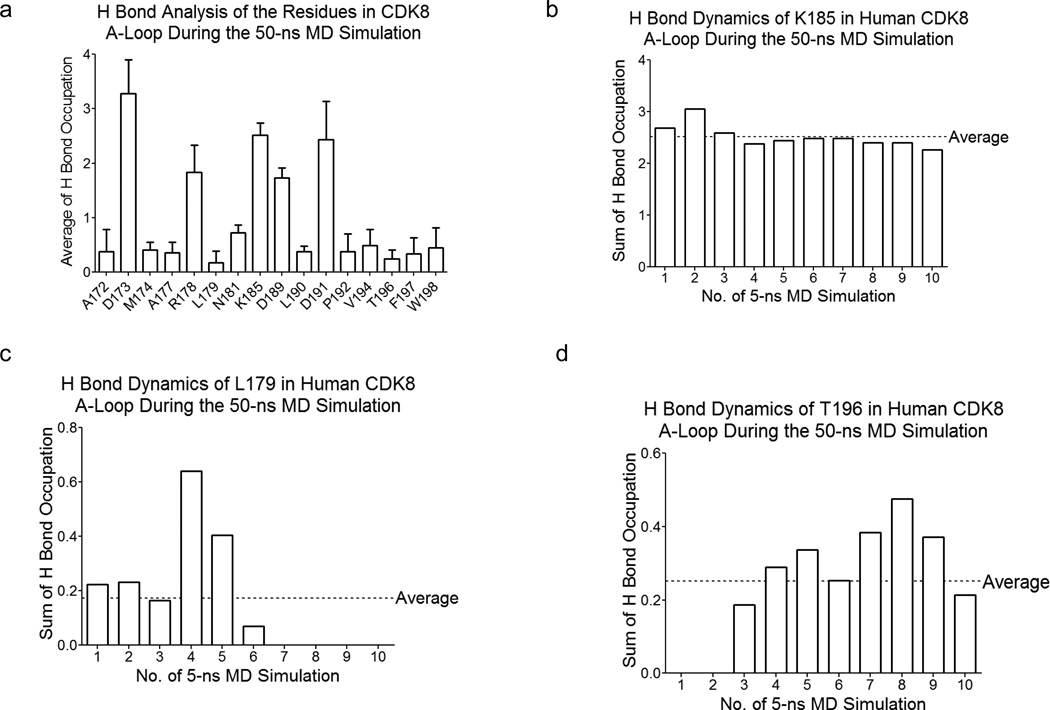
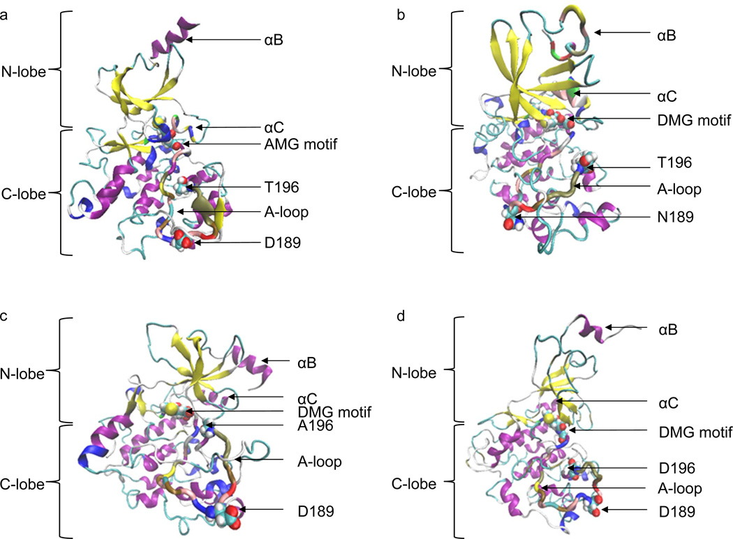
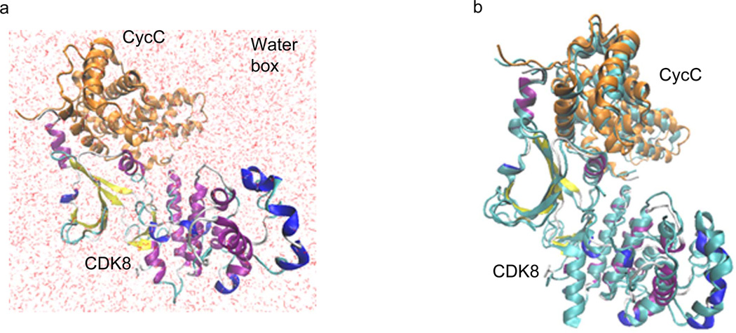
The Mediator, a conserved multisubunit protein complex in eukaryotic organisms, regulates gene expression by bridging sequence-specific DNA-binding transcription factors to the general RNA polymerase II machinery. In yeast, Mediator complex is organized in three core modules (head, middle and tail) and a separable 'CDK8 submodule' consisting of four subunits including Cyclin-dependent kinase CDK8 (CDK8), Cyclin C (CycC), MED12, and MED13. The 3-D structure of human CDK8-CycC complex has been recently experimentally determined. To take advantage of this structure and the improved theoretical calculation methods, we have performed molecular dynamic simulations to study dynamics of CDK8 and two CDK8 point mutations (D173A and D189N), which have been identified in human cancers, with and without full length of the A-loop, as well as the binding between CDK8 and CycC. We found that CDK8 structure gradually loses two helical structures during the 50-ns molecular dynamic simulation, likely due to the presence of the full-length A-loop. In addition, our studies showed the hydrogen bond occupation of the CDK8 A-loop increases during the first 20-ns MD simulation and stays stable during the later 30-ns MD simulation. Four residues in the A-loop of CDK8 have high hydrogen bond occupation, while the rest residues have low or no hydrogen bond occupation. The hydrogen bond dynamic study of the A-loop residues exhibits three types of changes: increasing, decreasing, and stable. Furthermore, the 3-D structures of CDK8 point mutations D173A, D189N, T196A and T196D have been built by molecular modeling and further investigated by 50-ns molecular dynamic simulations. D173A has the highest average potential energy, while T196D has the lowest average potential energy, indicating that T196D is the most stable structure. Finally, we calculated theoretical binding energy of CDK8 and CycC by MM/PBSA and MM/GBSA methods, and the negative values obtained from both methods demonstrate stability of CDK8-CycC complex. Taken together, these analyses will improve our understanding of the exact functions of CDK8 and the interaction with its partner CycC.
Keywords: CDK8; CycC; Molecular dynamics.
Published by Elsevier Ltd.
Figures
Similar articles
-
Highlighting the Major Role of Cyclin C in Cyclin-Dependent Kinase 8 Activity through Molecular Dynamics Simulations.Int J Mol Sci. 2024 May 15;25(10):5411. doi: 10.3390/ijms25105411. Int J Mol Sci. 2024. PMID: 38791449 Free PMC article.
-
A molecular dynamics investigation of CDK8/CycC and ligand binding: conformational flexibility and implication in drug discovery.J Comput Aided Mol Des. 2018 Jun;32(6):671-685. doi: 10.1007/s10822-018-0120-3. Epub 2018 May 8. J Comput Aided Mol Des. 2018. PMID: 29737445 Free PMC article.
-
Mediator kinase module and human tumorigenesis.Crit Rev Biochem Mol Biol. 2015;50(5):393-426. doi: 10.3109/10409238.2015.1064854. Epub 2015 Jul 16. Crit Rev Biochem Mol Biol. 2015. PMID: 26182352 Free PMC article. Review.
-
The structure of CDK8/CycC implicates specificity in the CDK/cyclin family and reveals interaction with a deep pocket binder.J Mol Biol. 2011 Sep 16;412(2):251-66. doi: 10.1016/j.jmb.2011.07.020. Epub 2011 Jul 23. J Mol Biol. 2011. PMID: 21806996
-
Dysregulation of CDK8 and Cyclin C in tumorigenesis.J Genet Genomics. 2011 Oct 20;38(10):439-52. doi: 10.1016/j.jgg.2011.09.002. Epub 2011 Sep 16. J Genet Genomics. 2011. PMID: 22035865 Free PMC article. Review.
Cited by
-
Hit discovery of potential CDK8 inhibitors and analysis of amino acid mutations for cancer therapy through computer-aided drug discovery.BMC Chem. 2024 Apr 13;18(1):73. doi: 10.1186/s13065-024-01175-6. BMC Chem. 2024. PMID: 38615023 Free PMC article.
-
Highlighting the Major Role of Cyclin C in Cyclin-Dependent Kinase 8 Activity through Molecular Dynamics Simulations.Int J Mol Sci. 2024 May 15;25(10):5411. doi: 10.3390/ijms25105411. Int J Mol Sci. 2024. PMID: 38791449 Free PMC article.
-
A molecular dynamics investigation of CDK8/CycC and ligand binding: conformational flexibility and implication in drug discovery.J Comput Aided Mol Des. 2018 Jun;32(6):671-685. doi: 10.1007/s10822-018-0120-3. Epub 2018 May 8. J Comput Aided Mol Des. 2018. PMID: 29737445 Free PMC article.
-
Development of a TSR-Based Method for Protein 3-D Structural Comparison With Its Applications to Protein Classification and Motif Discovery.Front Chem. 2021 Jan 13;8:602291. doi: 10.3389/fchem.2020.602291. eCollection 2020. Front Chem. 2021. PMID: 33520934 Free PMC article.
-
Mediator kinase module and human tumorigenesis.Crit Rev Biochem Mol Biol. 2015;50(5):393-426. doi: 10.3109/10409238.2015.1064854. Epub 2015 Jul 16. Crit Rev Biochem Mol Biol. 2015. PMID: 26182352 Free PMC article. Review.
References
-
- Shapiro GI. Cyclin-dependent kinase pathways as targets for cancer treatment. J Clin Oncol. 2006;24(11):1770–1783. - PubMed
-
- Schwartz GK, Shah MA. Targeting the cell cycle: a new approach to cancer therapy. J Clin Oncol. 2005;23(36):9408–9421. - PubMed
-
- Fisher RP. Secrets of a double agent: CDK7 in cell-cycle control and transcription. J Cell Sci. 2005;118(Pt 22):5171–5180. - PubMed
Publication types
MeSH terms
Substances
Grants and funding
LinkOut - more resources
Full Text Sources
Other Literature Sources
Miscellaneous
