Review
. 2014 Jul:19:95-107.
doi: 10.1016/j.dnarep.2014.03.022. Epub 2014 Apr 19.

The cutting edges in DNA repair, licensing, and fidelity: DNA and RNA repair nucleases sculpt DNA to measure twice, cut once

Affiliations
Review

The cutting edges in DNA repair, licensing, and fidelity: DNA and RNA repair nucleases sculpt DNA to measure twice, cut once

Susan E Tsutakawa et al. DNA Repair (Amst). 2014 Jul.

Abstract

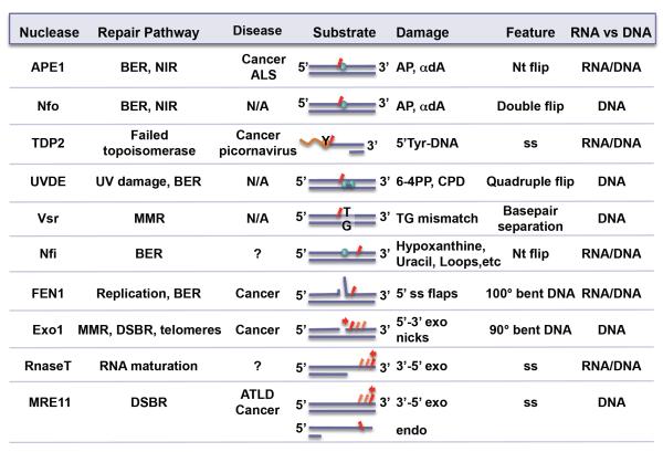
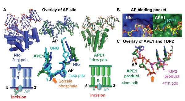
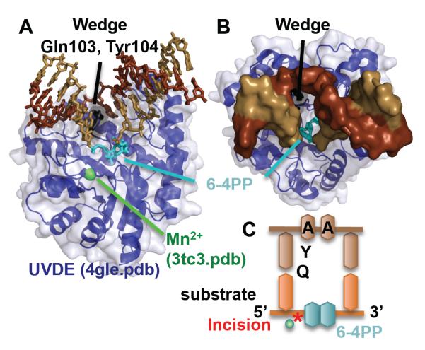
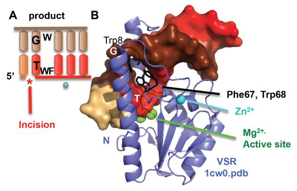
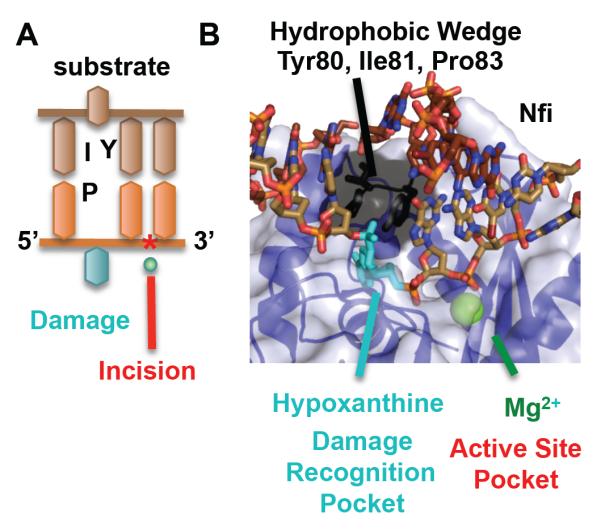
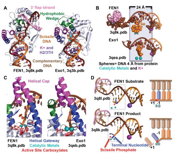
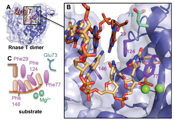
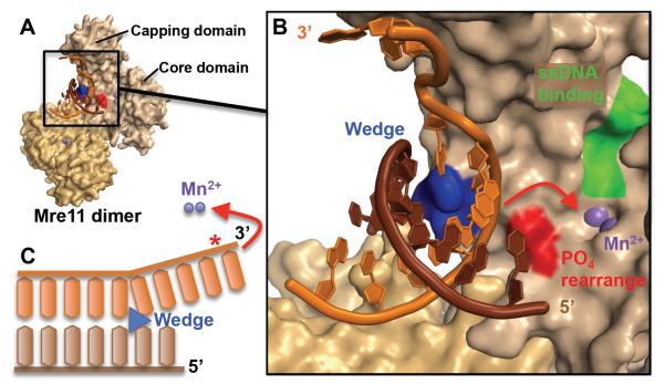
To avoid genome instability, DNA repair nucleases must precisely target the correct damaged substrate before they are licensed to incise. Damage identification is a challenge for all DNA damage response proteins, but especially for nucleases that cut the DNA and necessarily create a cleaved DNA repair intermediate, likely more toxic than the initial damage. How do these enzymes achieve exquisite specificity without specific sequence recognition or, in some cases, without a non-canonical DNA nucleotide? Combined structural, biochemical, and biological analyses of repair nucleases are revealing their molecular tools for damage verification and safeguarding against inadvertent incision. Surprisingly, these enzymes also often act on RNA, which deserves more attention. Here, we review protein-DNA structures for nucleases involved in replication, base excision repair, mismatch repair, double strand break repair (DSBR), and telomere maintenance: apurinic/apyrimidinic endonuclease 1 (APE1), Endonuclease IV (Nfo), tyrosyl DNA phosphodiesterase (TDP2), UV Damage endonuclease (UVDE), very short patch repair endonuclease (Vsr), Endonuclease V (Nfi), Flap endonuclease 1 (FEN1), exonuclease 1 (Exo1), RNase T and Meiotic recombination 11 (Mre11). DNA and RNA structure-sensing nucleases are essential to life with roles in DNA replication, repair, and transcription. Increasingly these enzymes are employed as advanced tools for synthetic biology and as targets for cancer prognosis and interventions. Currently their structural biology is most fully illuminated for DNA repair, which is also essential to life. How DNA repair enzymes maintain genome fidelity is one of the DNA double helix secrets missed by James Watson and Francis Crick, that is only now being illuminated though structural biology and mutational analyses. Structures reveal motifs for repair nucleases and mechanisms whereby these enzymes follow the old carpenter adage: measure twice, cut once. Furthermore, to measure twice these nucleases act as molecular level transformers that typically reshape the DNA and sometimes themselves to achieve extraordinary specificity and efficiency.

Keywords: APE1; Base excision repair; Crystallography; DNA; DNA repair; DNase; Double strand break repair; EndoIV; EndoV; Endonucleases; Enzyme–DNA complex; Exo1; Exonuclease; FEN1; Genome maintenance; Magnesium; Manganese; Metals; Mismatch repair; Mre11; Nfi; Nfo; Nucleases; Nucleotide incision repair; RNA; RNase; Structure-specific nuclease; TDP2; Telomere; UVDE; Vsr; Zinc.


Figures

Fig. 1
Fig. 1
The ten nucleases discussed in this review, highlighting their biological roles and differences in substrate specificity and features. Under diseases, not applicable (N/A) and unknown (?) are denoted. Relative incision position(s) on substrate is shown in red.
Fig. 2
Fig. 2
Nfo and APE1 share a common DNA sculpting to select for AP sites. A) Overlay of the AP site in substrate structures highlights the common DNA structure, despite the differences in protein structure and catalytic metals. The closeup highlights that this is not a default flipped out AP nucleotide conformation, as the abasic site in UNG shows small but significant shifts in phosphate and sugar positions (cyan arrows). Below each enzyme, a DNA schematic shows nucleotide flip and incision position (*). B) The shallow binding pocket (protein shown as a surface) selects for abasic sites. For clarity, Arg177 was shown in cyan. C) After overlay of the protein in APE1 and TDP2 DNA-bound structures, the scissile phosphate (orange arrow) matches, but the nucleotide is oriented inversely.
Fig. 3
Fig. 3
UVDE, which incises the phosphodiester backbone 5′ to the 6-4 PP, quadruple nucleotide flips the covalently linked base damage and the two nucleotides opposite. A) Side and B) top views of UVDE, inserts a wedge (Gln103 and Tyr104), as it double flips two nucleotides on both strands. C) Schematic of DNA and relative position of wedge residues. It is notable how accessible is the bound 6-4 PP.
Fig. 4
Fig. 4
Vsr breaks the helical stacking of its substrate as part of its recognition mechanism. A) DNA schematic shows how Vsr endonuclease incises 5′ to a TG mismatch. B) The structure unexpectedly reveals that Vsr inserts three residues (Phe67, Trp68, and Trp8) to break the stacking on the 3′ side of T in a T/G mismatch. Incision occurs on the 5′ side of the T. Product DNA is shown as surface, colored by chain. The N-terminal helix (N) clamps the product DNA down onto the main catalytic core.
Fig. 5
Fig. 5
Two pockets, one for recognition and one for incision, underlie Nfi mechanism. A) DNA schematic shows the relative position of the damage and the phosphodiester incision 3′ to the damage. B) The substrate structure (2w36.pdb) overlaid with the Mg2+ from the product structure (2w35.pdb) shows two pockets, one for damage recognition (shown for hypoxanthine) and one for incision with a single Mg2+. A helical wedge separates the two pockets.
Fig. 6
Fig. 6
EN1 and Exo1 use multiple mechanisms for substrate specificity. A) FEN1 and Exo1 use a hydrophobic wedge to block the path of the duplex DNA. B) FEN1 and Exo1 bind primarily to the complementary strand in two sections, the active site and the K+/H2/3TH. These regions are separated by 24 Å or a helical turn apart. C) The active sites, with seven invariantly conserved carboxylates and two catalytic metals, are protected by a helical gateway and a helical cap. The helical gateway could select for ssDNA or ssRNA. The terminal nucleotide stacks against an aromatic residue and is contacted by two conserved basic residues. D) A double base unpairing in the mechanisms is suggested by the observation of +1 and −1 paired nucleotides in the substrate and a −1 unpaired nucleotide in the product. The scissile phosphate is distant (5 Å) from the catalytic metals when the nucleotides are paired in the substrate structure, but is within catalytic distance (2.2 Å) in the product structure.
Fig. 7
Fig. 7
Rnase T dimer selects for ssDNA or ssRNA or 3′ overhangs by blocking the complementary strand (brown) with a steric helical wedge and Phe29. A) Global and B) closeup of the duplex structure (3va3.pdb) with Mg2+ atoms from an overlaid RnaseT/ssDNA structure (3v9w.pdb) to show relative position of active site metals and scissile phosphate (*). C) DNA schematic showing how the 3′ overhang is then selected through intercalation by Phe146, Phe124, and Phe77 and positioning of the phosphodiester bond for catalysis. For terminal cytosines with reduced incision activity, Glu73 will flip and pull on the cytosine, disrupting the positioning of the phosphodiester backbone and one of the two Mg2+ ions.
Fig. 8
Fig. 8
The Mre11 exonuclease mechanism involves sculpting the DNA end to direct the scissile strand to the active site. A) Both subunits of the Mre11 dimer interact with a branched DNA end (3dsd.pdb). In this structure, the substrate was not cleaved because the catalytic His85 is mutated into a serine. B) Closeup of the active site (shown by purple Mn2+ ions) and its surroundings. The complementary strand (brown) is unmodified while the scissile strand (orange) is sculpted by the helical wedge (blue). The postulated phosphate rearrangement residues involved in the rotation of the 3′ strand into the active site are shown in red (includes His52). On the other side of the active site, a pocket (green) is hypothesized to bind ssDNA for the endonuclease activity of Mre11. C) Schematic of the phosphate rearrangement mechanism (red) in order to sculpt the scissile strand into the active site. The scissile bond is shown with a red star (*).

References

    1. Crick F. The double helix: a personal view. Nature. 1974;248:766–769. - PubMed
    1. Arber W, Linn S. DNA modification and restriction. Annual Review of Biochemistry. 1969;38:467–500. - PubMed
    1. Linn S, Arber W. Host specificity of DNA produced by Escherichia coli, X. In vitro restriction of phage fd replicative form. Proceedings of the National Academy of Sciences of the United States of America. 1968;59:1300–1306. - PMC - PubMed
    1. Hanawalt PC. The Awakening of DNA Repair at Yale. Yale J Biol Med. 2013;86:517–523. - PMC - PubMed
    1. Harrington JJ, Lieber MR. Functional domains within FEN-1 and RAD2 define a family of structure-specific endonucleases: implications for nucleotide excision repair. Genes Dev. 1994;8:1344–1355. - PubMed

Publication types

MeSH terms

LinkOut - more resources