. 2014 Sep 15:41:120-30.
doi: 10.1016/j.niox.2014.04.008. Epub 2014 Apr 19.

AP39, a novel mitochondria-targeted hydrogen sulfide donor, stimulates cellular bioenergetics, exerts cytoprotective effects and protects against the loss of mitochondrial DNA integrity in oxidatively stressed endothelial cells in vitro

Affiliations

AP39, a novel mitochondria-targeted hydrogen sulfide donor, stimulates cellular bioenergetics, exerts cytoprotective effects and protects against the loss of mitochondrial DNA integrity in oxidatively stressed endothelial cells in vitro

Bartosz Szczesny et al. Nitric Oxide. .

Abstract

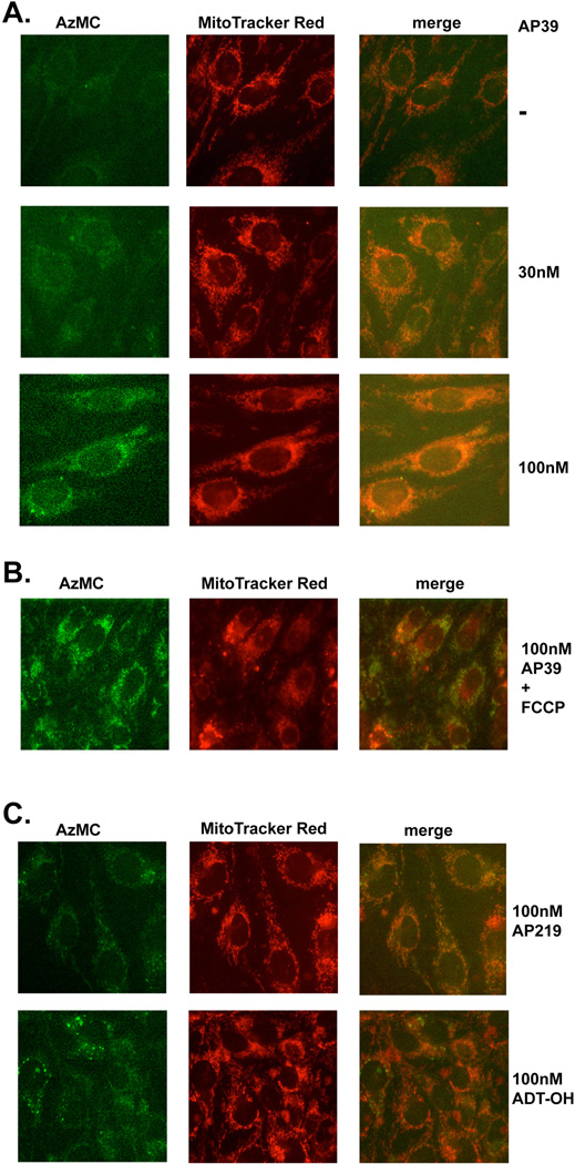
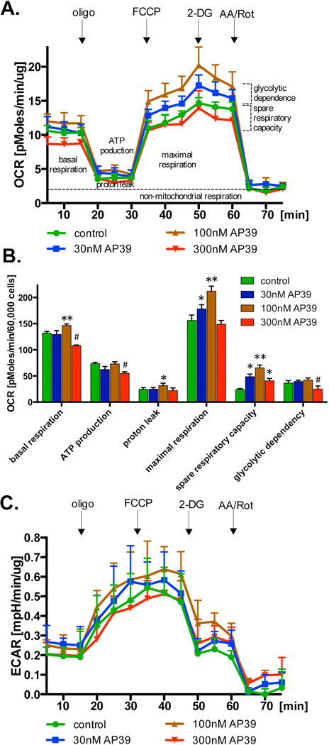
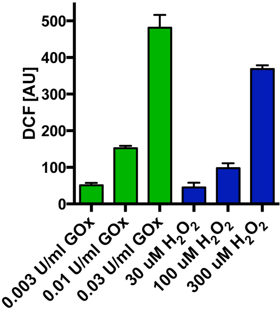
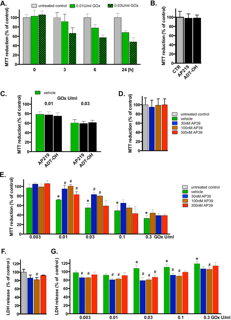
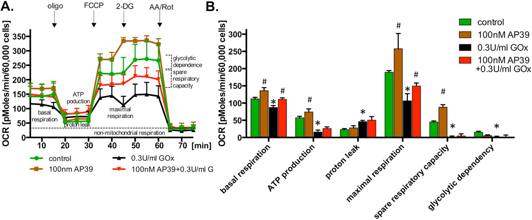
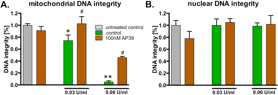
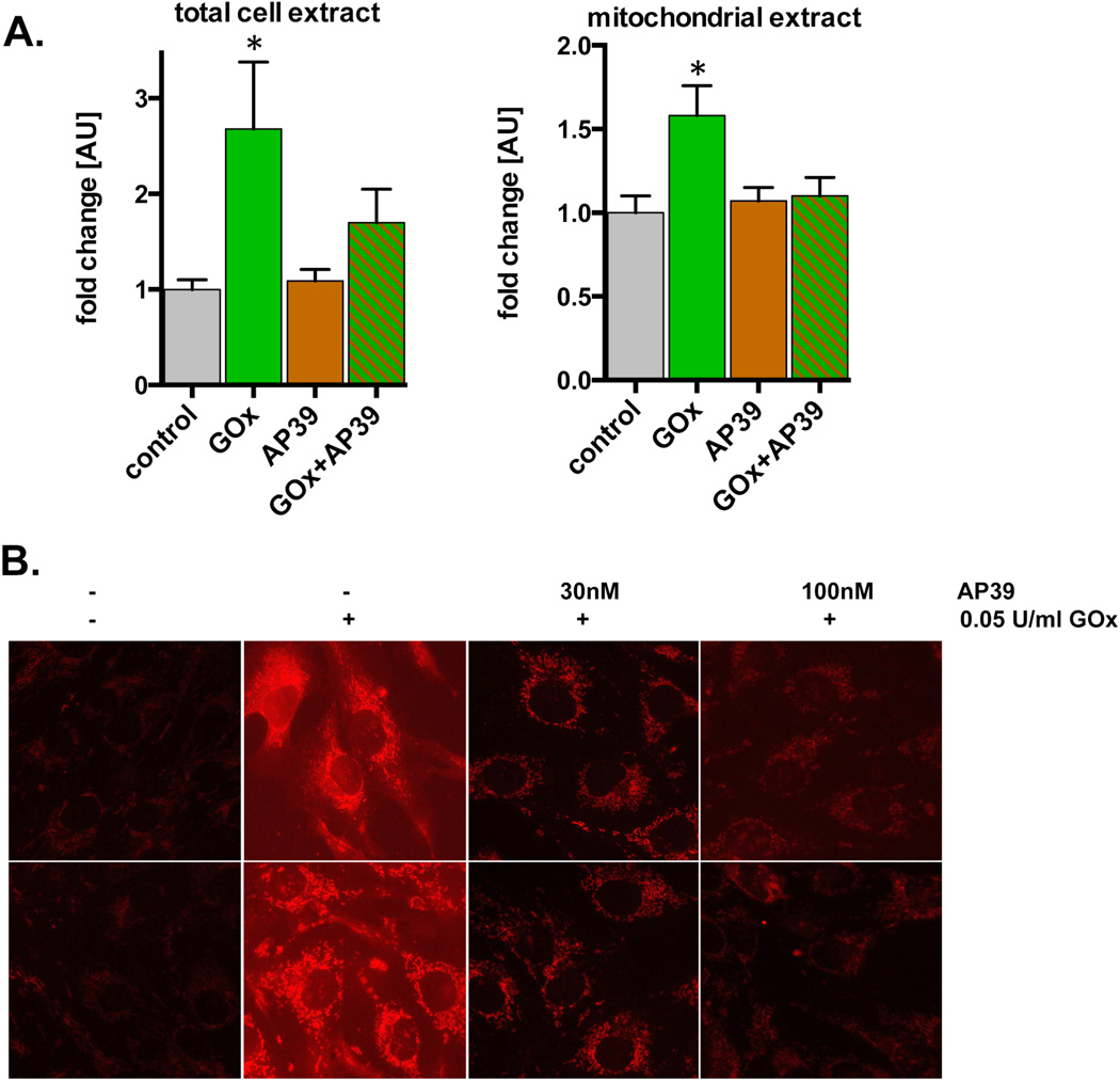
The purpose of the current study was to investigate the effect of the recently synthesized mitochondrially-targeted H2S donor, AP39 [(10-oxo-10-(4-(3-thioxo-3H-1,2-dithiol-5yl)phenoxy)decyl) triphenylphosphonium bromide], on bioenergetics, viability, and mitochondrial DNA integrity in bEnd.3 murine microvascular endothelial cells in vitro, under normal conditions, and during oxidative stress. Intracellular H2S was assessed by the fluorescent dye 7-azido-4-methylcoumarin. For the measurement of bioenergetic function, the XF24 Extracellular Flux Analyzer was used. Cell viability was estimated by the combination of the MTT and LDH methods. Oxidative protein modifications were measured by the Oxyblot method. Reactive oxygen species production was monitored by the MitoSOX method. Mitochondrial and nuclear DNA integrity were assayed by the Long Amplicon PCR method. Oxidative stress was induced by addition of glucose oxidase. Addition of AP39 (30-300 nM) to bEnd.3 cells increased intracellular H2S levels, with a preferential response in the mitochondrial regions. AP39 exerted a concentration-dependent effect on mitochondrial activity, which consisted of a stimulation of mitochondrial electron transport and cellular bioenergetic function at lower concentrations (30-100 nM) and an inhibitory effect at the higher concentration of 300 nM. Under oxidative stress conditions induced by glucose oxidase, an increase in oxidative protein modification and an enhancement in MitoSOX oxidation was noted, coupled with an inhibition of cellular bioenergetic function and a reduction in cell viability. AP39 pretreatment attenuated these responses. Glucose oxidase induced a preferential damage to the mitochondrial DNA; AP39 (100 nM) pretreatment protected against it. In conclusion, the current paper documents antioxidant and cytoprotective effects of AP39 under oxidative stress conditions, including a protection against oxidative mitochondrial DNA damage.

Keywords: Bioenergetics; Cytoprotection; DNA repair; Mitochondria; Oxidative stress.


Conflict of interest statement

Conflict of Interest Statement

MW, MEW and AP have filed for patent protection for AP39 WO2013045951 on 01/10/12.

Figures

Fig. 1
Fig. 1. Chemical structures of AP39 and the two control molecules AP219 and ADT-OH
AP219 is an AP39-like scaffold that does not have the H2S donor group and is the predicted product of AP39, if the compound undergoes hydrolysis by intracellular esterases. ADT-OH is the H2S donor moiety used in AP39, without the mitochondrially targeted TPP+ group.
Fig. 2
Fig. 2. AP39 generates H2S mainly in mitochondria
(A) Endothelial cells (bEnd.3) were treated with various concentration of AP39 (as indicated) for 1 hour and intracellular H2S was detected using AzMC fluorescent probe as described in Materials and Methods. Mitochondrial localization was monitored by MitoTracker Red. Note the concentration-dependent increase in H2S signal in response to AP39 treatment, which, at least in part, co-localized within the mitochondrial regions. (B) Lack of increase in mitochondrial H2S signal in response to AP39, when cells were pretreated with the mitochondrial uncoupling agent FCCP. (C) Lack of increase in H2S fluorescence with AP219 and lack of increase in mitochondrial H2S fluorescence with ADT-OH. The figures are representative of three independent experiments that were run in duplicates for each end-point.
Fig. 3
Fig. 3. Effect of AP39 on cellular bioenergetics in resting cells
Endothelial cells (bEnd.3) were incubated with various concentration of AP39 for 1 hour and bioenergetics parameters were determined using Extracellular Flux Analyzer as described in the Materials and Methods. Part (A) shows representative tracings; part (B) shows calculated bioenergetic parameters. * and ** show significant enhancement of a bioenergetic parameter, compared to control (no AP39) (p<0.05 and p<0.01, respectively); # shows significant inhibition of a bioenergetic parameter, compared to control (p<0.05). The results shown in part (B) show mean±SEM values from three independent experiments with 5 replicates for each end-point.
Fig. 4
Fig. 4. Generation of H2O2 by glucose oxidase in culture medium
Comparison of the increase in DCF fluorescence in response to glucose oxidase and H2O2 at 1 hour after incubation in tissue culture medium. The figure shows mean±SEM values from three independent experiments that were run in duplicates.
Fig. 5
Fig. 5. Cytoprotective effects of AP39 in oxidatively stressed cells
(A) Time-course of the change in MTT reduction at various time points after glucose oxidase exposure in bEnd.3 endothelial cells. Cells were exposed to glucose oxidase (0.01 or 0.03 U/ml) for 1h, followed by a washout and replacement of the medium with fresh tissue culture medium. Cells were incubated for a subsequent 23h period, followed by the measurement of MTT conversion. (B) Lack of effect of AP219 or ADT-OH (100 nM) on MTT reduction in bEnd.3 endothelial cells, as measured at 24 hours. (C) Lack of protective effect of AP219 or ADT-OH (100 nM) on the glucose oxidase-induced decrease in MTT reduction, as measured in bEnd.3 endothelial cells at 24 hours. (D) Lack of effect of AP39 (30–300 nM) on the mitochondrial conversion of MTT to formazan, an index of mitochondrial function/cell viability in bEnd.3 endothelial cells at 24 hours. Note that AP39 alone does not affect MTT conversion. (E) Effect of various concentrations of AP39 on MTT conversion in cells exposed to various concentration of oxidative stress induced by increasing concentration of glucose oxidase (GOx). Note that there is a decrease in MTT conversion in GOx treated cells; these effects are attenuated by AP39. Please also note that AP39-mediated protection was only observed at intermediate concentrations of GOx; the protective effects were no longer observed at the highest concentrations (0.1 – 0.3 U/ml) of GOx used. (F) Effect of AP39 on the breakdown of the integrity of the plasma membrane, as measured by LDH release into the extracellular medium. Please note that an intermediate concentration of AP39 attenuates basal LDH release in cells not treated with oxidants, perhaps indicative of improved viability or protection against a small degree of baseline cell dysfunction/cell death. (G) Effect of AP39 on LDH release in cells exposed to various concentration of GOx. Please note that AP39 decreased the release of LDH at intermediate concentrations of GOx; the protective effects were no longer observed at the highest concentration (0.3 U/ml) of GOx used. * p<0.05 shows a significant decrease in MTT or a significant increase in LDH in response to GOx treatment, when compared to baseline control (in the absence of GOx or AP39). # p<0.05 shows a significant enhancement of MTT or a significant reduction of LDH by AP39, when compared to its corresponding control at the same concentration of GOx, or in the absence of GOx. Results shown in parts A–G show mean±SEM values from three independent experiments with 4–8 replicates for each end-point.
Fig. 6
Fig. 6. Protection by AP39 against the oxidative stress-induced loss of cellular bioenergetics
(A) Endothelial cells (bEnd.3) were incubated with 100 nM AP39, 0.3 U/ml GOx or their combination for 1 hour and bioenergetics parameters were determined using Extracellular Flux Analysis. Various calculated bioenergetics parameters are shown in panel (B). Please note, that similarly to the results shown in Figure 3, 100 nM AP39 increased basal respiration, maximal respiration and spare respiratory capacity. The bioenergetic parameters were markedly reduced by oxidative stress. Treatment of oxidatively stressed endothelial cells with AP39 attenuated partially maintained basal and maximal respiration, but did not affect the adverse effect of GOx on the other bioenergetic parameters evaluated. * and ** shows significant enhancement of a bioenergetic parameter, compared to control (no AP39) (p<0.05 and p<0.01, respectively); # shows significant inhibition of a bioenergetic parameter, compared to control (p<0.05). The results show mean±SEM values from three independent experiments with 5 replicates for each end-point.
Fig. 7
Fig. 7. AP39 protects mitochondrial DNA from oxidatively induced damage
The bEnd.3 cells were exposed to two different concentrations of glucose oxidase (as indicated) in the presence or absence of 100 nM AP39 for 1 hour. DNA integrity (A) mitochondrial and (B) nuclear genome was estimated using PCR of long DNA fragments. Note, that 100nM of AP39 does not decrease integrity of the mitochondrial or nuclear DNA. The concentration of GOx used in this study induces only reduction of mitochondrial DNA integrity, in dose-dependent manner. AP39 significantly protects mitochondrial DNA from GOx challenged. * and ** shows significant enhancement of a mitochondrial DNA integrity, compared to GOx treated samples (p<0.05 and p<0.01, respectively). The results show mean±SEM values from two independent experiments that were run in triplicates for each end-point.
Fig. 8
Fig. 8. Reduction of oxidative stress by AP39 in endothelial cells exposed to glucose oxidase
(A) The effect of AP39 on the level of oxidized carbonyl groups induced by 0.03 U/ml GOx was measured in total cell extracts or mitochondrial preparations of bEnd.3 cells (oxyblot assay). Note that there was increase in oxidative carbonylation in response to GOx, and this was attenuated in the AP39 treated samples. *shows a significant increase in protein oxidation compared to the baseline control; p<0.05. (B) The effect of AP39 is shown on the level of MitoSOX fluorescence. Note the concentration-dependent reduction of superoxide generation in AP39 treated cells. The results show mean±SEM values from two independent experiments that were run in triplicates for each end-point.

References

    1. Szabo C. Hydrogen sulphide and its therapeutic potential. Nat. Rev. Drug Discov. 2007;6:917–935. - PubMed
    1. Szabo C, Papapetropoulos A. Hydrogen sulphide and angiogenesis: mechanisms and applications. Br. J. Pharmacol. 2011;164:853–865. - PMC - PubMed
    1. Whiteman M, Le Trionnaire S, Chopra M, Fox B, Whatmore J. Emerging role of hydrogen sulfide in health and disease: critical appraisal of biomarkers and pharmacological tools. Clin. Sci. (Lond) 2011;121:459–488. - PubMed
    1. Wang R. Physiological implications of hydrogen sulfide: a whiff exploration that blossomed. Physiol. Rev. 2012;92:791–896. - PubMed
    1. Elrod JW, Calvert JW, Morrison J, Doeller JE, Kraus DW, Tao L, Jiao X, Scalia R, Kiss L, Szabo C, Kimura H, Chow CW, Lefer DJ. Hydrogen sulfide attenuates myocardial ischemia-reperfusion injury by preservation of mitochondrial function. Proc. Natl. Acad. Sci. USA. 2007;104:15560–15565. - PMC - PubMed

Publication types

MeSH terms