

Diagnostic effectiveness of quantitative [¹⁸F]flutemetamol PET imaging for detection of fibrillar amyloid β using cortical biopsy histopathology as the standard of truth in subjects with idiopathic normal pressure hydrocephalus
- PMID: 24755237
- PMCID: PMC4003513
- DOI: 10.1186/2051-5960-2-46
Diagnostic effectiveness of quantitative [¹⁸F]flutemetamol PET imaging for detection of fibrillar amyloid β using cortical biopsy histopathology as the standard of truth in subjects with idiopathic normal pressure hydrocephalus
Abstract
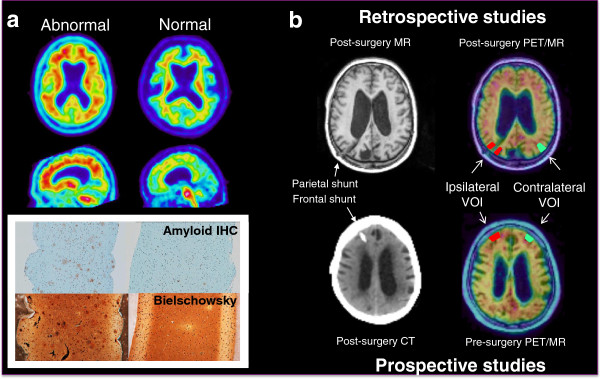
Introduction: PET imaging of amyloid-β (Aβ) in vivo holds promise for aiding in earlier diagnosis and intervention in Alzheimer's disease (AD) and mild cognitive impairment. AD-like Aβ pathology is a common comorbidity in patients with idiopathic normal pressure hydrocephalus (iNPH). Fifty patients with iNPH needing ventriculo-peritoneal shunting or intracranial pressure monitoring underwent [18F]flutemetamol PET before (N = 28) or after (N = 22) surgery. Cortical uptake of [18F]flutemetamol was assessed visually by blinded reviewers, and also quantitatively via standard uptake value ratio (SUVR) in specific neocortical regions in relation to either cerebellum or pons reference region: the cerebral cortex of (prospective studies) or surrounding (retrospective studies) the biopsy site, the contralateral homolog, and a calculated composite brain measure. Aβ pathology in the biopsy specimen (standard of truth [SoT]) was measured using Bielschowsky silver and thioflavin S plaque scores, percentage area of grey matter positive for monoclonal antibody to Aβ (4G8), and overall pathology impression. We set out to find (1) which pair(s) of PET SUVR and pathology SoT endpoints matched best, (2) whether quantitative measures of [18F]flutemetamol PET were better for predicting the pathology outcome than blinded image examination (BIE), and (3) whether there was a better match between PET image findings in retrospective vs. prospective studies.
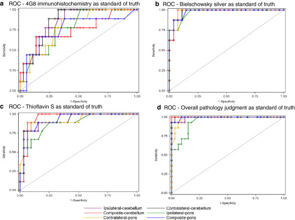
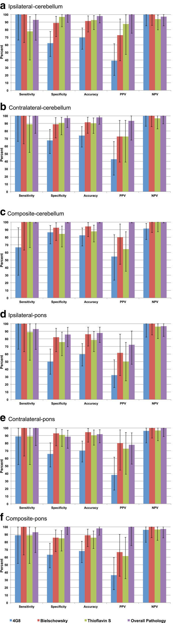
Results: Of the 24 possible endpoint/SoT combinations, the one with composite-cerebellum SUVR and SoT based on overall pathology had the highest Youden index (1.000), receiver operating characteristic area under the curve (1.000), sensitivity (1.000), specificity (1.000), and sum of sensitivity and specificity for the pooled data as well as for the retrospective and prospective studies separately (2.00, for all 3). The BIE sum of sensitivity and specificity, comparable to that for quantitation, was highest using Bielschowsky silver as SoT for all SUVRs (ipsilateral, contralateral, and composite, for both reference regions). The composite SUVR had a 100% positive predictive value (both reference regions) for the overall pathology diagnosis. All SUVRs had a 100% negative predictive value for the Bielschowsky silver result.
Conclusion: Bielschowsky silver stain and overall pathology judgment showed the strongest associations with imaging results.
Figures
References
-
- Blümke I. History of Neuroscience:Alois Alzheimer's First Report on Cortical Neurodegeneration. IBRO History of Neuroscience. Accessed: 11 Oct 2013 at http://www.ibro.info/Pub/Pub_Main_Display.asp?LC_Docs_ID=3531.
-
- McKhann G, Drachman D, Folstein M, Katzman R, Price D, Stadlan EM. Clinical diagnosis of Alzheimer’s disease: report of the NINCDS-ADRDA Work Group under the auspices of Department of Health and Human Services Task Force on Alzheimer’s Diseases. Neurology. 1984;2:939–944. doi: 10.1212/WNL.34.7.939. - DOI - PubMed
-
- Albert MS, DeKosky ST, Dickson D, Dubois B, Feldman HH, Fox NC, Gamst A, Holtzman DM, Jagust WJ, Peteresen RC, Snyder PJ, Carrillo MC, Thies B, Phelps CH. The diagnosis of mild cognitive impairment due to Alzheimer’s disease: Recommendations from the National Institute on Aging-Alzheimer’s Association workgroups on diagnostic guidelines for Alzheimer’s disease. Alzheimers Dement. 2011;2:270–279. doi: 10.1016/j.jalz.2011.03.008. - DOI - PMC - PubMed
-
- Jack CR Jr, Albert MS, Knopman DS, McKhann GM, Sperling RA, Carrillo MC, Thies B, Phelps CH. Introduction to the recommendations from the National Institute on Aging-Alzheimer’s Association workgroups on diagnostic guidelines for Alzheimer’s disease. Alzheimers Dement. 2011;2:257–262. doi: 10.1016/j.jalz.2011.03.004. - DOI - PMC - PubMed
Publication types
MeSH terms
Substances
Grants and funding
LinkOut - more resources
Full Text Sources
Other Literature Sources
Medical
Research Materials
