. 2014 Oct 15;210(8):1239-47.
doi: 10.1093/infdis/jiu240. Epub 2014 Apr 21.

Increased susceptibility to vaginal simian/human immunodeficiency virus transmission in pig-tailed macaques coinfected with Chlamydia trachomatis and Trichomonas vaginalis

Affiliations

Increased susceptibility to vaginal simian/human immunodeficiency virus transmission in pig-tailed macaques coinfected with Chlamydia trachomatis and Trichomonas vaginalis

Tara R Henning et al. J Infect Dis. .

Abstract

Background: Sexually transmitted infections (STIs) are associated with an increased risk of human immunodeficiency virus (HIV) infection, but their biological effect on HIV susceptibility is not fully understood.

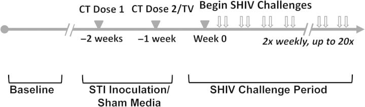
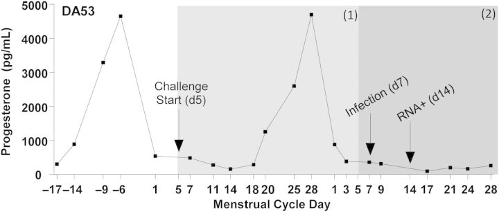
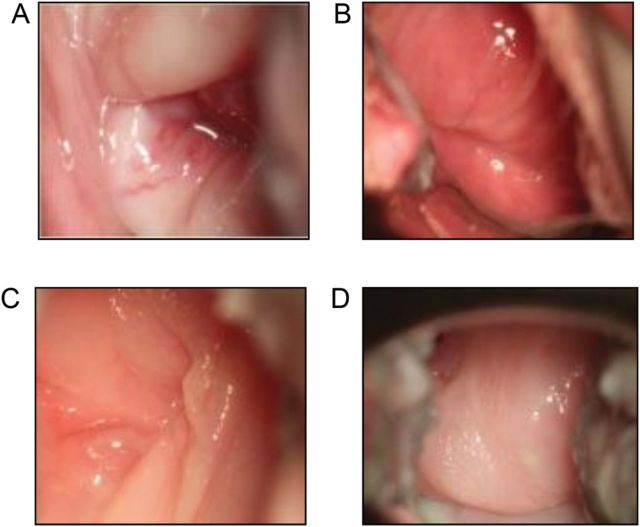
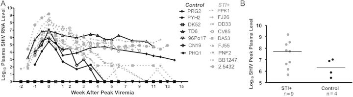
Methods: Female pig-tailed macaques inoculated with Chlamydia trachomatis and Trichomonas vaginalis (n = 9) or medium (controls; n = 7) were repeatedly challenged intravaginally with SHIVSF162p3. Virus levels were evaluated by real-time polymerase chain reaction, plasma and genital cytokine levels by Luminex assays, and STI clinical signs by colposcopy.

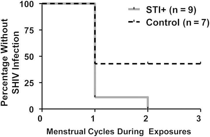
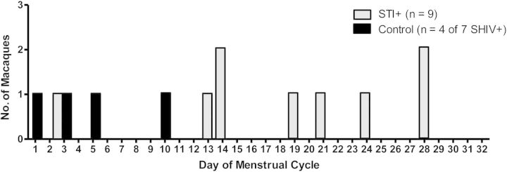
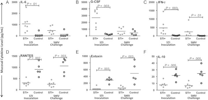
Results: Simian/HIV (SHIV) susceptibility was enhanced in STI-positive macaques (P = .04, by the log-rank test; relative risk, 2.5 [95% confidence interval, 1.1-5.6]). All STI-positive macaques were SHIV infected, whereas 3 controls (43%) remained uninfected. Moreover, relative to STI-negative animals, SHIV infections occurred earlier in the menstrual cycle in STI-positive macaques (P = .01, by the Wilcoxon test). Levels of inflammatory cytokines (interferon γ, interleukin 6, and granulocyte colony-stimulating factor [G-CSF]) were higher in STI-positive macaques during STI inoculation and SHIV exposure periods (P ≤ .05, by the Wilcoxon test).

Conclusions: C. trachomatis and T. vaginalis infection increase the susceptibility to SHIV, likely because of prolonged genital tract inflammation. These novel data demonstrate a biological link between these nonulcerative STIs and the risk of SHIV infection, supporting epidemiological associations of HIV and STIs. This study establishes a macaque model for studies of high-risk HIV transmission and prevention.

Keywords: Chlamydia; HIV risk; HIV susceptibility model; STI or STD; Trichomonas; macaque; menstrual cycle.


Figures

Figure 1.
Figure 1.
Study design. Analyses focused on 3 primary study stages: baseline, sexually transmitted pathogen (or sham media) inoculation period, and simian/human immunodeficiency virus (SHIV) challenge period. Abbreviations: CT, Chlamydia trachomatis; TV, Trichomonas vaginalis. Progesterone levels were measured once or twice weekly for 8–12 weeks during baseline (and throughout SHIV challenge period) to accurately assess menstrual cycle phase.
Figure 2.
Figure 2.
Representative menstrual cycle time line, relative to simian/human immunodeficiency virus (SHIV) susceptibility periods­. Progesterone levels were plotted and used to determine menstrual cycle day, with day 1 of each cycle designated as the day following the steep decline in the hormone level. For analysis purposes, 1 menstrual cycle equals 1 SHIV susceptibility period, and during SHIV exposures, a single susceptibility period was defined from the menstrual cycle day on which SHIV challenges began until the same respective day in the following cycle, as indicated by gray shading. Macaques which became infected within 1 menstrual cycle had a deduced viral eclipse–corrected day of infection within the first susceptibility period, or in the case of DA53 (as shown), SHIV infection occurred in the second menstrual cycle, requiring 2 susceptibility periods for infection. For uninfected macaques in the control arm, up to 3 susceptibility periods were observed without infection (Supplementary Materials).
Figure 3.
Figure 3.
Representative colposcopy images. Tissue edema, erythema, vascularization (A), and petechiae (B) of 2 representative sexually transmitted infection–positive animals (DA53 and FJ55). Normal cervical mucosa of 2 representative control macaques (DK52 and PYH2; C and D).
Figure 4.
Figure 4.
Susceptibility periods during simian/human immunodeficiency virus challenges/exposures. Kaplan–Meier survival analysis comparing the number of susceptibility periods, or completed menstrual cycles, required for infection among sexually transmitted infection (STI)–positive (gray line) versus control (dashed black line) animals. STI-positive animals were infected within fewer menstrual cycles, compared with controls (P = .04, log–rank test).
Figure 5.
Figure 5.
For each simian/human immunodeficiency virus (SHIV)–positive animal (9 of 9 in the sexually transmitted infection [STI]–positive group and 4 of 7 in the control group), the sampling time point at which plasma SHIVSF162p3 RNA was first detected was plotted by the respective day of the menstrual cycle. Plasma progesterone levels were monitored throughout the study course, and day 1 was defined as the time point immediately following the steep progesterone decline (as described in Figure 2). Plasma SHIVSF162p3 RNA in STI-positive animals (grey bars) was detected later in the menstrual cycle, compared with controls (black bars; P = .01, by the Wilcoxon test).
Figure 6.
Figure 6.
A, Longitudinal plasma simian/human immunodeficiency virus (SHIV) RNA levels. Log10 SHIV RNA levels plotted (y-axis) for sexually transmitted infection (STI)–positive (grey dashed) and control (black solid) animals, relative to the time of peak viremia (x-axis). Differences in levels in SHIV-infected animals between the 2 study groups were not statistically significant (96Po17, DK52, and PHQ1 were excluded from analyses as uninfected macaques). B, Peak plasma SHIV RNA levels. No significant difference was noted between SHIV-infected, STI-positive (grey) and control macaques. Hash indicates median levels.
Figure 7.
Figure 7.
Median per-subject levels of cervicovaginal (mucosal) cytokines. Analytes measured from available samples of 7 sexually transmitted infection (STI)–positive and 5 control macaques. Median levels of the inflammatory markers interleukin 6 (IL-6; A), granulocyte colony-stimulating factor (G-CSF; B), and interferon γ (IFN-γ; C) were significantly higher during the sexually transmitted pathogen inoculation period in STI-positive animals, compared with control animals (P = ≤ .01, by the Wilcoxon test); levels of IFN-γ remained higher during the SHIV challenge period (P = .01). Median levels of the human immunodeficiency virus–inhibitory chemokines RANTES (D) and eotaxin (E) and the downregulatory cytokine interleukin 10 (IL-10; F) were significantly higher in control animals versus STI-positive animals during both the STI inoculation and SHIV challenge phases (P ≤ .002, by the Wilcoxon test).

References

    1. Cohen MS. Sexually transmitted diseases enhance HIV transmission: no longer a hypothesis. Lancet. 1998;351(suppl 3):5–7. - PubMed
    1. Gray RH, Wawer MJ, Sewankambo NK, et al. Relative risks and population attributable fraction of incident HIV associated with symptoms of sexually transmitted diseases and treatable symptomatic sexually transmitted diseases in Rakai District, Uganda. Rakai Project Team. AIDS. 1999;13:2113–23. - PubMed
    1. Laga M, Manoka A, Kivuvu M, et al. Non-ulcerative sexually transmitted diseases as risk factors for HIV-1 transmission in women: results from a cohort study. AIDS. 1993;7:95–102. - PubMed
    1. Quinn TC. STDs, viral load, circumcision, and the transmission of HIV. In: Braun JF, Horn T, editors. The PRN handbook. New York City: Physicians Research Network; 2000.
    1. Sexton J, Garnett G, Rottingen JA. Metaanalysis and metaregression in interpreting study variability in the impact of sexually transmitted diseases on susceptibility to HIV infection. Sex Transm Dis. 2005;32:351–7. - PubMed

Publication types

MeSH terms