. 2014 Apr 22;9(4):e95370.
doi: 10.1371/journal.pone.0095370. eCollection 2014.

miR-200c regulates IL8 expression by targeting IKBKB: a potential mediator of inflammation in leiomyoma pathogenesis

Affiliations

miR-200c regulates IL8 expression by targeting IKBKB: a potential mediator of inflammation in leiomyoma pathogenesis

Tsai-Der Chuang et al. PLoS One. .

Abstract

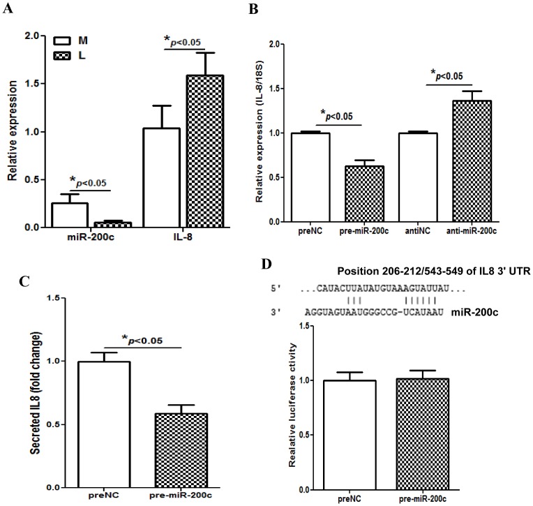
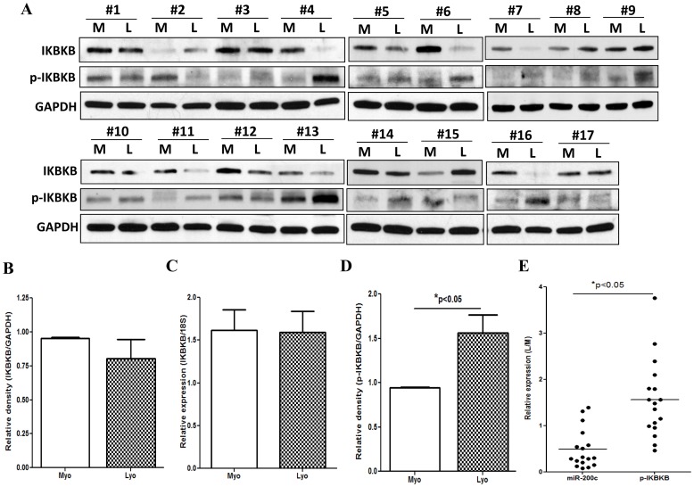
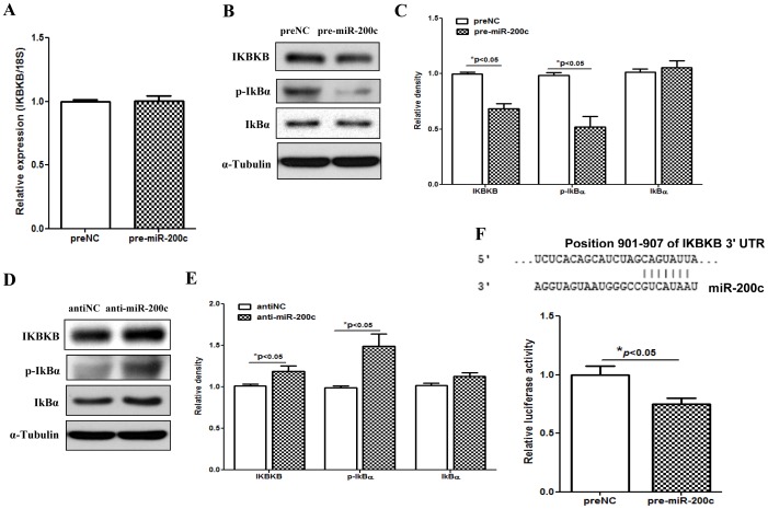
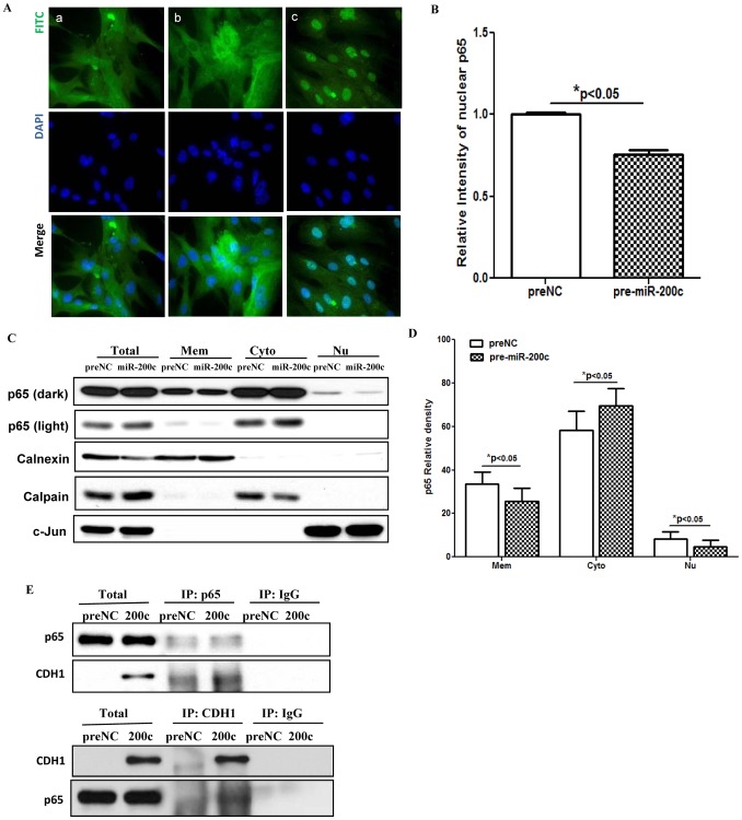
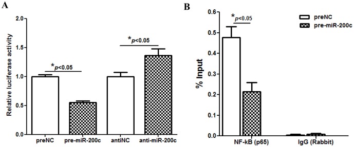
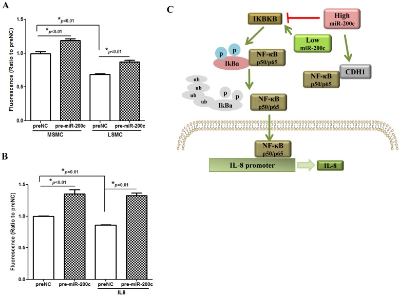
We have previously reported that leiomyoma expressed lower levels of miR-200c and elevated IL8 as compared to paired myometrium. Here we addressed the regulatory functions of miR-200c on the expression of inflammatory mediators and cellular viability using leiomyomas and paired myometrium and their isolated primary smooth muscle cells. Our results indicated that gain-of function or knockdown of miR-200c in leiomyoma smooth muscle cells (LSMC) regulated IL8 mRNA and protein expression through direct targeting of IKBKB and alteration of NF-kB activity. Additionally, leiomyoma expressed higher levels of phosphorylated IKBKB with no significant difference in the level of IKBKB mRNA and protein as compared to matched myometrium. Gain-of function of miR-200c in LSMC resulted in decreased IkBα phosphorylation and p65 nuclear translocation, which led to decreased p65 transcriptional activity of IL8 promoter, and increased caspase 3/7 activity which was not reversible following IL8 restoration. Collectively, our results suggest that NF-κB signaling pathway is a target of miR-200c regulatory function, and low level of miR-200c expression in leiomyoma by transcriptional regulation of inflammatory mediators such as IL8, in part account for development of leiomyomas.


Conflict of interest statement

Competing Interests: The authors have declared that no competing interests exist.

Figures

Figure 1
Figure 1. Gain-of function of miR-200c led to down-regulation of IL8.
Bar graphs in figure 1A show the relative expression of miR-200c and IL8 in leiomyoma (L) and matched myometrium (M) (N = 49). Relative expression of IL8 mRNA (Fig. 1B) and IL8 content (Fig. 1C) determined by QRT-PCR and ELISA of culture conditioned media of LSMC transfected with pre-miR-200c, anti-miR-200c or negative control (preNC or antiNC) for 48 hrs and 72 hrs respectively. Figure 1D shows relative luciferase activity in LSMC co-transfected with pGL3 construct carrying a 3′UTR fragment of IL8, firefly luciferase reporters, pRL-TK and pre-miR-200c or preNC. The ratio of firefly:Renilla was determined and reported as relative luciferase activity as compared to preNC. Sequence alignment of miR-200c seed regions and IL8 mRNAs target sites at their 3′UTRs with the coordinated positions are shown at the top of graph. The data are reported as mean ± SEM of experiments performed using 3 to 5 sets of isolated LSMC prepared from leiomyoma from three different patients. The results were analyzed using non-parametric student t-test and corresponding lines with asterisks on the bars denote statistical significance.
Figure 2
Figure 2. phosphorylated IKBKB displayed an inverse relationship with miR-200c expression.
Figure 2A shows western blot analysis of IKBKB and phosphorylated IKBKB (p-IKBKB) (Ser 177/181) of tissue extracts (N = 17) from leiomyomas (L) and paired myometrium (M) with GAPDH was used as loading control. The relative band densities of IKBKB (Fig. 2B) and phosphorylated IKBKB (Fig. 2D) in leiomyoma and paired myometrium were determined and compared with values in myometrium independently set as 1 for each pair. Figure 2C shows relative expression of IKBKB mRNA level in leiomyoma and paired myometrium from untreated group (N = 49). The data were analyzed using nonparametric student t-test. Figure 2E shows the relative expression miR-200c as compared to the level of phosphorylated IKBKB (p-IKBKB) in above 17 leiomyomas as compared to matched myometrium. The results were analyzed using paired student t-test and corresponding lines with asterisks denote statistical significance.
Figure 3
Figure 3. IKBKB is a direct target of miR-200c.
Figure 3A shows the influence of gain-of function of miR-200c in LSMC on the expression of IKBKB after 48 hrs transfection as determined by QRT-PCR. Figures B, C, D and E show western blots and band intensity analysis of IKBKB, IkBα and phosphorylated IkBα (p-IkBα) (Ser 32/36) in LSMC following transfection with preNC, pre-miR-200c, antiNC or anti-miR-200c for 48 hrs with α-tubulin used as loading control. The relative band intensities were determined and the values for preNC or antiNC were independently set as 1 for comparative analysis. Figure 3F shows luciferase reporter assay following co-transfection of LSMC with firefly luciferase reporter carrying a 3′UTR fragment of IKBKB, pRL-TK, and pre-miR-200c or preNC. The ratio of Firefly:Renilla was determined after 48 hrs and reported as relative luciferase activity as compared to preNC independently set as 1 for each assessment. The results presented as mean ± SEM of three sets of independent experiments using LSMC isolated from 3 patients and analyzed using unpaired student t test. Asterisks denote statistical significance indicated by corresponding lines. Sequence alignment of miR-200c seed regions and IKBKB mRNAs target sites at their 3′UTRs with the coordinated positions are shown at the top of graph.
Figure 4
Figure 4. Gain-of function of miR-200c suppressed NF-kB signaling pathway.
Figure 4A shows immunofluorescence staining of LSMC transfected with pre-NC, (panel a), pre-miR-200c (panel b) for 48 hrs, or treated with IL-1b (5 ng/ml) for 1 h as positive control (panel c). The cells were fixed and immunostained for p65 subunit of NF-kB (green, p65) using p65 antibody and counter-stained with DAPI (blue, nucleus) with arrows indicating to NF-kB p65 nuclear immunostaining. Figure B shows mean ± SEM of relative nuclear immunostaining intensity of p65 (*p<0.05 compared to preNC). Figure 4C shows immunoblot analysis of NF-kB p65 in LSMC (3×10⁵/100 mm dish) transfected with pre-miR-200c or preNC for 48 hrs. The cells were harvested and subfractionated into membrane (Mem), cytoplasmic (Cyto), and nuclear (Nu) proteins and subjected to immunoblot analysis, with Calnexin, Calpain and c-Jun served as markers for the respective subcellular fractions. The NF-kB p65 band intensity (Fig. D) was semiquantified and reported as mean ± SEM of percentage of total p65 associated with Mem, Cyto and Nu fractions in cells transfected with pre-miR-200c or preNC (*p<0.05 when compared to preNC). Figure 4E shows immunoprecipitation and immunoblotting of LSMC total cell lysates after transfection with pre-miR-200c or preNC using p65 or CDH1 (E-cadherin) antibodies. Similar results were obtained from three sets of independent experiments.
Figure 5
Figure 5. Gain-of function of miR-200c inhibited NF-kB p65 binding activity in IL8 promoter.
Figure 5A shows the level of NF-kB activity in LSMC transfected with a luciferase reporter construct containing preserved NF-kB binding sites, pRL-TK, pre-miR-200c, anti-miR-200c, preNC or antiNC. The ratio of Firefly:Renilla was determined after 48 hrs and reported as relative luciferase activity as compared to preNC which was independently set as 1 for each cell. (Fig. 5B) NF-kB binding ability in endogenous IL8 promoter accessed by CHIP assay. LSMC were transfected with pre-miR-200c or preNC. After incubation for 48 hrs, cells were harvested and processed for CHIP assay using p65 antibody. Immunoprecipitated chromatin was analyzed by PCR using the specific primer for IL8 promoter and presented by percent input method. The results are presented as mean ± SEM of three sets of independent experiments using primary LSMC isolated from 3 patients and analyzed using non-parametric student unpaired t test with asterisks denote statistical significance indicated by corresponding lines.
Figure 6
Figure 6. Gain-of function of miR-200c induced caspase 3/7 activity, but the effect could not be reversed by IL8 restoration.
Figure 6A shows influence of gain-of function of miR-200c and preNC on caspase 3/7 activity in MSMC and LSMC after 96 hrs of incubation with culture media changed every two days. The results are reported as mean ± SEM of experiments performed in six replicates from three independent cell preparations using the same paired of primary MSMC and LSMC isolated from 3 patients and analyzed using nonparametric student t-test with asterisks denote statistical significance as compared to preNC. Figure 6B shows caspase 3/7 activity in LSMC transfected with pre-miR-200c or preNC treated with or without IL8 (50 ng/ml) for 96 hrs. The results are reported as mean ± SEM of experiments performed in six replicates, using three independent cell preparations and analyzed using nonparametric student t-test with asterisks denote statistical significance as compared to preNC. Figure 6C shows a schematic representation of aberrant expression and regulatory function of miR-200c on the expression of IL8 through NF-kB signaling pathway involving IKBKB in LSMC. Our results suggest that aberrant expression of miR-200c through functional regulation of specific target genes serve as mediator of inflammatory and tissue turnover play a key role in leiomyoma development, growth and associated symptoms.

References

    1. Chegini N (2010) Proinflammatory and profibrotic mediators: principal effectors of leiomyoma development as a fibrotic disorder. Semin Reprod Med 28: 180–203. - PMC - PubMed
    1. Schafer M, Werner S (2008) Cancer as an overhealing wound: an old hypothesis revisited. Nat Rev Mol Cell Biol 9: 628–638. - PubMed
    1. Wynn TA (2008) Cellular and molecular mechanisms of fibrosis. J Pathol 214: 199–210. - PMC - PubMed
    1. Fabian MR, Sonenberg N, Filipowicz W (2010) Regulation of mRNA translation and stability by microRNAs. Annu Rev Biochem 79: 351–379. - PubMed
    1. Huntzinger E, Izaurralde E (2011) Gene silencing by microRNAs: contributions of translational repression and mRNA decay. Nat Rev Genet 12: 99–110. - PubMed

Publication types

MeSH terms