. 2014 May;6(5):393-403.
doi: 10.1038/nchem.1919. Epub 2014 Apr 20.

Optimized orthogonal translation of unnatural amino acids enables spontaneous protein double-labelling and FRET

Affiliations

Optimized orthogonal translation of unnatural amino acids enables spontaneous protein double-labelling and FRET

Kaihang Wang et al. Nat Chem. 2014 May.

Erratum in

  • Nat Chem. 2014 Feb;7(2):178. Wilf, Nabil W [corrected to Wilf, Nabil M]

Abstract

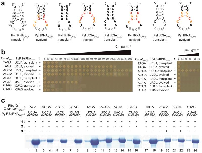
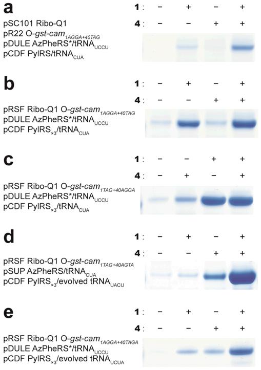
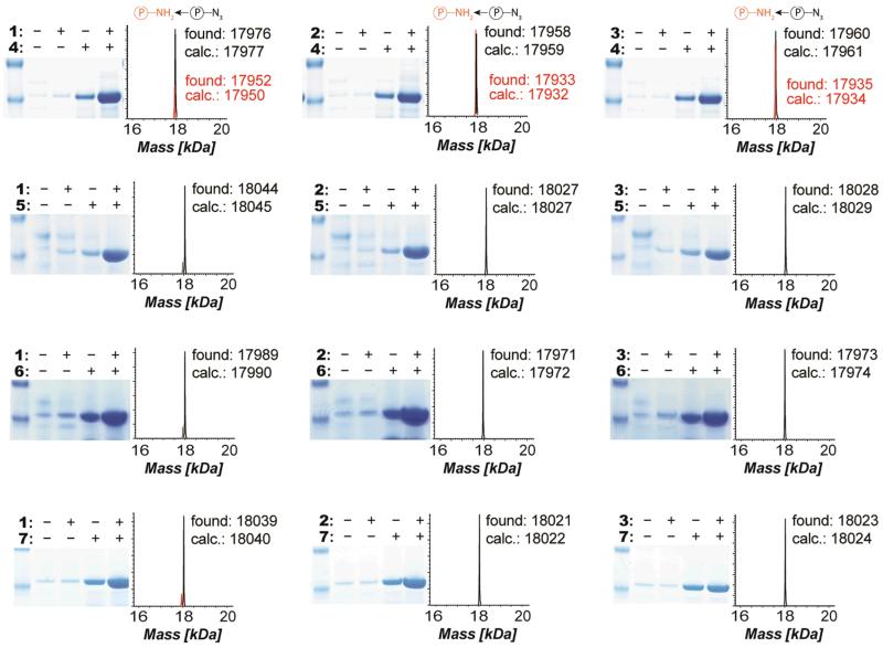
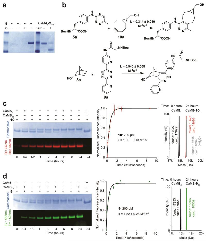
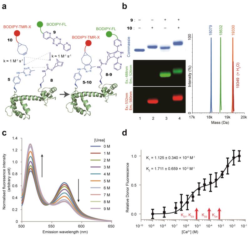
The ability to introduce different biophysical probes into defined positions in target proteins will provide powerful approaches for interrogating protein structure, function and dynamics. However, methods for site-specifically incorporating multiple distinct unnatural amino acids are hampered by their low efficiency. Here we provide a general solution to this challenge by developing an optimized orthogonal translation system that uses amber and evolved quadruplet-decoding transfer RNAs to encode numerous pairs of distinct unnatural amino acids into a single protein expressed in Escherichia coli with a substantial increase in efficiency over previous methods. We also provide a general strategy for labelling pairs of encoded unnatural amino acids with different probes via rapid and spontaneous reactions under physiological conditions. We demonstrate the utility of our approach by genetically directing the labelling of several pairs of sites in calmodulin with fluorophores and probing protein structure and dynamics by Förster resonance energy transfer.


Figures

Figure 1
Figure 1. Optimizing Pyl tRNA(N8)XXXX for incorporating unnatural amino acids in response to quadruplet codons decoded by ribo-Q1
a. Nucleotides that are targeted for mutagenesis in the anticodon stem loop of Pyl tRNA(N8)XXXX are represented in orange on tRNA bound to the ribosome , and the rest of the tRNA is in yellow. rRNA in pale green with ribo-Q1 mutation sites in red, and mRNA in purple. The structural image is based on PDB ID 2J00, created with Pymol (www.pymol.org). b. The anticodon stem loop of Pyl tRNA(N8)XXXX. The nucleotides in orange are randomized in each library. Codon sequences and mRNAs are in purple, and anticodons in grey. c. Two-step selection procedure for identifying specific and efficient Pyl tRNA(N8)XXXX library members in orthogonal translation. A negative O-barnase selection followed by a positive O-cat selection. Negative selection in the absence of unnatural amino acids eliminates Pyl tRNA(N8)XXXX library members that are mis-aminoacylated with natural amino acids by endogenous aminoacyl synthetases. Subsequent positive selection enriches evolved Pyl tRNA(N8)XXXX library members that are aminoacylated with the added unnatural amino acid by PylRS and efficiently decoded at quadruplet codons by ribo-Q1. a.a, amino acid; O-mRNA, orthogonal mRNA; cat, chloramphenicol acetyl transferase.
Figure 2
Figure 2. Evolved Pyl tRNA(N8)XXXX direct the efficient unnatural amino acids incorporation in response to quadruplet codons decoded by ribo-Q1
a. The selected anticodon stem-loop sequences of evolved Pyl tRNAXXXX, and the corresponding transplant sequences. The anticodons are in gray, and the nucleotides mutated in the library are shown in colour. Positions where parental sequence is selected are in orange, and positions where new nucleotides are selected are in red. b, Evolved Pyl tRNAXXXX substantially enhance the incorporation of unnatural amino acids by ribo-Q1 in response to quadruplet codons when compared to the corresponding transplant Pyl tRNAXXXX. The unnatural amino acid-dependent decoding of quadruplet codons in the O-cat111XXXX was measured by survival on increasing concentrations of chloramphenicol (Cm). c. Diverse unnatural amino acids are efficiently incorporated in recombinant proteins in response to quadruplet codons using PylRS/Pyl tRNAXXXX with orthogonal translation. Full gels are given in Supplementary Fig. S4.
Figure 3
Figure 3. Efficient incorporation of multiple distinct unnatural amino acids into a single polypeptide
a. Site-specific incorporation of 1 and 4. AGGA replaces the 1st codon and UAG replaces the 40th codon in the cam open reading frame of O-gst-cam to make O-gst-cam1AGGA+40TAG. Decoding of both the AGGA and TAG codons by ribo-Q1 produces full length Gst-CaM, and failure to decode these codons leads to premature termination of the polypeptide. b. and c. The site-specific incorporation efficiency of 1 and 4 is improved by reducing the number of plasmids. d. The newly evolved PylRS/tRNAUACU pair substantially increases the efficiency of double incorporation e. Incorporating two distinct unnatural amino acids using two distinct quadruplet codons. Full gels are given in Supplementary Fig. S7.
Figure 4
Figure 4. Efficient incorporation of a matrix of pairs of unnatural amino acids, including photocrosslinkers and chemical handles (azides, alkenes, alkynes, tetrazines) demonstrates generality
Cells contained O-gst-cam1TAG+40AGTA and ribo-Q1 expressed from an RSF plasmid, the pSUP MjAzPheRS/tRNACUA plasmid (or a variant specific for the relevant substrate) and the pCDF PylRS/evolved tRNAUACU plasmid. All combinations of PylRS substrates (1-3) and MjTyrRS active site variant substrates (4-7) were incorporated in 3×4 matrix. We further confirmed the incorporation of distinct unnatural amino acids by ESI and MALDI mass spectrometry (Supplementary Fig. S8). Full gels are given in Supplementary Fig. S8. We observed an additional peak for protein samples with 4 corresponding to the reduction of the azide to an amine.
Figure 5
Figure 5. 5 and 8 do not react with each other in a protein, but can be efficiently labelled with 10 and 9
a. 5 and 8 do not react when placed in proximity within a protein. CaM51-8149 was purified from cells bearing pRSF ribo-Q1 O-gst-cam1TAG+149AGTA, pSUP MjTetPheRS/tRNACUA, pCDF NorKRS×3/evolved tRNAUACU. CaM41-2149 undergoes a Cu(I) catalyzed click reaction to cyclize the protein (right gel panel). CaM51-8149 does not cyclize, as judged by mobility shift (compare left and right panels) and ESI-MS (Supplementary Fig. S10). b. Rate constants for the indicated reactions. c. CaM51 and CaM840 were incubated with 100 molar equivalents of 10 at 25°C. Only CaM51 was labelled with 10, yielding CaM5-101. Labelling was visualized using a Typhoon Imager and the resulting time-dependent fluorescence was used to calculate the on-protein labelling rate constant. All measurements were repeated twice and the error bars represent the standard deviation. ESI-MS confirmed that protein labelling is quantitative. d. CaM51 and CaM840 were incubated with 100 molar equivalents of 9 at 25°C. Only CaM840 was labelled with 9, which yielded CaM8-940. The labelling reaction was analysed as described in c. ESI-MS confirmed that labelling was quantitative.
Figure 6
Figure 6. Site-specific double-labelling of CaM with a FRET pair to follow changes in protein conformation
a. Strategy for protein double-labelling via inverse electron-demand Diels-Alder reactions. b. Quantitative and site-specific double labelling of CaM51-840. Purified CaM51-840 (Lane 1) was labelled with 100 equivalents (200μM) of 10, which yielded CaM5-101-840 (Lane 2) or 100 equivalents 9, which yielded CaM51-8-940 (Lane 3). CaM51-8-940 was labelled with 100 equivalents of 10, which yielded CaM5-101-8-940 (Lane 4). Labelling was visualised by fluorescence imaging and led to a mobility shift. All labelling reactions were quantitative, as confirmed by ESI-MS. CaM51-840 (blue peak; calculated Mass=18081 Da, observed Mass=18079 Da), CaM51-8-940 (green peak; calculated Mass=18635, observed Mass=18632), CaM5-101-8-940 (orange peak; calculated Mass=19336, observed Mass=19330). c. Fluorescence spectra of CaM5-101-8-9149 (following donor excitation at 485 nm) in the presence of increasing concentrations of urea. d. The relative donor-fluorescence intensity from doubly labelled CaM5-101-8-940 as a function of Ca2+ concentration. All measurements were repeated at least six times and the error bars represent the standard deviation. K1, K2 are for the observed transitions. R2= 0.9005. KD1, KD2, KD3 and KD4 are the reported KD (dissociation constants) values for sequential Ca2+ binding .

Comment in

References

    1. Zhang J, Campbell RE, Ting AY, Tsien RY. Creating new fluorescent probes for cell biology. Nature Reviews Molecular Cell Biology. 2002;3:906–918. - PubMed
    1. Kajihara D, et al. FRET analysis of protein conformational change through position-specific incorporation of fluorescent amino acids. Nat Methods. 2006;3:923–929. - PubMed
    1. Scott CP, Abel-Santos E, Wall M, Wahnon DC, Benkovic SJ. Production of cyclic peptides and proteins in vivo. Proc Natl Acad Sci U S A. 1999;96:13638–13643. - PMC - PubMed
    1. Li P, Roller PP. Cyclization strategies in peptide derived drug design. Curr Top Med Chem. 2002;2:325–341. - PubMed
    1. Wang K, Schmied WH, Chin JW. Reprogramming the genetic code: from triplet to quadruplet codes. Angew Chem Int Ed Engl. 2012;51:2288–2297. - PubMed

Publication types