. 2014 Apr 22;9(4):e95846.
doi: 10.1371/journal.pone.0095846. eCollection 2014.

NF-kappa B modulation is involved in celastrol induced human multiple myeloma cell apoptosis

Affiliations

NF-kappa B modulation is involved in celastrol induced human multiple myeloma cell apoptosis

Haiwen Ni et al. PLoS One. .

Abstract

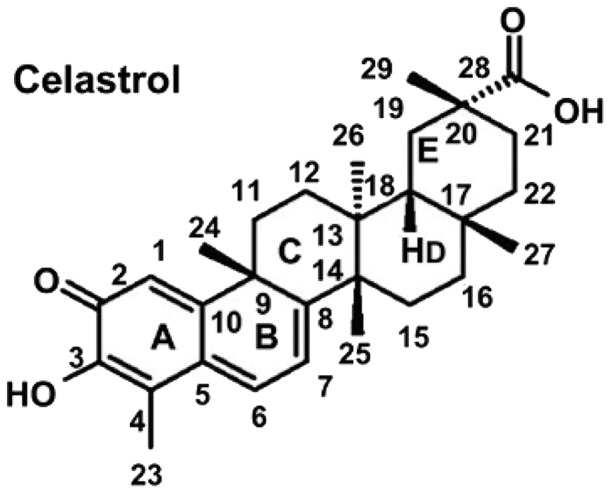
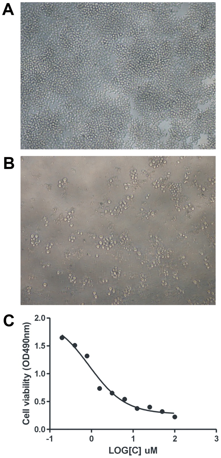
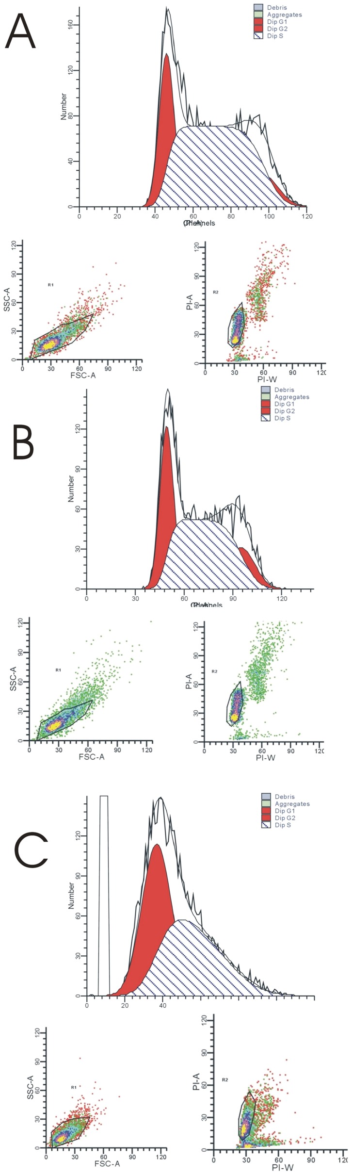
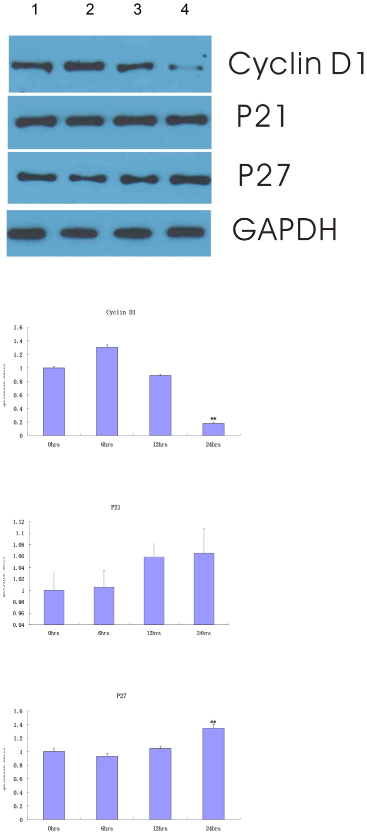
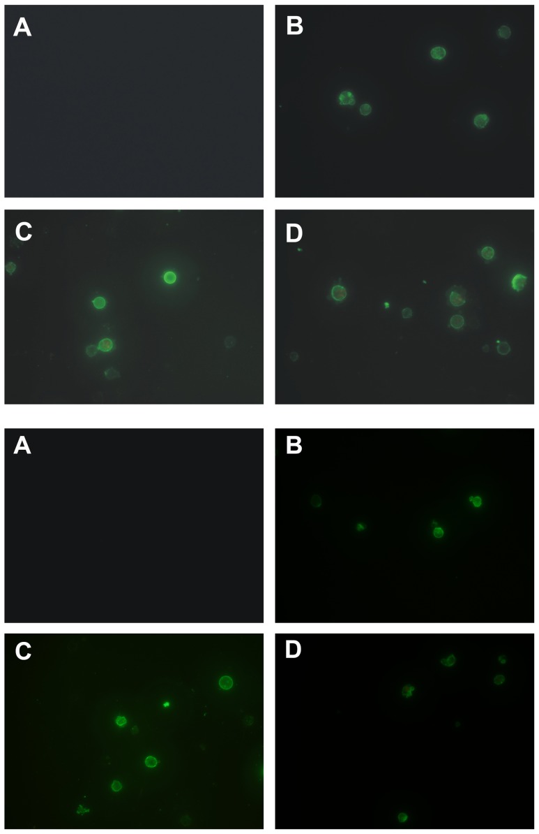
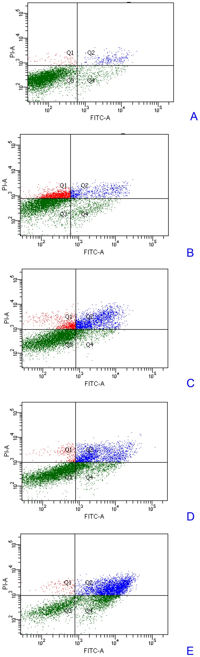
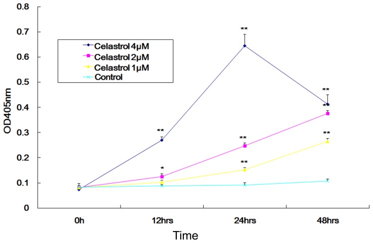
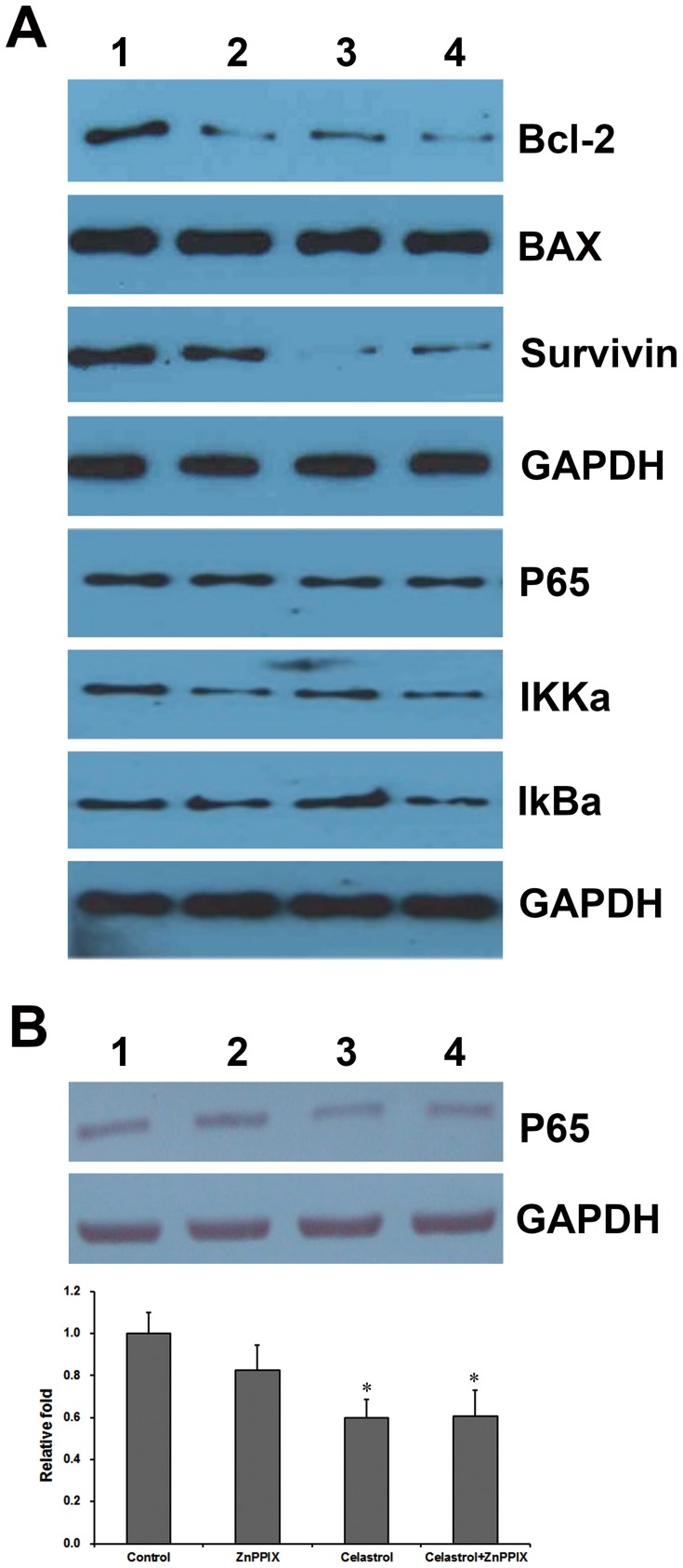
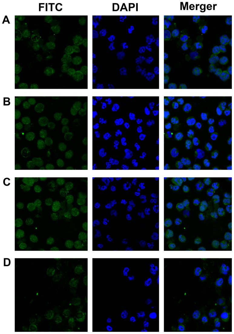
Celastrol is an active compound extracted from the root bark of the traditional Chinese medicine Tripterygium wilfordii Hook F. To investigate the effect of celastrol on human multiple myeloma cell cycle arrest and apoptosis and explore its molecular mechanism of action. The activity of celastrol on LP-1 cell proliferation was detected by WST-8 assay. The celastrol-induced cell cycle arrest was analyzed by flow cytometry after propidium iodide staining. Nuclear translocation of the nuclear factor kappa B (NF-κB) was observed by fluorescence microscope. Celastrol inhibited cell proliferation of LP-1 myeloma cell in a dose-dependent manner with IC50 values of 0.8817 µM, which was mediated through G1 cell cycle arrest and p27 induction. Celastrol induced apoptosis in LP-1 and RPMI 8226 myeloma cells in a time and dose dependent manner, and it involved Caspase-3 activation and NF-κB pathway. Celastrol down-modulated antiapoptotic proteins including Bcl-2 and survivin expression. The expression of NF-κB and IKKa were decreased after celastrol treatment. Celastrol effectively blocked the nuclear translocation of the p65 subunit and induced human multiple myeloma cell cycle arrest and apoptosis by p27 upregulation and NF-kB modulation. It has been demonstrated that the effect of celastrol on NF-kB was HO-1-independent by using zinc protoporphyrin-9 (ZnPPIX), a selective heme oxygenase inhibitor. From the results, it could be inferred that celastrol may be used as a NF-kB inhibitor to inhibit myeloma cell proliferation.


Conflict of interest statement

Competing Interests: Wanzhou Zhao is employed by Sino-EU Biomedical Innovation Center (SEBIC), OG Pharma Corporation. This does not alter the authors' adherence to all the PLOS ONE policies on sharing data and materials.

Figures

Figure 1
Figure 1. Chemical structures of Celastrol.
Celastrol is a natural triterpenoid quinone methide isolated from the Chinese plant genuses of celastrus, maytenus, and tripterygium.
Figure 2
Figure 2. Celastrol inhibited LP-1 human multiple myeloma cell proliferation.
Morphological signatures of celastrol growth inhibitory effects. Cells were treated with various doses of celastrol and cell viability was determined by MTT assay. Computer-assisted phase-contrast microscopy illustrations of control LP-1 human multiple myeloma cells (A) and treated with celastrol at concentration 0.78125 µM for 72 h (B). IC50 was shown as calculated by Prism 5.0. The IC50 growth inhibitory concentration for 72 h is 0.8817 µM (C).
Figure 3
Figure 3. Celastrol causes accumulation of LP-1 human multiple myeloma cells in the G1 phase.
LP-1 cells (2×10⁶ mL-1) were treated with 0.5 µM celastrol for 0 (A), 12 (B) or 24 h (C), after which the cells were washed, fixed, stained with PI, and analyzed for DNA content by flow cytometry. Cell population in each cell cycle phase was numerically depicted. The G1 phase cell population was 24.15%, 29.79% and 49.20%, respectively. Data represent one of three independent experiments.
Figure 4
Figure 4. Celastrol up-modulated p27 protein expression in LP-1 cells.
LP-1 cells were treated with 0.5 µM celastrol for 0 h (lane 1), 6 h (lane 2), 12 h (lane 3), and 24 h (lane 4), respectively. Whole cell lysates were prepared, separated on SDS-PAGE, and subjected to Western blot using antibodies against Cyclin D1, p27, and p21. The same blots were stripped and reprobed with GAPDH antibody to show equal protein loading. Expression fold was the ratio of protein expression in the celastrol-treated group to 0 h. Columns, mean; bars, SD (n = 3). *, p<0.05; **, p<0. 01.
Figure 5
Figure 5. Celastrol induced apoptosis in LP-1 cells (up) and RPMI 8226 cells (down).
Cells were treated with 1 µM celastrol for 0 h (A), 12 h (B), 24 (C), and 36 h (D) incubated with Annexin V conjugated with EGFP and analyzed with a fluorescence microscope for early apoptotic effects.
Figure 6
Figure 6. Celastrol induced apoptosis in a LP-1 cells.
LP-1 cells were treated with 1 µM celastrol for 0 h (A), 12 h (B), 24 h (C), 36 h (D), and 48 h (E) incubated with Annexin V conjugated with EGFP and analyzed with a flow cytometer for apoptotic effects. The apoptotic cell percentage was 7.8±0.4%, 16.1±1.2% (p<0.05 vs 0 h), 18.0±3.1% (p<0.05 vs 0 h), 39.5±5.8% (p<0.01 vs 0 h), and 71.2±8.6% (p<0.01 vs 0 h), respectively. The results shown are representative of three independent experiments.
Figure 7
Figure 7. Celastrol induced caspase-3 activation.
The cells were treated with 0 µM, 1 µM, 2 µM, and 4 µM celastrol for the indicated times. The whole cell extracts were prepared and subjected to enzymatic activity of caspase-3 by colorimetric substrate Ac-DEVD-pNA. The amount of yellow color at 405 nm indicating Caspase-3 activation fold was compared in the celastrol treated group to 0 h. point, mean; bars, SD (n = 3). *, p<0.05; **, p<0. 01.
Figure 8
Figure 8. Celastrol down-modulated antiapoptotic proteins expression in LP-1 cells.
(A). LP-1 cells were treated with 1 µM celastrol for 0 h (lane 1), 12 h (lane 2), 24 h (lane 3), and 48 h (lane 4), respectively. The whole cell lysates were prepared, separated on SDS-PAGE, and subjected to Western blot using antibodies against Bcl-2, Bax, Survivin, P65, IKKa, and IkBa. (B). LP-1 cells were treated with medium (lane 1), 1 µM ZnPPIX (lane 2), 1 µM celastrol (lane 3), and 1 µM celastrol combined with 1 µM ZnPPIX (lane 4) for 24 h, respectively. The same blots were stripped and reprobed with GAPDH antibody to show equal protein loading. Relative expression fold was first normalized by GADPH and then the ratio of protein expression in the celastrol/ZnPPIX -treated group was compared to control. Columns, mean; bars, SD (n = 3). *, p<0.05. No significance was observed from cells treated with Celastrol combined with ZnPPIX and those treated with only Celastrol (p>0.05).
Figure 9
Figure 9. Celastrol inhibited constitutively active NF-kB in LP-1 MM cells.
An altered subcellular distribution of NF-κb was observed. LP-1 cells were incubated with medium (A), 1 µM celastrol for 30 min (B), 1 µM Zinc Protoporphyrin-9 (ZnPPIX) (C), 1 µM celastrol combined with 1 µM ZnPPIX (D) and then analysed for the intracellular distribution of p65 by fluorescence microscope. Green indicates p65, and blue indicates nuclei (original magnification ×400). The results shown are representative of three independent experiments.

References

    1. Richardson PG, Mitsiades C, Schlossman R, Ghobrial I, Hideshima T, et al. (2008) Bortezomib in the front-line treatment of multiple myeloma. Expert Rev Anticancer Ther 8: 1053–1072. - PubMed
    1. Hideshima T, Chauhan D, Shima Y, Raje N, Davies FE, et al. (2000) Thalidomide and its analogs overcome drug resistance of human multiple myeloma cells to conventional therapy. Blood 96: 2943–2950. - PubMed
    1. Glasmacher A, Hahn C, Hoffmann F, Naumann R, Goldschmidt H, et al. (2006) A systematic review of phase-II trials of thalidomide monotherapy in patients with relapsed or refractory multiple myeloma. Br J Haematol 132: 584–593. - PubMed
    1. Raab MS, Podar K, Breitkreutz I, Richardson PG, Anderson KC (2009) Multiple myeloma. Lancet 374: 324–339. - PubMed
    1. Kumar S, Rajkumar SV (2006) Thalidomide and lenalidomide in the treatment of multiple myeloma. Eur J Cancer 42: 1612–1622. - PubMed

MeSH terms