. 2014 Apr 22;9(4):e95192.
doi: 10.1371/journal.pone.0095192. eCollection 2014.

Homogenous 96-plex PEA immunoassay exhibiting high sensitivity, specificity, and excellent scalability

Affiliations

Homogenous 96-plex PEA immunoassay exhibiting high sensitivity, specificity, and excellent scalability

Erika Assarsson et al. PLoS One. .

Abstract

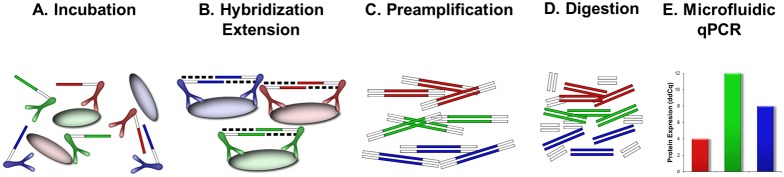
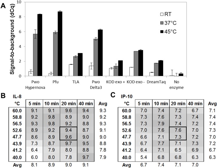
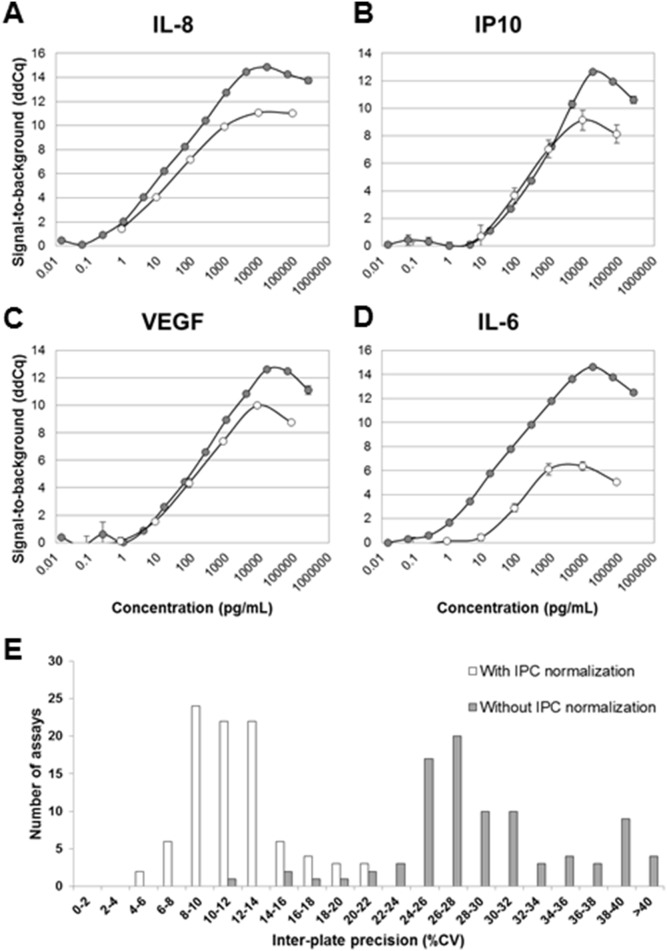
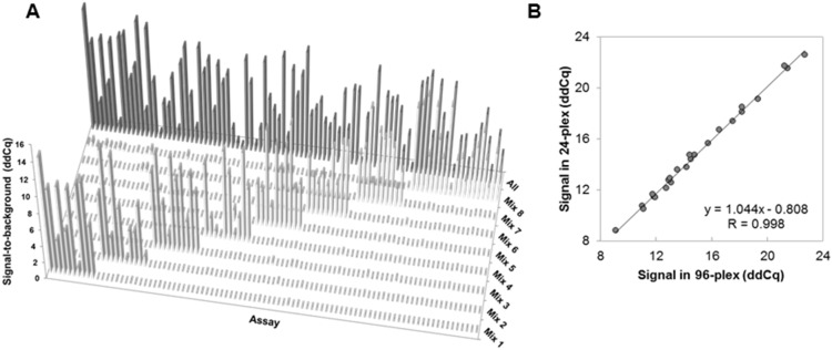
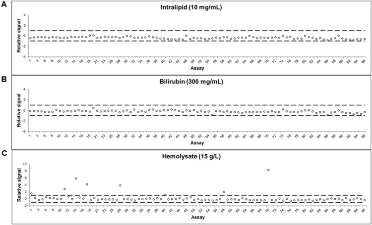
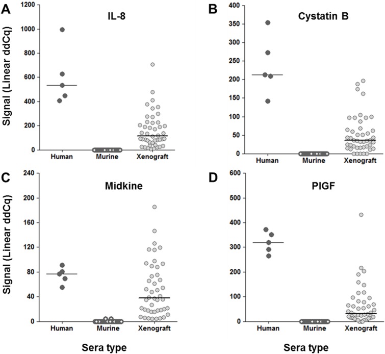
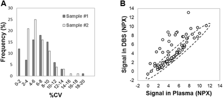
Medical research is developing an ever greater need for comprehensive high-quality data generation to realize the promises of personalized health care based on molecular biomarkers. The nucleic acid proximity-based methods proximity ligation and proximity extension assays have, with their dual reporters, shown potential to relieve the shortcomings of antibodies and their inherent cross-reactivity in multiplex protein quantification applications. The aim of the present study was to develop a robust 96-plex immunoassay based on the proximity extension assay (PEA) for improved high throughput detection of protein biomarkers. This was enabled by: (1) a modified design leading to a reduced number of pipetting steps compared to the existing PEA protocol, as well as improved intra-assay precision; (2) a new enzymatic system that uses a hyper-thermostabile enzyme, Pwo, for uniting the two probes allowing for room temperature addition of all reagents and improved the sensitivity; (3) introduction of an inter-plate control and a new normalization procedure leading to improved inter-assay precision (reproducibility). The multiplex proximity extension assay was found to perform well in complex samples, such as serum and plasma, and also in xenografted mice and resuspended dried blood spots, consuming only 1 µL sample per test. All-in-all, the development of the current multiplex technique is a step toward robust high throughput protein marker discovery and research.


Conflict of interest statement

Competing Interests: All authors (besides SBT and JS) are employees of Olink AB commercializing the described method under the name Proseek Multiplex. Patent name: “Exonuclease enabled proximity extension assays” and number: WO2012104261A1. There are no new patents, products in development or marketed products to declare. This does not alter our adherence to all the PLOS ONE policies on sharing data and materials.

Figures

Figure 1
Figure 1. Design and description of 96-plex PEA.
(A) 94 pairs of specific antibodies are equipped with oligonucletotides (PEA probes) and mixed with an antigen-containinig sample. (B) Upon sample incubation, all proximity probe pairs bind their specific antigens, which brings the probe oligonucleotides in close proximity to hybridize. The oligonucleotides have unique annealing sites that allows pair-wise binding of matching probes. Addition of a DNA polymerase leads to an extension and joining of the two oligonucleotides and formation of a PCR template. (C) Universal primers are utilized to preamplify all 96 different DNA templates in paralell. (D) Uracil-DNA glycosylase partly digests the DNA templates and remove all unbound primers. (E) Finally each individual DNA sequence is detected and quantified using specific primers in by microfluidic qPCR.
Figure 2
Figure 2. Improved enzymatic performance using a hyper-thermostable DNA polymerase.
(A) Seven hyper-thermostable DNA polymerases were compared using an IL-8 assay (10 pM Ag) with regards to their ability to remain inactive during RT addition (low background signal) while efficient at either RT (white bars), 37°C (gray bars), or 45°C (black bars) extension. Pwo Hypernova generated high signal-to-noise values (dCq) and chosen as the most suitable candidate. (B–C) Assessment of optimal time and temperature for the extension reaction using an IL-8 and an IP-10 assay (100 pM Ag). 20 min at about 50°C generated the highest signal-to-noise values (dCq) while retaining robust signals.
Figure 3
Figure 3. New preamplification and normalization protocols resulting in improved precision and sensitivity.
The optimized 96-plex PEA protocol (gray series) was used to analyze standard curves generated for (A) IL-8, (B) IP-10, (C) VEGF, and (D) IL-6 and the results compared that of the initial singleplex PEA protocol (white series) (E) A new normalization procedure was introduced, which significantly reduced the inter-assay precison from average 29 to 12%CV. Histograms show the distribution of CV% across 92 assays and 7 analyzed plasma/serum samples with (white), or without (gray) IPC normalization.
Figure 4
Figure 4. Demonstrating high specificity and scalability of 96-plex PEA.
(A) Submixes of antigens were analyzed and demonstrated that the different assays only responded to the submix containing the corresponding antigen and at signal-to-noise levels similar to that of the mix containing all antigens. (B) Scalability was assessed by analyzing 24 PEA assays either in 24-plex or 96-plex, and the normalized levels plotted and compared in an xy scatter. This demonstrated a Pearson correlation value (R) as high as 0.998.
Figure 5
Figure 5. Low interference in biological samples.
To determine whether PEA was affected by interference, three known interfering substances were spiked in plasma and measured by 96-plex PEA. No interference was seen with plasma containing (A) bilirubin or (B) intralipid, while a few assays showed increased signal in plasma containing (C) hemolysate, most likely due to actual analytes leaking out from the disrupted blood cells.
Figure 6
Figure 6. Detection of human proteins in sera of xenografted mice.
Nude mice were grafted with human tumor cells (SK-N-FI). Before (white symbols) and on day 30 after inoculation (gray symbols), the sera were analyzed with 96-plex PEA and compared to that of normal human sera (black symbols). Shown are ddCq-values for 4 examples of protein assays: (A) IL-8, (B) Cystatin B, (C) Midkine, and (D) PlGF, for which significant levels of human protein were detected in xenografted mice while being undetected in normal mice.
Figure 7
Figure 7. Applying multiplex PEA to the analysis of dried blood spots (DBS).
DBS samples were analyzed by 96-plex PEA. (A) Inter-spot precision was assessed in duplicate samples. Shown is the distribution of %CV across all assays for two individuals (gray and white). (B) EDTA DBS and EDTA plasma samples from the same individual were analyzed. The normalized protein expression es (NPX) is shown as an XY scatter plot demonstrating a Pearson correlation value (R) of 0.75 between the two sample types (gray series).

Similar articles

Cited by

References

    1. Mor G, Visintin I, Lai Y, Zhao H, Schwartz P, et al. (2005) Serum protein markers for early detection of ovarian cancer. Proceedings of the National Academy of Sciences of the United States of America 102: 7677–7682. - PMC - PubMed
    1. Xiao T, Ying W, Li L, Hu Z, Ma Y, et al. (2005) An approach to studying lung cancer-related proteins in human blood. Molecular & cellular proteomics: MCP 4: 1480–1486. - PubMed
    1. Brand RE, Nolen BM, Zeh HJ, Allen PJ, Eloubeidi MA, et al. (2011) Serum biomarker panels for the detection of pancreatic cancer. Clinical cancer research: an official journal of the American Association for Cancer Research 17: 805–816. - PMC - PubMed
    1. Chang ST, Zahn JM, Horecka J, Kunz PL, Ford JM, et al. (2009) Identification of a biomarker panel using a multiplex proximity ligation assay improves accuracy of pancreatic cancer diagnosis. Journal of translational medicine 7: 105. - PMC - PubMed
    1. Pla-Roca M, Leulmi RF, Tourekhanova S, Bergeron S, Laforte V, et al. (2012) Antibody colocalization microarray: a scalable technology for multiplex protein analysis in complex samples. Molecular & cellular proteomics: MCP 11: M111 011460. - PMC - PubMed

Publication types