. 2014 Jul;34(7):1223-32.
doi: 10.1038/jcbfm.2014.75. Epub 2014 Apr 23.

Repeated mild traumatic brain injury causes chronic neuroinflammation, changes in hippocampal synaptic plasticity, and associated cognitive deficits

Affiliations

Repeated mild traumatic brain injury causes chronic neuroinflammation, changes in hippocampal synaptic plasticity, and associated cognitive deficits

Stephanie L Aungst et al. J Cereb Blood Flow Metab. 2014 Jul.

Abstract

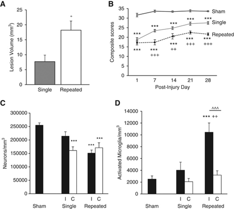
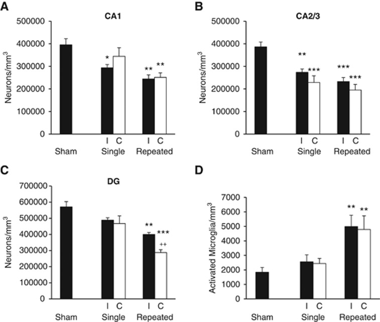
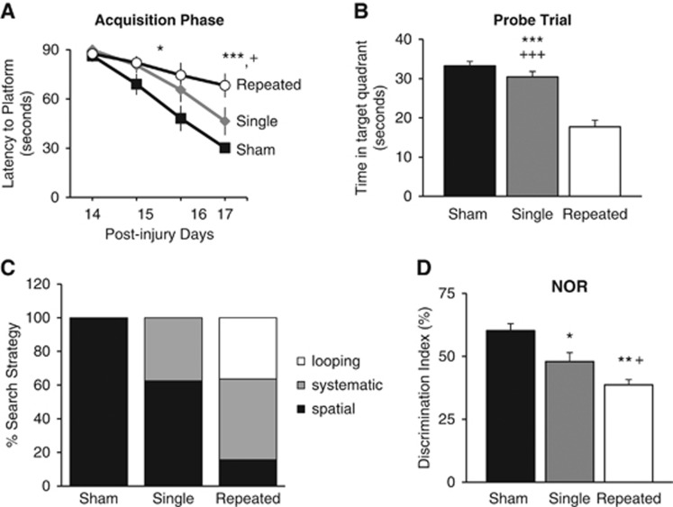
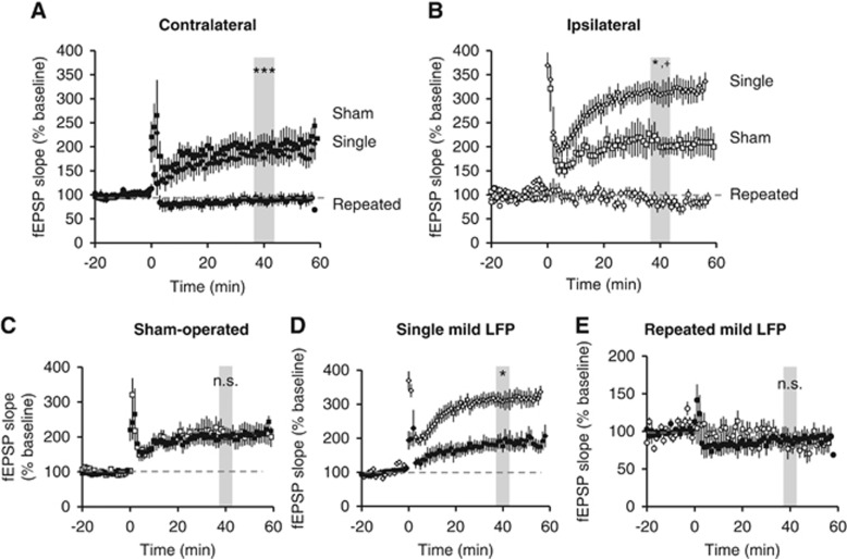
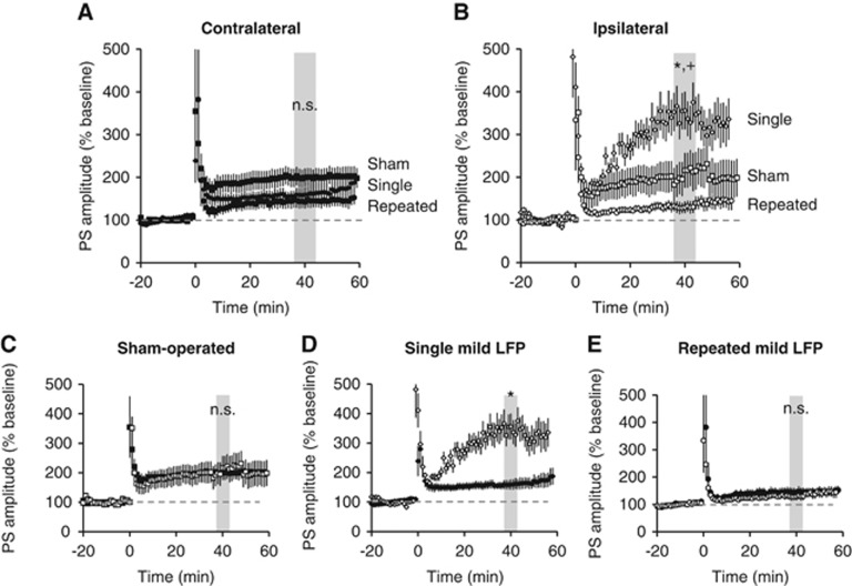
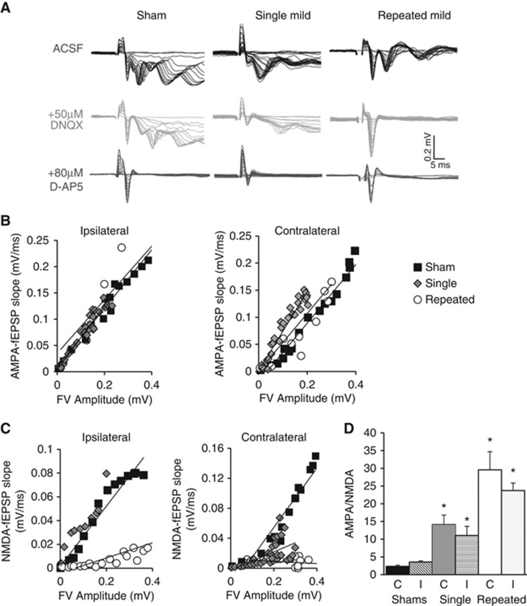
Repeated mild traumatic brain injury (mTBI) can cause sustained cognitive and psychiatric changes, as well as neurodegeneration, but the underlying mechanisms remain unclear. We examined histologic, neurophysiological, and cognitive changes after single or repeated (three injuries) mTBI using the rat lateral fluid percussion (LFP) model. Repeated mTBI caused substantial neuronal cell loss and significantly increased numbers of activated microglia in both ipsilateral and contralateral hippocampus on post-injury day (PID) 28. Long-term potentiation (LTP) could not be induced on PID 28 after repeated mTBI in ex vivo hippocampal slices from either hemisphere. N-Methyl-D-aspartate (NMDA) receptor-mediated responses were significantly attenuated after repeated mTBI, with no significant changes in α-amino-3-hydroxy-5-methyl-4-isoxazolepropionic acid (AMPA) receptor-mediated responses. Long-term potentiation was elicited in slices after single mTBI, with potentiation significantly increased in ipsilateral versus contralateral hippocampus. After repeated mTBI, rats displayed cognitive impairments in the Morris water maze (MWM) and novel object recognition (NOR) tests. Thus, repeated mTBI causes deficits in the hippocampal function and changes in excitatory synaptic neurotransmission, which are associated with chronic neuroinflammation and neurodegeneration.


Figures

Figure 1
Figure 1
Cortical lesion volume, sensorimotor function, and bilateral cortical neurodegeneration and microglial activation. Quantitative analysis of lesion volume and unbiased stereological quantification of surviving neurons and activated microglial phenotypes were analyzed on PID 28. (A) Analysis of lesion volume show repeated mild traumatic brain injury (mTBI) significantly increased the cortical lesion volume compared with single mTBI (+P<0.05, student t-test). (B) Repeated mTBI rats had significant sensorimotor impairment on all post-injury days (PIDs) compared with sham rats (***P<0.001) and compared with single mTBI rats on PIDs 7, 14, 21, and 28 (+++P<0.001, ++P<0.01). Single mTBI rats had significant motor impairment compared with sham rats on all PIDs (***P<0.001). Repeated measures one-way analysis of variance (ANOVA), Tukey post hoc. (C) There was significant neurodegeneration after repeated mTBI in both ipsilateral (I) and contralateral (C) cortex compared with sham tissue (***P<0.001) and in single mTBI contralateral cortex compared with sham tissue (***P<0.001). There was no significant difference between single and repeated mTBI or between I and C within the injury groups. (D) There was a significant increase in the number of activated microglia in the ipsilateral cortex after repeated mTBI compared with shams (***P<0.001) and to single mTBI (++P<0.01). There was also a difference within the repeated mTBI group between I and C hemispheres (^^^P<0.001). One-way ANOVA, Tukey post hoc. Mean±s.e.m., n=5 to 8 rats per group.
Figure 2
Figure 2
Bilateral hippocampal neurodegeneration and microglial activation. Unbiased stereological quantification of surviving neurons and activated microglial phenotypes was performed on post-injury day (PID) 28. (A) Neuronal survival in the ipsilateral (I) hippocampus was significantly decreased after single and repeated mild traumatic brain injury (mTBI) in areas CA1 (*P<0.05 versus sham; **P<0.01 versus sham) and in contralateral (C) CA1 after repeated mTBI (**P<0.01 versus sham). There was no significant difference between neuronal numbers in single and repeated mTBI. (B) Neuronal survival in CA2/3 after single mTBI was significantly decreased (I, **P<0.01 versus sham; C, ***P<0.001 versus sham) as was neuronal survival after repeated mTBI in both hemispheres (***P<0.001 versus sham). There was no significant difference between single and repeated mTBI neuronal survival. (C) Repeated mTBI caused significant bilateral neuronal cell loss in the dentate gyrus (DG) (I, P<0.01 versus sham; C, ***P<0.001 versus sham, ++P<0.01 versus single mTBI). There was no significant difference between single mTBI and sham neuronal numbers. (D) Repeated mTBI caused a significant increase in the number of activated microglial phenotypes in both the I and C hippocampus (**P<0.01 versus sham). There was no significant difference between single mTBI and sham tissue, or single and repeated mTBI. There was no significant difference between hemispheres within the injury groups in any hippocampal subregion for neuronal cell numbers or activated microglial numbers. Analyzed by one-way analysis of variance, Tukey post hoc; Mean±s.e.m., n=4–6 rats per group.
Figure 3
Figure 3
Repeated mild traumatic brain injuries (mTBI) impairs performance on cognitive tasks. (A) Spatial learning was tested using the acquisition phase of the Morris water maze (MWM). Rats with repeated mTBI had a significantly longer latency to the platform on post-injury day (PID) 16 compared with sham rats (*P<0.05) and on PID 17 compared with rats with single mTBI (+P<0.05) and sham rats (***P<0.001). Repeated measures one-way analysis of variance (ANOVA), Tukey post hoc. (B) Reference memory was assessed using the probe trial of MWM on PID 18. Repeated mTBI caused significant cognitive impairment compared with single mTBI (+++P<0.001) and shams (***P<0.001). (C) Analysis of search strategy revealed repeated mTBI rats exhibited significantly higher reliance on systematic and looping strategies than spatial behavior (P<0.001, χ2=40.05), whereas single mTBI relied mostly on spatial and systematic and shams relied only on spatial. (D) Retention memory was evaluated using the novel object recognition (NOR) task on PID 21. Repeated mTBI caused significant impairment in discrimination of the novel object compared with shams (**P<0.01) and single mTBI (+P<0.05). Single mTBI showed significant impairment compared with shams (*P<0.05). Morris water maze and NOR-one-way ANOVA, Tukey post hoc. Mean±s.e.m., n=8–9 rats/injury group, n=5 rats/shams. Search strategy-λ2.
Figure 4
Figure 4
Mild traumatic brain injury (mTBI) alters potentiation at SC-CA1 synapses. Field excitatory post-synaptic potentials were recorded from stratum radiatum in area CA1 from acute ipsilateral and contralateral hippocampal slices. Long-term potentiation was induced after 20 minutes of stable baseline using high frequency stimulation. (A) Potentiation was observed in slices taken from sham-operated and single mTBI rats in the contralateral hippocampus with sham versus single mild not significantly different (P=0.9); however, there was no potentiation observed in any slices from repeated mTBI slices in the contralateral hippocampus (***P<0.001 versus sham and single mTBI). (B) In the ipsilateral hippocampus, potentiation was observed in sham and single mTBI rats. However, there was greater potentiation in single mTBI slices compared with sham slices (*P<0.05), while there was no potentiation observed in the repeated mTBI slices (*P<0.05 versus sham, +P<0.001 versus single). (C) The amount of potentiation achieved in the contralateral (closed squares) versus ipsilateral (open squares) hippocampal slices in sham animals was not significantly different (P=0.9). (D) Potentiation in ipsilateral (open diamonds) hippocampus after single mTBI was significantly more than the contralateral side (closed diamonds) (*P<0.05). (E) There was no potentiation achieved in either the ipsilateral or contralateral hippocampus after repeated mTBI (P=0.9). One-way analysis of variance, Tukey post hoc. Mean±s.e.m. n=5 to 8 rats/group; 3 to 5 slices per rat.
Figure 5
Figure 5
Mild traumatic brain injury (mTBI) alters potentiation in CA1 pyramidal cells. Population spikes (PS) were recorded from stratum pyramidale in area CA1 from acute ipsilateral and contralateral hippocampal slices. Long-term potentiation was induced after 20 minutes of stable baseline using high frequency stimulation (4 × 100 Hz). (A) Potentiation was observed in slices taken from all injury groups in the contralateral hippocampus and the level of potentiation was not significantly different (P=0.9). (B) In the ipsilateral hippocampus, potentiation was observed in all injury groups. However, there was greater potentiation in single mTBI slices compared with sham slices (*P<0.05) and repeated mTBI slices (+P<0.01). (C) There was no significant difference between ipsilateral and contralateral slices within the sham-operated group. (D) Ipsilateral slices had significantly higher levels of potentiation compared with contralateral slices from single mTBI rats (*P<0.05). (E) There was no significant difference in the level of potentiation achieved in the contralateral and ipsilateral slices taken from repeated mTBI rats. One-way analysis of variance, Tukey post hoc. Mean±s.e.m. n=5 to 8 rats/group; 3 to 5 slices per rat.
Figure 6
Figure 6
NMDAR-mediated synaptic responses in SC-CA1. Recordings from stratum radiatum were done in 0 mmol/L Mg2+ artificial cerebrospinal fluid with 0.1 mmol/L picrotoxin. (A) Traces are representative from each injury group. To determine AMPAR-mediated responses, an input/output (I/O) curve was generated by increasing the stimulus intensity by 0.5 mV steps (black traces). To determine NMDAR-mediated responses 50 μmol/L of DNQX was perfused into the bath for 15 minutes, then a second I/O curve was generated in the same manner (gray traces). To confirm the NMDAR-dependent nature of the signal in DNQX, 80 μmol/L D-AP5 was washed into the bath for 5 minutes to abolish NMDAR-mediated responses (red traces). (B) AMPAR-mediated field excitatory post-synaptic potentials (fEPSP) slopes were measured and plotted against fiber volley (FV) amplitudes and have a linear relationship for both the ipsilateral and contralateral hippocampal slices (C) NMDAR-mediated fEPSP slopes were measured and plotted against FV amplitudes (D) AMPA/NMDA ratios were calculated using the mid-stimulus range with fiber volleys from before and after DNQX being the same amplitude (101±0.01% similar, data not shown); there was a significant increase in AMPA/NMDA ratio after single mild compared with shams (P<0.05 versus sham) and repeated mild compared with sham and single mild (P<0.05 versus sham, P<0.05 versus single mild). One-way analysis of variance, Student–Newman–Keuls post hoc. Mean±s.e.m., n=4 to 6 rats, 2 to 5 slices.

Similar articles

Cited by

References

    1. Report to Congress on mild traumatic brain injury in the United States: steps to prevent a serious public health problem . Centers for Disease Control and Prevention (CDC) National Center for Injury Prevention and Control: Atlanta (GA); 2003.
    1. Omalu B, Hammers JL, Bailes J, Hamilton RL, Kamboh MI, Webster G, et al. Chronic traumatic encephalopathy in an Iraqi war veteran with posttraumatic stress disorder who committed suicide. Neurosurg Focus. 2011;31:E3. - PubMed
    1. Omalu B, Bailes J, Hamilton RL, Kamboh MI, Hammers J, Case M, et al. Emerging histomorphologic phenotypes of chronic traumatic encephalopathy in American athletes Neurosurgery 201169173–183.discussion 183. - PubMed
    1. Shultz SR, Bao F, Omana V, Chiu C, Brown A, Cain DP. Repeated mild lateral fluid percussion brain injury in the rat causes cumulative long-term behavioral impairments, neuroinflammation, and cortical loss in an animal model of repeated concussion. J Neurotrauma. 2012;29:281–294. - PubMed
    1. Shultz SR, Bao F, Weaver LC, Cain DP, Brown A. Treatment with an anti-CD11d integrin antibody reduces neuroinflammation and improves outcome in a rat model of repeated concussion. J Neuroinflammation. 2013;10:26. - PMC - PubMed

Publication types