

doi: 10.1038/jcbfm.2014.73.
Epub 2014 Apr 23.
Cortical microinfarcts on 7T MRI in patients with spontaneous intracerebral hemorrhage
Affiliations
- PMID: 24756079
- PMCID: PMC4083387
- DOI: 10.1038/jcbfm.2014.73
Item in Clipboard
Cortical microinfarcts on 7T MRI in patients with spontaneous intracerebral hemorrhage
J Cereb Blood Flow Metab.
2014 Jul.
Erratum in
-
Cortical microinfarcts on 7 T MRI in patients with spontaneous intracerebral hemorrhage.J Cereb Blood Flow Metab. 2015 Jul;35(7):1222. doi: 10.1038/jcbfm.2015.34. J Cereb Blood Flow Metab. 2015. PMID: 26122138 Free PMC article. No abstract available.
Abstract
In patients with spontaneous intracerebral hemorrhage (ICH) coexisting abnormalities on brain imaging can provide clues on the etiology of the underlying small vessel disease. We examined cortical cerebral microinfarcts as a novel marker of coexistent vascular damage in ICH. Twelve patients with spontaneous ICH and 15 controls underwent 7Tesla magnetic resonance imaging (MRI). Microinfarcts were present in 9 of 12 patients with spontaneous ICH, and in 5 of 15 controls. This explorative study shows, for the first time, that microinfarcts appear to be a very common vascular comorbidity in spontaneous ICH. Future larger studies should further assess the etiological significance of these lesions.
Figures
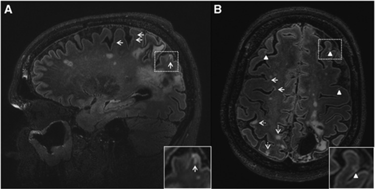
Multiple cortical microinfarcts on the fluid-attenuated inversion recovery (FLAIR) scan of a 52-year-old woman (patient 8) with a left occipito-parietal intracerebral hemorrhage (ICH). A sagittal view (A) and a transversal view (B) of the brain are depicted. Numerous cortical microinfarcts (arrows, insert A) were present throughout the brain, including multiple with cavitation (arrowheads, insert B). In the cavitated cortical microinfarcts (insert B), note the interruption of the cortex and the hyperintense rim surrounding the hypointense cavity. Also many cortical infarcts (>3 mm) were present (dashed arrows).
References
-
- Van Asch CJ, Luitse MJ, Rinkel GJ, van der Tweel I, Algra A, Klijn CJ. Incidence, case fatality, and functional outcome of intracerebral haemorrhage over time, according to age, sex, and ethnic origin: a systematic review and meta-analysis. Lancet Neurol. 2010;9:167–176. - PubMed
-
- Itoh Y, Yamada M, Hayakawa M, Otomo E, Miyatake T. Cerebral amyloid angiopathy: a significant cause of cerebellar as well as lobar cerebral haemorrhage in the elderly. J Neurol Sci. 1993;116:135–141. - PubMed
Publication types
MeSH terms
LinkOut - more resources
Full Text Sources
Other Literature Sources
