. 2014 Sep;159(9):2329-37.
doi: 10.1007/s00705-014-2070-y. Epub 2014 Apr 23.

Identification and genetic characterization of porcine hemagglutinating encephalomyelitis virus from domestic piglets in China

Affiliations

Identification and genetic characterization of porcine hemagglutinating encephalomyelitis virus from domestic piglets in China

Bo Dong et al. Arch Virol. 2014 Sep.

Abstract

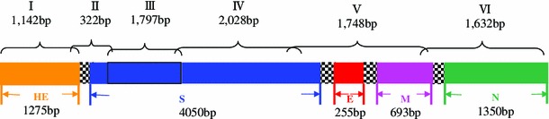
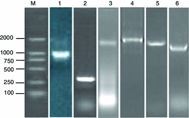
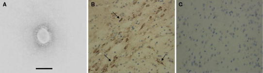
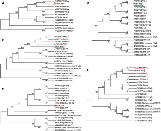
In this study, we investigated an acute outbreak of porcine hemagglutinating encephalomyelitis on a farm of 127 pigs in Jilin province, China. Porcine hemagglutinating encephalomyelitis virus (PHEV) was detected in suckling and weaning pigs by RT-PCR assays. Coronavirus-like particles were observed by electron microscopy. The virus isolate was designated PHEV-JT06. The clinical signs, nervous symptoms and positive labeling of neurons in the cerebral cortex with an immunohistochemical stain in PHEV-JT06-infected BALB/c mice supported the diagnosis of PHEV infection. The five full-length PHEV-JT06 structural genes were cloned, sequenced and analyzed. Phylogenetic studies based on the nucleotide and amino acid sequences of the five genes in the outbreak showed that PHEV remained genetically stable. PHEV shares 95.3-99.3% amino acid sequence identity with American strains (AY078417), suggesting that the Chinese isolate is most likely derived from the North American strain. Additionally, PHEV, HCoV-OC43 and BCoV were genetically close. These results may provide some insights into the genotype of the etiological agent responsible for the porcine hemagglutinating encephalomyelitis outbreak and could also provide a comparative view of the genomics of the five structural proteins of PHEV.


Figures

Fig. 1
Fig. 1
Schematic illustration of the organization of the targeted genes coding for the five structural proteins, showing the locations of the binding sites for the designed primers (reference virus HEV-67N, accession no. AY078417). The upper numbers represent the sizes of the PCR products, specifically, the PHEV HE, S, E, M and N genes
Fig. 2
Fig. 2
Amplification of the HE, S, E, M and N genes by PCR. Lane 1, PCR product (1142 bp) containing the major region of the PHEV-JT06 HE gene; lane 2, PCR product (322 bp) containing the conjunctive region of the PHEV-JT06 HE and S genes; lane 3 and 4, PCR products (1797 bp, 2028 bp) containing the major region of the PHEV-JT06 S gene; lane 5, PCR product (1748 bp) containing the region of PHEV-JT06 S, E, and M genes and their conjunctive regions; lane 6, PCR product (1632 bp) containing the region of the PHEV-JT06 M and N genes and their conjunctive region; M, DL2000 DNA marker (bp). The six gene products were identified by DNA sequencing
Fig. 3
Fig. 3
Identification of porcine hemagglutinating encephalomyelitis virus by morphologic and immunological assays. A Electron microphotograph showing the characteristic morphology of the PHEV isolate from PK-15 cells inoculated with the brain tissue of dead piglets (bar = 100 nm). B A dark brown positive signal (arrow) can be seen in a nerve cell of the cerebral cortex from a PHEV-JT06-infected BALB/c mouse by IHC (magnification ×400). C Negative control treated with normal goat serum (magnification ×400)
Fig. 4
Fig. 4
Phylogenetic analysis of betacoronaviruses based on amino acid sequences of the five major structural proteins. The phylogenetic tree was constructed using the neighbor-joining program of MEGA version 5.2, and bootstrap analysis was performed with 1000 trials. The scale bar beneath the tree indicates amino acid substitution per site. A, B, C, D and E represent trees constructed with sequences of HE, S, E, M and N proteins, respectively. The sequence identified in this study is underlined and is clustered with PHEV sequences. The scale bar beneath the tree indicates amino acid substitution per site

References

    1. Hasoksuz M, Alekseev K, Vlasova A, Zhang XS, Spiro D, Halpin R, Wang SL, Ghedin E, Saif LJ. Biologic, antigenic and full-length genomic characterization of a bovine-like coronavirus isolated from a giraffe. J Virol. 2007;81(10):4981–4990. doi: 10.1128/JVI.02361-06. - DOI - PMC - PubMed
    1. Hirano N, Tohyama K, Taira H. Spread of swine hemagglutinating encephalomyelitis virus from peripheral nerves to the CNS. Adv Exp Med Biol. 1998;440:601–607. doi: 10.1007/978-1-4615-5331-1_78. - DOI - PubMed
    1. Li YC, Bai WZ, Hirano N, Hayashida T, Taniguchi T, Sugita Y, Tohyama K, Hashikawa T. Neurotropic virus tracing suggests a membranous-coating-mediated mechanism for transsynaptic communication. J Comp Neurol. 2013;521(1):203–212. doi: 10.1002/cne.23171. - DOI - PMC - PubMed
    1. Masters PS. The molecular biology of coronaviruses. Adv Virus Res. 2006;66:193–292. doi: 10.1016/S0065-3527(06)66005-3. - DOI - PMC - PubMed
    1. Roe CK, Alexander TJ. A disease of nursing pigs previously unreported in ontario. Can J Comp Med Vet Sci. 1958;22(9):305–307. - PMC - PubMed

Publication types

MeSH terms

Associated data

LinkOut - more resources