Review
. 2014 Jun 14;12(22):3544-61.
doi: 10.1039/c4ob00447g. Epub 2014 Apr 23.

Self-assembly of peptides to nanostructures

Affiliations
Review

Self-assembly of peptides to nanostructures

Dindyal Mandal et al. Org Biomol Chem. .

Abstract

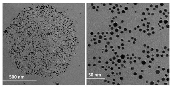
The formation of well-ordered nanostructures through self-assembly of diverse organic and inorganic building blocks has drawn much attention owing to their potential applications in biology and chemistry. Among all organic building blocks, peptides are one of the most promising platforms due to their biocompatibility, chemical diversity, and resemblance to proteins. Inspired by the protein assembly in biological systems, various self-assembled peptide structures have been constructed using several amino acids and sequences. This review focuses on this emerging area, the recent advances in peptide self-assembly, and formation of different nanostructures, such as tubular structures, fibers, vesicles, and spherical and rod-coil structures. While different peptide nanostructures have been discovered, potential applications are explored in drug delivery, tissue engineering, wound healing, and surfactants.


Figures

Figure 1
Figure 1
Cyclic peptide structures with alternating D- and L-amino acids adopted flat ring-shaped conformations and assembled into ordered parallel arrays of solid-state nanotubes. The illustration emphasizes the antiparallel ring stacking and the presence of extensive hydrogen-bonding interactions between subunits (for clarity most side chains are omitted). Reprinted with permission from ref. 10. Copyright 2013 American Chemical Society.
Figure 2
Figure 2
Molecular modeling of cut-away structures formed from the peptides with negatively charged heads and glycine tail. Left picture shows peptide nanotube with an area sliced away. Right picture exhibits a peptide nanovesicle. Color code: red, negatively charged aspartic acid heads; green, nonpolar glycine tail. The modeled dimension is 50-100 nm in diameter. Reprinted with permission from ref. 14. Copyright 2013 American Chemical Society.
Figure 3
Figure 3
Proposed plausible self-assembly process of the nanodoghnut structure. (A) Randomly oriented and distributed peptides at low concentration; (B) Micelle formation above the critical aggregation concentration (CAC); (C) Fusion or elongation of the micelles for the formation of a nanopipe; (D) Bending of the nanopipe for the formation of a nanodoughnut structure. Reprinted with permission from ref. 16. Copyright 2013 American Chemical Society.
Figure 4
Figure 4
Schematic illustration of peptide surfactant self-assembly. (a) A3K had the shortest chain and had no apparent critical aggregation concentration (CAC) detected, giving rise to the lowest effective ae (equilibrium area occupied by each surfactant at the curved interface in the aggregate) and the highest packing parameter; consequently, stacked A3K bilayers were formed. (b) With increasing hydrophobic tail length and decreasing CAC of A6K, the electrostatic repulsion between the head groups increased. This, together with the packing and entropic effect, led to the lowering of the packing parameter and the formation of nanofibers. (c) A9K had the lowest CAC, the highest electrostatic repulsion between head groups, and the largest entropic effect arising from the longest tail, resulting in the lowest p and the formation of nanorods. In each case, lysine (K) groups remained at the outer surface of the nanostructures formed. Reprinted with permission from ref. 19. Copyright 2013 American Chemical Society.
Figure 5
Figure 5
Cleavage of 3-nitrobenzyl group from a PA molecule consisting of a 2-nitrobenzyl group, a palmitoyl tail, and an oligopeptide segment GV3A3E3. Reprinted with permission from ref. 28. Copyright 2013 American Chemical Society.
Figure 6
Figure 6
Nanobelts assembled from a PA containing four amino acids and an alkyl tail. (a) Chemical structure of the PA. (b-d) AFM images of peptide nanobelts at different scanning sizes. The assembled nanobelts are the dominant structures in the assembly system (almost artifact free). Reprinted with permission from ref. 26. Copyright 2013 American Chemical Society.
Figure 7
Figure 7
Cryo-TEM image of 0.1 wt % C16H31OVVEE aqueous solution clearly demonstrates cylindrical nanofibers. Reprinted with permission from ref. 26. Copyright 2013 American Chemical Society.
Figure 8
Figure 8
Synthesis of a C3-symmetric β-sheet peptide conjugate and schematic illustration of the self-assembly. Reprinted with permission from ref. 31. Copyright 2013 American Chemical Society.
Figure 9
Figure 9
Cyclic to linear peptide conformational switch using a reductive trigger. Reprinted with permission from ref. 32a. Copyright 2013 American Chemical Society.
Figure 10
Figure 10
Spontaneously formed aggregates by peptide guided assembly of PEO-peptide conjugate. Light microscopy (left) and SEM micrograph (right). Reprinted with permission from ref. 35. Copyright 2013 American Chemical Society.
Figure 11
Figure 11
Structure and sequence of TβP peptides. Reprinted with permission from ref. 36. Copyright 2013 Wiley Publishing Group.
Figure 12
Figure 12
Schematic representation of the self-assembly into vesicles of the diblock copolymer PGA15-b-PLys15. Reprinted with permission from ref. 41. Copyright 2013 American Chemical Society.
Figure 13
Figure 13
Structures of peptide rod–coil building blocks and their self-assembly into nanocapsule structures. Reprinted with permission from ref. 43. Copyright 2013 RSC Publishing Group.
Figure 14
Figure 14
Chemical structures of synthesized cyclic peptides (F = phenylalanine, R = arginine, K = lysine, W = tryptophan, C = cysteine, A = alanine). Reprinted with permission from ref. 45.
Figure 15
Figure 15
(a) and (b) FE-SEM image of self-assembled structure of [WR]5 (2 mM) in water after 20 days; (c) TEM images of self-assembled structures of [WR]3 in water after 2 weeks (without negative staining); (d) TEM images of self-assembled structures of [WR]4 in water after 2 weeks (without negative staining); (e) TEM images of self-assembled structures of [WR]5 in water after 2 weeks (without negative staining). Reprinted with permission from ref. 45.
Figure 16
Figure 16
TEM images of l(KW)5-AuNPs. Reprinted with permission from ref. 47. Copyright 2013 American Chemical Society.
Figure 17
Figure 17
TEM images of c[KW]5-AuNPs. Reprinted with permission from ref. 47. Copyright 2013 American Chemical Society.
Figure 18
Figure 18
Negatively stained TEM images of [WR]4 (1 mM), PEpYLGLD (1 mM), and PEpYLGLD-loaded [WR]4 (1 mM) in water after one day. Reprinted with permission from ref. 48. Copyright 2013 American Chemical Society.
Figure 19
Figure 19
Proposed mechanism of the self-assembly and interactions between PEpYLGLD and [WR]4. Reprinted with permission from ref. 48. Copyright 2013 American Chemical Society.
Figure 20
Figure 20
The formation of amyloid fibrils. Folded monomer undergoes a conformational transition into a b-sheet-rich state, usually through a partial unfolded state. Self-assembly of these intermediates into ladders of b strands results in the formation of ordered filaments that aggregate into the well-known amyloid fibrils. Reprinted with permission from ref. 55. Copyright 2013 Wiley Publishing Group.
Figure 21
Figure 21
Schematic drawing showing proposed self-assembly of K60L20 into vesicles. Reprinted from ref. 63. Copyright 2013 American Chemical Society.
Figure 22
Figure 22
Schematic representation of the supradendrimer design. SD-1 (1) and SD-2 (2) peptide sequences are shown with the one-letter code and are simplified into helical wheels with 3.5 residues per turn. The sequences show heptad repeats characteristic of canonical coiled coils, commonly labeled a–g. Residues at sites a and d shape helix–helix interfaces. These are hydrophobic and made up of isoleucine and leucine, respectively, to ensure the formation of dimers. A) SD-1-type of electrostatic interactions, g–c–e’: e’ and g positions are occupied by glutamates that form salt bridges with arginine residues at c; g–c interactions are intrahelical, whereas c–e’ are interhelical and are between two SD-1 copies of different dimers. Dimers trimerize (I) to give a noncovalent dendrimeric architecture, with a branching cell formed by six dimers (II). A single cysteine residue in the f position that replaces a glutamine is shown in brackets. B) Mixed SD-1/SD-2 (SD-1,2)-type of electrostatic interactions, g–c–e’(1)/b–e, g– e’(2): intrahelical b–e interactions in 2 inhibit interhelical e–g’ interactions; g–e’ interactions contribute to the 1–2 interface. Because the width of a superhelix is approximately 2 nm, the diameter of an extended branching cell (II’) is estimated to not exceed 4–6 nm. To form the supradendrimer II’, each copy of SD-2 should be arranged with at least nine copies of SD-1 to round the equimolar SD-1,2 ratio to 10:1. SD-2 wheels are highlighted in black around an ac-KKK-am hub containing an e-aminohexanoic acid (eAhx) as spacer. The circles of helical wheels are black for arginines, gray for glutamates, and outlined in gray for lysines. Arrows represent salt bridges. Reprinted with permission from ref. 65. Copyright 2013 Wiley Publishing Group.

References

    1. Lehn JM. Proc. Natl. Acad. Sci. USA. 2002;99:4763–4768. - PMC - PubMed
    1. Whitesides GM, Mathias JP, Set CT. Science. 1991;254:1312–1319. - PubMed
    1. Sacchettini JC, Kelly JW. Nat. Rev. Drug Discov. 2002;1:267–275. - PubMed
    1. Lim YB, Moon KS, Lee M. Chem. Soc. Rev. 2009;38:925–934. - PubMed
    2. Ulijn RV, Smith AM. Chem. Soc. Rev. 2008;37:664–675. - PubMed
    1. Zelzer M, Ulijn RV. Chem. Soc. Rev. 2010;39:3351–3357. - PubMed
    2. Rica RDL, Matsui H. Chem. Soc. Rev. 2010;39:3499–3509. - PMC - PubMed

Publication types