Review
. 2014 Jun 15;592(12):2491-500.
doi: 10.1113/jphysiol.2014.271858. Epub 2014 Apr 22.

Autonomic neural control of heart rate during dynamic exercise: revisited

Affiliations
Review

Autonomic neural control of heart rate during dynamic exercise: revisited

Daniel W White et al. J Physiol. .

Abstract

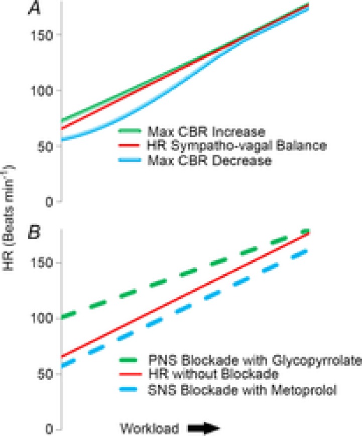
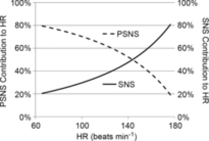
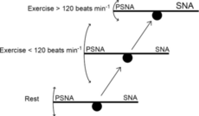
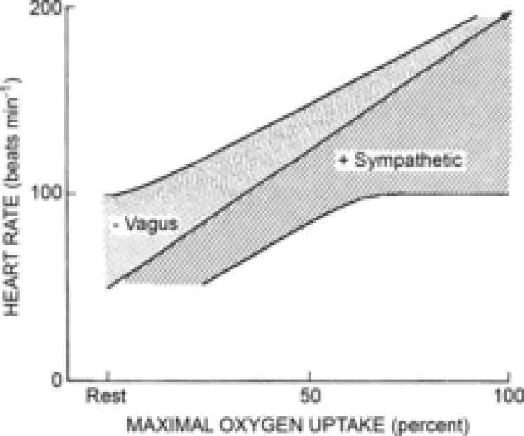
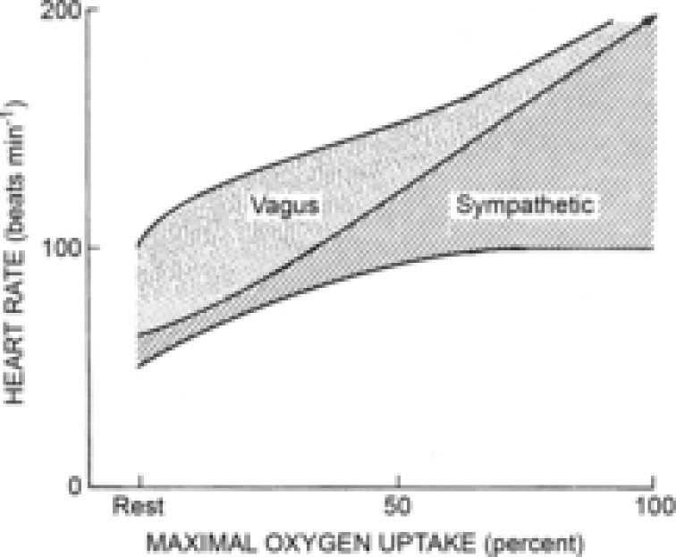
The accepted model of autonomic control of heart rate (HR) during dynamic exercise indicates that the initial increase is entirely attributable to the withdrawal of parasympathetic nervous system (PSNS) activity and that subsequent increases in HR are entirely attributable to increases in cardiac sympathetic activity. In the present review, we sought to re-evaluate the model of autonomic neural control of HR in humans during progressive increases in dynamic exercise workload. We analysed data from both new and previously published studies involving baroreflex stimulation and pharmacological blockade of the autonomic nervous system. Results indicate that the PSNS remains functionally active throughout exercise and that increases in HR from rest to maximal exercise result from an increasing workload-related transition from a 4 : 1 vagal-sympathetic balance to a 4 : 1 sympatho-vagal balance. Furthermore, the beat-to-beat autonomic reflex control of HR was found to be dependent on the ability of the PSNS to modulate the HR as it was progressively restrained by increasing workload-related sympathetic nerve activity.

In conclusion: (i) increases in exercise workload-related HR are not caused by a total withdrawal of the PSNS followed by an increase in sympathetic tone; (ii) reciprocal antagonism is key to the transition from vagal to sympathetic dominance, and (iii) resetting of the arterial baroreflex causes immediate exercise-onset reflexive increases in HR, which are parasympathetically mediated, followed by slower increases in sympathetic tone as workloads are increased.


Figures

Figure 1
Figure 1. Autonomic reflex and steady-state influence on heart rate with increasing exercise
A, modelling of the reflexive control of heart rate (HR) by the carotid baroreflex (CBR). Steady state HR at each workload target HR (Workload) represents the sympatho–vagal balance (red line). Increases and decreases in HR by simulated hypo- and hypertension induced by neck pressure/suction (green and blue lines, respectively) are plotted against workload. These data indicate that the parasympathetic influence on the reflex control of HR is diminished but never absent as the exercise workload increases from rest to maximal exercise. B, modelling of pharmacological blockade studies of each branch of the autonomic nervous system from rest to maximal exercise showing the steady state HR at each workload target HR (Workload) without blockade (solid red line). Steady state HRs at each workload with muscarinic blockade by glycopyrrolate (green dashed line) and β1-adrenergic blockade by metoprolol (blue dashed line) represent the un-opposed sympathetic HR and un-opposed parasympathetic HR, respectively. As HR increases with workload, the sympatho–vagal balance shows greater sympathetic dominance. PSNS, parasympathetic nervous system; SNS, sympathetic nervous system.
Figure 2
Figure 2. Contributions to heart rate by each branch of the autonomic nervous system determined by autonomic blockade studies
The parasympathetic nervous system (PSNS) contributes 80% influence to resting heart rate (HR) and the sympathetic nervous system (SNS) contributes the other 20%. Both branches make an equal contribution at close to 140 beats min−1, after which the ratio changes quickly to a more sympathetically dominant system. The respective lines indicate the change in HR from a single branch's selective autonomic blockade as a percentage change in HR from the sum of the absolute values of HR change of both branches.
Figure 3
Figure 3. See-saw model of autonomic beat-to-beat reflex control of heart rate
At rest as well as throughout exercise, there is a sympatho–vagal balance. The parasympathetic functional control is related to the sympathetic tone and maintains a balance. The relative amount of sympathetic nervous activity (SNA) on the right side of the see-saw affects the reflexive control of heart rate (HR) by the parasympathetic nervous activity (PSNA) on the left side as depicted by the size of the arrows. As exercise is initiated, there is a slight decrease in sympathetic tone caused by the loading of the cardiopulmonary baroreceptors, which allows for changes in the parasympathetic nervous outflow to exert more influence on the heart. Along with this initial decrease in sympathetic tone is a transient decrease in blood pressure so that HR is increased reflexively by the lessening of parasympathetic tone to maintain and increase cardiac output. As exercise workload increases, central command and exercise pressor reflexes force increases in sympathetic tone, causing a further rise in HR and a depression of parasympathetically mediated HR reflex response.
Figure 4
Figure 4. Modified version of the diagram proposed by Rowell (
The modified diagram (Fig. 5–4, Rowell 1993) depicts the steady state continuum of autonomic influence from both branches of the autonomic nervous system throughout exercise. The shaded area under the central line represents the sympathetic influence at all exercise workloads. The dotted area represents the parasympathetic influence of heart rate (HR) at all exercise workloads. The centre line indicates the relative HR (i.e. the dynamic sympatho–vagal balance).
Figure 5
Figure 5. Modified version of the diagram proposed by Rowell (1993)
The modified diagram (Fig. 5–4, Rowell 1993) depicts the reflex continuum of autonomic influence from both branches of the autonomic nervous system throughout exercise. The shaded area under the central line represents the sympathetic influence at all exercise workloads. The dotted area represents the functional parasympathetic modulation of heart rate (HR) at all exercise workloads. As the area of the sympathetic portion of the graph increases, the area of the parasympathetic modulation portion of the graph decreases to show the inverse relationship between sympathetic tone and parasympathetic modulation. The centre line indicates the relative HR (i.e. dynamic sympatho–vagal balance).

References

    1. Adlam D, Herring N, Douglas G, De Bono JP, Li D, Danson EJ, Tatham A, Lu CJ, Jennings KA, Cragg SJ, Casadei B, Paterson DJ, Channon KM. Regulation of β-adrenergic control of heart rate by GTP-cyclohydrolase 1 (GCH1) and tetrahydrobiopterin. Cardiovasc Res. 2012;93:694–701. - PMC - PubMed
    1. Armour JA. Cardiac neuronal hierarchy in health and disease. Am J Physiol Regul Integr Comp Physiol. 2004;287:R262–R271. - PubMed
    1. Bainbridge FA. On some cardiac reflexes. J Physiol. 1914;48:332–340. - PMC - PubMed
    1. Basnayake SD, Hyam JA, Pereira EA, Schweder PM, Brittain JS, Aziz TZ, Green AL, Paterson DJ. Identifying cardiovascular neurocircuitry involved in the exercise pressor reflex in humans using functional neurosurgery. J Appl Physiol (1985) 2011;110:881–891. - PubMed
    1. Bevegard BS, Shepherd JT. Circulatory effects of stimulating the carotid arterial stretch receptors in man at rest and during exercise. J Clin Invest. 1966;45:132–142. - PMC - PubMed

Publication types

MeSH terms