. 2014 Jul;34(1):24-34.
doi: 10.3892/ijmm.2014.1747. Epub 2014 Apr 22.

Geldanamycin attenuates 3‑nitropropionic acid‑induced apoptosis and JNK activation through the expression of HSP 70 in striatal cells

Affiliations

Geldanamycin attenuates 3‑nitropropionic acid‑induced apoptosis and JNK activation through the expression of HSP 70 in striatal cells

Yong-Joon Choi et al. Int J Mol Med. 2014 Jul.

Abstract

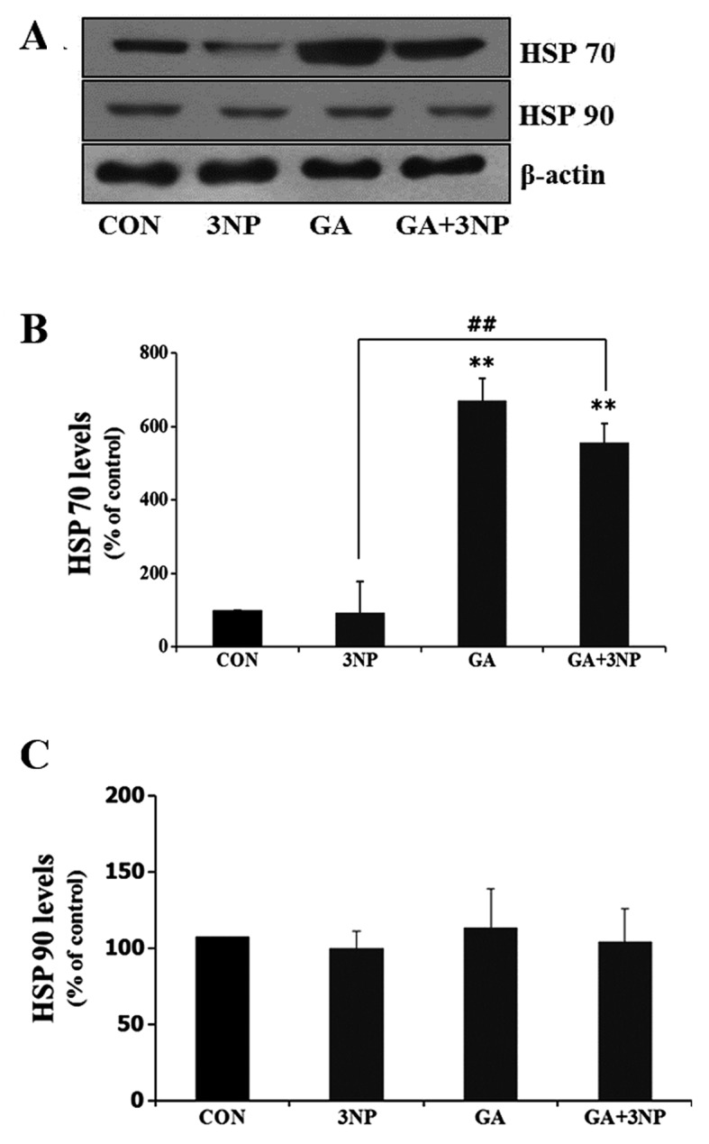
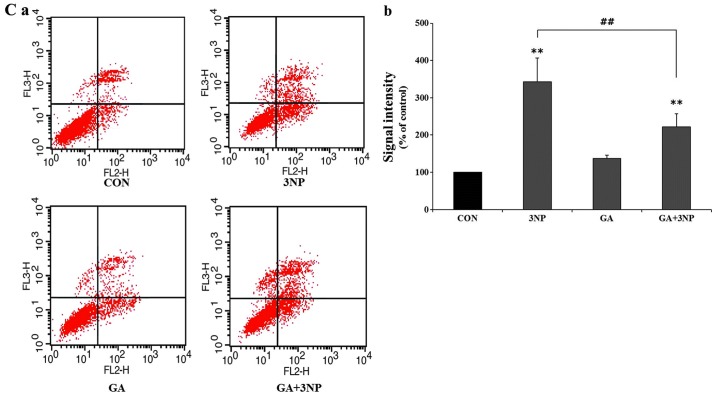
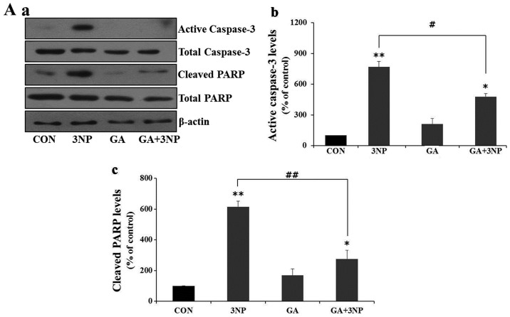
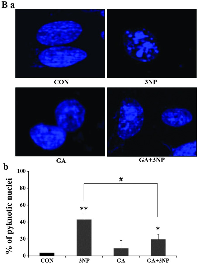
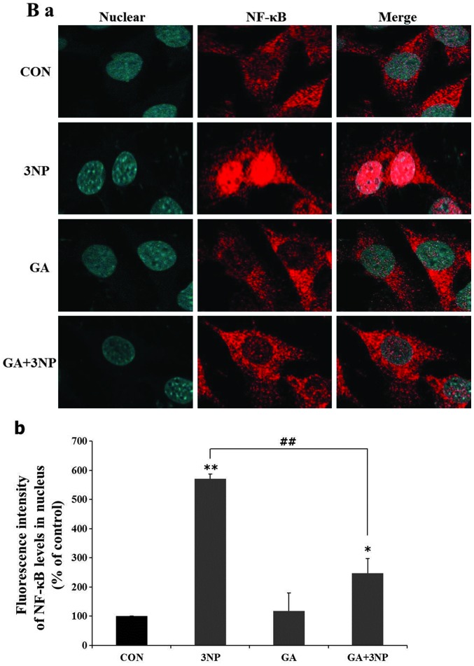
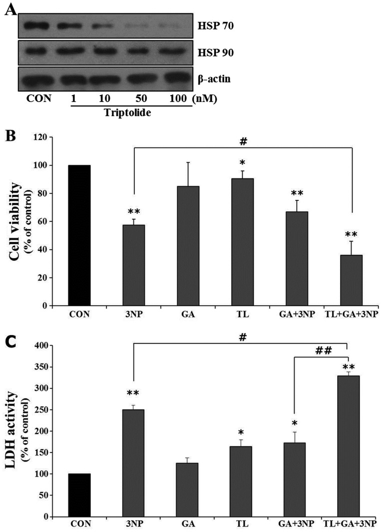
Although selective striatal cell death is a characteristic hallmark in the pathogenesis of Huntington's disease (HD), the underlying mechanism of striatal susceptibility remains to be clarified. Heat shock proteins (HSPs) have been reported to suppress the aggregate formation of mutant huntingtin and concurrent striatal cell death. In a previous study, we observed that heat shock transcription factor 1 (HSF1), a major transcription factor of HSPs, significantly attenuated 3‑nitropropionic acid (3NP)‑induced reactive oxygen species (ROS) production and apoptosis through the expression of HSP 70 in striatal cells. To investigate the differential roles of HSPs in 3NP‑induced striatal cell death, the effect of geldanamycin (GA), an HSP 90 inhibitor, was examined in 3NP‑stimulated striatal cells. GA significantly attenuated 3NP‑induced striatal apoptosis and ROS production with an increased expression of HSP 70. Triptolide (TL), an HSP 70 inhibitor, abolished GA‑mediated protective effects in 3NP‑stimulated striatal cells. To understand the underlying mechanism by which GA‑mediated HSP 70 protects striatal cells against 3NP stimulation, the involvement of various signaling pathways was examined. GA significantly attenuated 3NP‑induced c‑Jun N‑terminal kinase (JNK) phosphorylation and subsequent c‑Jun phosphorylation in striatal cells. Taken together, the present study demonstrated that GA exhibits protective properties against 3NP‑induced apoptosis and JNK activation via the induction of HSP 70 in striatal cells, suggesting that expression of HSP 70 may be a valuable therapeutic target in the treatment of HD.


Figures

Figure 1
Figure 1
Geldanamycin (GA)-induced expression of heat shock protein (HSP) 70 in striatal cells. (A) Striatal cells were treated with 500 nM GA and incubated in the presence or absence of 10 μM 3-nitropropionic acid (3NP). After 24 h of incubation, HSP 70 and HSP 90 were assessed by immunoblotting. HSP 70 expression was increased by GA, whereas expression of HSP 90 did not change. Loading control was confirmed by β-actin. (B and C) Quantitative analysis was obtained from three individual experiments (n=3). **P<0.01 indicates significant differences compared to the control. ##P<0.05 indicates significant differences between the indicated groups.
Figure 2
Figure 2
Protective effects of geldanamycin (GA) on the cytotoxicity of 3-nitropropionic acid (3NP). (A and B) Cell viability was examined with 3-(4,5-dimethylthiazol-2-yl)-2,5-diphenyltetrazolium bromide (MTT) assay and lactate dehydrogenase leakage (LDH) assay. Striatal cells were treated with 10 μM 3NP, 500 nM and their combination as indicated. After 24 h, MTT and LDH assays were performed. GA significantly attenuated 3NP-induced cytotoxicity in striatal cells, albeit not completely. Data are shown as mean ± SD from three individual experiments (n=3). (Ca) Representative FACS images and (Cb) quantitative analysis of FACS data. Striatal cells were incubated in the presence or absence of 3NP after treatment of GA. FACS assay was carried out to determine initiation and amplitude of apoptotic dell death. Only Annexin V-positive cells were indicated as early apoptotic cells and Annexin V- and 7-AAD-positive cells were considered as late apoptotic cells. The number of total apoptotic cells, which are positive to both Annexin-5 and 7-AAD, was significantly decreased in presence of GA compared to only 3NP. Data were obtained from four independent experiments (n=4). *P<0.05 and **p<0.01 indicate differences compared to the control. #P<0.05 and ##p<0.01 indicates significant differences between the indicated groups.
Figure 2
Figure 2
Protective effects of geldanamycin (GA) on the cytotoxicity of 3-nitropropionic acid (3NP). (A and B) Cell viability was examined with 3-(4,5-dimethylthiazol-2-yl)-2,5-diphenyltetrazolium bromide (MTT) assay and lactate dehydrogenase leakage (LDH) assay. Striatal cells were treated with 10 μM 3NP, 500 nM and their combination as indicated. After 24 h, MTT and LDH assays were performed. GA significantly attenuated 3NP-induced cytotoxicity in striatal cells, albeit not completely. Data are shown as mean ± SD from three individual experiments (n=3). (Ca) Representative FACS images and (Cb) quantitative analysis of FACS data. Striatal cells were incubated in the presence or absence of 3NP after treatment of GA. FACS assay was carried out to determine initiation and amplitude of apoptotic dell death. Only Annexin V-positive cells were indicated as early apoptotic cells and Annexin V- and 7-AAD-positive cells were considered as late apoptotic cells. The number of total apoptotic cells, which are positive to both Annexin-5 and 7-AAD, was significantly decreased in presence of GA compared to only 3NP. Data were obtained from four independent experiments (n=4). *P<0.05 and **p<0.01 indicate differences compared to the control. #P<0.05 and ##p<0.01 indicates significant differences between the indicated groups.
Figure 3
Figure 3
Effects of 3-nitropropionic acid (3NP) and geldanamycin (GA) on apoptosis. Striatal cells were treated with GA, 3NP and GA+3NP. (Aa) After 24 h of treatment, cells were collected and protein extracts prepared from each samples were analyzed by immunoblotting using indicated antibodies. GA suppressed 3NP-induced caspase-3 activation and level of cleaved PARP. β-actin was used as a loading control. (Ab and c) Quantitative analysis was obtained from three individual experiments (n=3). The nuclear morphological changes are shown by 3NP-treated striatal cells. (Ba) The abrogation of nuclear damage by GA was measured using DAPI staining and (Bb) quantitative analysis of apoptotic cells. The more apoptotic cells were observed in only 3NP treated striatal cells compared to GA+3NP-treated striatal cells. Apoptotic cells were quantitatively counted using a microscope. In only 3NP-170-treated striatal cells, ~42% of cells showed DNA fragmentation whereas ~21% of cells included fragmentation in GA+3NP-treated striatal cells. Approximately 100 DAPI-positive cells were counted in each experiment. Quantitative analysis was obtained from three individual experiments (n=3). *P<0.05, **p<0.01 indicate significant differences compared to the control. #P<0.05, ##p<0.01 indicates significant differences between the indicated groups.
Figure 3
Figure 3
Effects of 3-nitropropionic acid (3NP) and geldanamycin (GA) on apoptosis. Striatal cells were treated with GA, 3NP and GA+3NP. (Aa) After 24 h of treatment, cells were collected and protein extracts prepared from each samples were analyzed by immunoblotting using indicated antibodies. GA suppressed 3NP-induced caspase-3 activation and level of cleaved PARP. β-actin was used as a loading control. (Ab and c) Quantitative analysis was obtained from three individual experiments (n=3). The nuclear morphological changes are shown by 3NP-treated striatal cells. (Ba) The abrogation of nuclear damage by GA was measured using DAPI staining and (Bb) quantitative analysis of apoptotic cells. The more apoptotic cells were observed in only 3NP treated striatal cells compared to GA+3NP-treated striatal cells. Apoptotic cells were quantitatively counted using a microscope. In only 3NP-170-treated striatal cells, ~42% of cells showed DNA fragmentation whereas ~21% of cells included fragmentation in GA+3NP-treated striatal cells. Approximately 100 DAPI-positive cells were counted in each experiment. Quantitative analysis was obtained from three individual experiments (n=3). *P<0.05, **p<0.01 indicate significant differences compared to the control. #P<0.05, ##p<0.01 indicates significant differences between the indicated groups.
Figure 4
Figure 4
Geldanamycin (GA) significantly reduced intracellular reactive oxygen species (ROS) generation by 3-nitropropionic acid (3NP). (A) Intracellular level of ROS was measured using confocal microscopy. Striatal cells were treated with GA and incubated in the presence or absence of 3NP. Cells were incubated with fluorescence probe H2DCFDA (10 μM). (B) H2DCFDA fluorescence was quantitatively analyzed with Fluoview 300 software. The production of 3NP-induced ROS was significantly attenuated by GA in striatal cells. Results are the means ± SD of three independent experiments performed in triplicate. *P<0.05, **p<0.01 indicate significant differences compared to the control. ##P<0.05 indicates significant differences between the indicated groups.
Figure 5
Figure 5
Geldanamycin (GA) inhibited 3-nitropropionic acid (3NP)-induced IκB degradation and nuclear translocation of NF-κB. Striatal cells were treated with GA, 3NP, GA+3NP or were untreated (control). (Aa) After 24 h of incubation, total proteins were extracted for immunoblotting of IκB-α. Degradation of IκB-α attenuated the GA+3NP group compared to the 3NP-only group. β-actin was used as a loading control. (Ba) Nuclear translocation of NF-κB was examined using immunocytochemistry assay. NF-κB (red panels) was mainly localized in the cytoplasm (control and GA). Nuclear translocation of NF-κB was facilitated only in 3NP compared to GA+3NP. Nuclei were visualized by Hoechst staining (Hoechst 33258). (Ab and Bb) Quantitative analysis was obtained from three individual experiments (n=3). *P<0.05, **p<0.01 indicate significant differences compared to the control. #P<0.05, ##p<0.01 indicates significant differences between the indicated groups.
Figure 5
Figure 5
Geldanamycin (GA) inhibited 3-nitropropionic acid (3NP)-induced IκB degradation and nuclear translocation of NF-κB. Striatal cells were treated with GA, 3NP, GA+3NP or were untreated (control). (Aa) After 24 h of incubation, total proteins were extracted for immunoblotting of IκB-α. Degradation of IκB-α attenuated the GA+3NP group compared to the 3NP-only group. β-actin was used as a loading control. (Ba) Nuclear translocation of NF-κB was examined using immunocytochemistry assay. NF-κB (red panels) was mainly localized in the cytoplasm (control and GA). Nuclear translocation of NF-κB was facilitated only in 3NP compared to GA+3NP. Nuclei were visualized by Hoechst staining (Hoechst 33258). (Ab and Bb) Quantitative analysis was obtained from three individual experiments (n=3). *P<0.05, **p<0.01 indicate significant differences compared to the control. #P<0.05, ##p<0.01 indicates significant differences between the indicated groups.
Figure 6
Figure 6
Triptolide (TL) inhibits the expression of heat shock protein (HSP) 70 and abrogates protective effect by geldanamycin (GA). (A) Striatal cells were treated with indicated concentration of TL. After 2 h, cells were harvested for immunoblotting of HSP 70 and HSP 90. Expression of HSP 70 was inhibited in a dose-dependent manner by TL, while no change was observed for HSP 90. β-actin was used as a loading control. TL significantly attenuated the protective effect of GA against 3-nitropropionic acid (3NP)-induced cell death. (B and C) Cell viability was examined with 3-(4,5-dimethylthiazol-2-yl)-2,5-diphenyltetrazolium bromide (MTT) assay and lactate dehydrogenase leakage (LDH) assay. Prior to treatment with 3NP, striatal cells were incubated for 2 h with TL, an inhibitor of HSP 70. TL did not cause cell death at a concentration of 50 nM used in the present study. Data were obtained from four independent experiments (n=4) and expressed as mean ± SD. *P<0.05 and **p<0.01 indicate significant differences compared to the control. #P<0.05 and ##p<0.01 indicates significant differences between the indicated groups.
Figure 7
Figure 7
Geldanamycin (GA) inhibits the phosphorylation of c-Jun N-terminal kinase (JNK)/c-Jun via mediation of heat shock protein (HSP) 70. Striatal cells were treated with GA, 3-nitropropionic acid (3NP) and GA+3NP. (A) After 4 h of treatment, cells were harvested and total proteins were extracted for immunoblotting of JNK and c-Jun. Treatment of GA resulted in the significant suppression of phosphorylation of JNK/c-Jun by 3NP. β-actin was used as a loading control. (B and C) Quantitative analysis of immunoblots was obtained from three individual experiments (n=3). *P<0.05, **p<0.01 indicate significant differences compared to the control. #P<0.05, ##p<0.01 indicates significant differences between the indicated groups.
Figure 7
Figure 7
Geldanamycin (GA) inhibits the phosphorylation of c-Jun N-terminal kinase (JNK)/c-Jun via mediation of heat shock protein (HSP) 70. Striatal cells were treated with GA, 3-nitropropionic acid (3NP) and GA+3NP. (A) After 4 h of treatment, cells were harvested and total proteins were extracted for immunoblotting of JNK and c-Jun. Treatment of GA resulted in the significant suppression of phosphorylation of JNK/c-Jun by 3NP. β-actin was used as a loading control. (B and C) Quantitative analysis of immunoblots was obtained from three individual experiments (n=3). *P<0.05, **p<0.01 indicate significant differences compared to the control. #P<0.05, ##p<0.01 indicates significant differences between the indicated groups.

References

    1. Andrew SE, Goldberg YP, Kremer B, et al. The relationship between trinucleotide (CAG) repeat length and clinical features of Huntington’s disease. Nat Genet. 1993;4:398–403. - PubMed
    1. Vonsattel JP, DiFiglia M. Huntington disease. J Neuropathol Exp Neurol. 1998;57:369–384. - PubMed
    1. Beal MF. Mitochondria take center stage in aging and neurodegeneration. Ann Neurol. 2005;58:495–505. - PubMed
    1. Oliveira JM, Jekabsons MB, Chen S, et al. Mitochondrial dysfunction in Huntington’s disease: the bioenergetics of isolated and in situ mitochondria from transgenic mice. J Neurochem. 2007;101:241–249. - PubMed
    1. Garcia M, Vanhoutte P, Pages C, Besson MJ, Brouillet E, Caboche J. The mitochondrial toxin 3-nitropropionic acid induces striatal neurodegeneration via a c-Jun N-terminal kinase/c-Jun module. J Neurosci. 2002;22:2174–2184. - PMC - PubMed

Publication types

MeSH terms