Review
. 2014 Apr 15;29(5):634-50.
doi: 10.1002/mds.25857.

The neurobiological basis of cognitive impairment in Parkinson's disease

Affiliations
Review

The neurobiological basis of cognitive impairment in Parkinson's disease

Glenda M Halliday et al. Mov Disord. .

Abstract

The recent formalization of clinical criteria for Parkinson's disease with dementia (PDD) codifies many studies on this topic, including those assessing biological correlates. These studies show that the emergence of PDD occurs on the background of severe dopamine deficits with, the main pathological drivers of cognitive decline being a synergistic effect between alpha-synuclein and Alzheimer's disease pathology. The presence of these pathologies correlates with a marked loss of limbic and cortically projecting dopamine, noradrenaline, serotonin, and acetylcholine neurons, although the exact timing of these relationships remains to be determined. Genetic factors, such as triplications in the α-synuclein gene, lead to a clear increased risk of PDD, whereas others, such as parkin mutations, are associated with a reduced risk of PDD. The very recent formalization of clinical criteria for PD with mild cognitive impairment (PD-MCI) allows only speculation on its biological and genetic bases. Critical assessment of animal models shows that chronic low-dose MPTP treatment in primates recapitulates PD-MCI over time, enhancing the current biological concept of PD-MCI as having enhanced dopamine deficiency in frontostriatal pathways as well as involvement of other neurotransmitter systems. Data from other animal models support multiple transmitter involvement in cognitive impairment in PD. Whereas dopamine dysfunction has been highlighted because of its obvious role in PD, the role of the other neurotransmitter systems, neurodegenerative pathologies, and genetic factors in PD-MCI remains to be fully elucidated.

Keywords: Parkinson's disease dementia; genetic risk; neuropathology; neurotransmitters; preclinical models.


Figures

Figure 1
Figure 1. Tissue histopathology of PD and PD-D
A Transverse section through the midbrain of a control (at left) showing the darkly pigmented substantia nigra in the ventral aspect of the midbrain, while the pigmented neurons in this structure are lost in patients with PD (at right). B Higher magnification (box in A) of a haematoxylin and eosin (H&E) stained section through the substantia nigra showing only a few pigmented neurons remaining with many smaller phagocytic microglia. C,D Higher magnification of a H&E stained (C) and an α-synuclein-immunoreactive (D) pigmented neuron in the substantia nigra of a PD patient containing a Lewy body. E,F α-Synuclein immunoreactive Lewy bodies and Lewy neurites in the amygdala (E) and anterior cingulate cortex (F) of a patient with PD-D. G Silver-stained neurofibrillary tangle in the cortex of a patient with PD-D. H β-amyloid-immunoreactive plaques in the cortex of a patient with PD-D. I Vascular ischaemic tissue damage identified in a H&E stained section of the globus pallidus in a patient with PD-D. J β-amyloid-immunoreactive congophilic angiopathy in the cortex of a patient with PD-D.
Figure 2
Figure 2. Dopamine pathways affected in PD and PD-D
Red outline=substantia nigra (SN) which contains both dopamine neurons in the pars compacta that give rise to the nigrostriatal projections, and GABA neurons in the pars reticulata which innervate the thalamus. Dotted red line=ventrolateral substantia nigra (VLa SN) which is selectively damaged in patients with PD. Yellow outline=ventral tegmental area (VTA) which contains both dopamine and non-dopamine neurons that project to limbic and cortical regions. Dotted orange outline=medial SN and VTA which give rise to mesolimbic projections affected in patients with PD-D. cp=cerebral peduncle, N. acc=nucleus accumbens, R=red nucleus.
Figure 3
Figure 3. Noradrenaline pathways affected in PD and PD-D
The A6 noradrenaline neurons innervate most of the brain including the substantia nigra (SN) and thalamus, pathways affected in patients with PD, as well as the cholinergic nucleus basalis (NBM), limbic and cortical regions, pathways affected in PD-D. N. acc=nucleus accumbens.
Figure 4
Figure 4. Serotonin pathways affected in PD and PD-D
The two major serotonin nuclei with projections to the forebrain are the dorsal raphe containing B6/7 neurons, located in the periaqueductal grey matter of the midbrain, and the median raphe nucleus containing B5/8 neurons, located in the midline of the upper pons. The dorsal raphe projects to the striatum and cortex, while the median raphe projects to limbic regions and cortex. Only the median raphe is affected in PD, but whether this is associated with dementia or not is not clear. aq=aqueduct, N. acc=nucleus accumbens, V=fourth ventricle.
Figure 5
Figure 5. Acetylcholine pathways affected in PD and PD-D
The two major acetylcholine nuclei with projections to the forebrain are the nucleus basalis containing Ch4 neurons, located in the basal forebrain, and the pedunculopontine nucleus containing Ch5 neurons, located at the junction of the midbrain and pons. The pedunculopontine nucleus projects to the thalamus and is affected in PD, while the nucleus basalis projects to limbic regions and cortex and is affected in PD-D. Caud=caudate nucleus, GPe=external globus pallidus, GPi=internal globus pallidus, ic=internal capsule, OT=optic tract, Put=putamen.
Figure 6
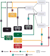
Figure 6. Summary of neurotransmitter pathways affected in PD and PD-D
Brain regions important for both movement and cognition are the neocortex, striatum, thalamus and limbic brain regions. These brain regions receive modulating neurotransmitter input from dopamine, noradrenaline, serotonin and acetylcholine nuclei located in the brainstem and basal forebrain. Patients with PD have significant degeneration of the dopamine neurons in the lateral substantia nigra (SN), acetylcholine neurons in the pedunculopontine nucleus (PPN) and noradrenaline neurons in the locus coeruleus (LC). In PD-D there is additional degeneration of acetylcholine neurons in the nucleus basalis (NBM), dopamine neurons in the medial SN, serotonin neurons in the median raphe (MR) and noradrenaline neurons in the LC. In addition, dysfunction without degeneration may occur in other neurotransmitter pathways (not shown).

References

    1. Dickson DW, Braak H, Duda JE, et al. Neuropathological assessment of Parkinson's disease: refining the diagnostic criteria. Lancet Neurol. 2009;8(12):1150–1157. - PubMed
    1. Halliday GM, McCann H. The progression of pathology in Parkinson's disease. Ann N Y Acad Sci. 2010;1184:188–195. - PubMed
    1. Singleton AB, Farrer MJ, Bonifati V. The genetics of Parkinson's disease: progress and therapeutic implications. Mov Disord. 2013;28(1):14–23. - PMC - PubMed
    1. Emre M, Aarsland D, Brown R, et al. Clinical diagnostic criteria for dementia associated with Parkinson's disease. Mov Disord. 2007;22(12):1689–1707. - PubMed
    1. Litvan I, Goldman JG, Troster AI, et al. Diagnostic criteria for mild cognitive impairment in Parkinson's disease: Movement Disorder Society Task Force guidelines. Mov Disord. 2012;27(3):349–356. - PMC - PubMed

Publication types