. 2014:2014:703653.
doi: 10.1155/2014/703653. Epub 2014 Mar 13.

Monocyte subsets in schistosomiasis patients with periportal fibrosis

Affiliations

Monocyte subsets in schistosomiasis patients with periportal fibrosis

Jamille Souza Fernandes et al. Mediators Inflamm. 2014.

Abstract

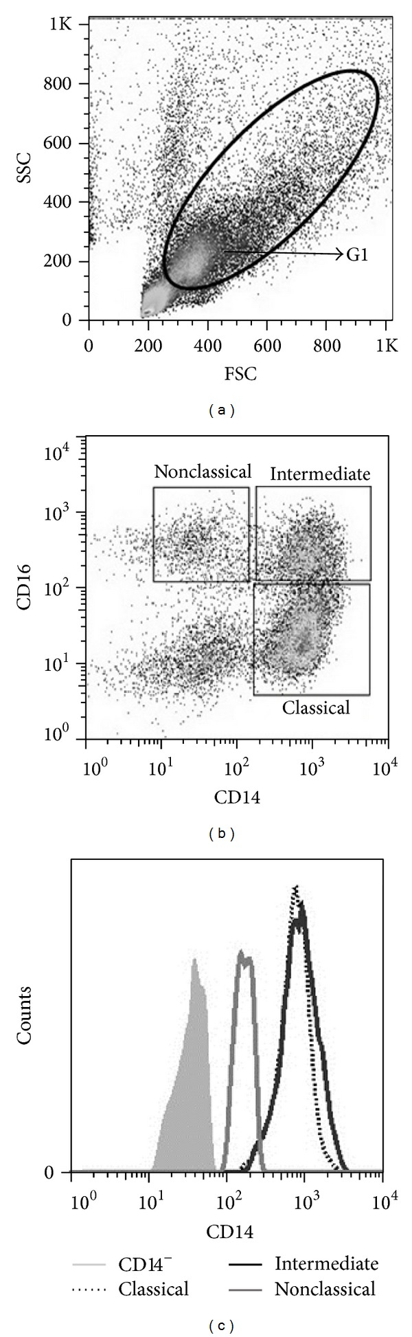
A major issue with Schistosoma mansoni infection is the development of periportal fibrosis, which is predominantly caused by the host immune response to egg antigens. Experimental studies have pointed to the participation of monocytes in the pathogenesis of liver fibrosis. The aim of this study was to characterize the subsets of monocytes in individuals with different degrees of periportal fibrosis secondary to schistosomiasis. Monocytes were classified into classical (CD14(++)CD16(-)), intermediate (CD14(++)CD16(+)), and nonclassical (CD14(+)CD16(++)). The expressions of monocyte markers and cytokines were assessed using flow cytometry. The frequency of classical monocytes was higher than the other subsets. The expression of HLA-DR, IL-6, TNF-α, and TGF-β was higher in monocytes from individuals with moderate to severe fibrosis as compared to other groups. Although no differences were observed in receptors expression (IL-4R and IL-10R) between groups of patients, the expression of IL-12 was lower in monocytes from individuals with moderate to severe fibrosis, suggesting a protective role of this cytokine in the development of fibrosis. Our data support the hypothesis that the three different monocyte populations participate in the immunopathogenesis of periportal fibrosis, since they express high levels of proinflammatory and profibrotic cytokines and low levels of regulatory markers.


Figures

Figure 1
Figure 1
The monocyte population was defined by nonspecific fluorescence from the forward scatter (FSC) and side scatter (SSC) as parameters of cell size and granularity, identifying the monocyte population, region 1 (G1) (a). Strategy for classification of monocyte subsets through the expression of CD14 and CD16 (b). A representative histogram of CD14 expression in monocyte subsets (c).
Figure 2
Figure 2
Representative histogram of HLA-DR expression (mean fluorescence intensity, MFI) on monocytes of patients with periportal fibrosis secondary to schistosomiasis ((a)–(c)). Expression of HLA-DR on classical (CD14++CD16), intermediate (CD14++CD16+), and nonclassical (CD14+CD16++) monocytes, respectively, in cultures without stimulation (white bar) and stimulated with 10 μg/mL of SEA (gray bar) ((d)–(f)). Without fibrosis (WF), incipient fibrosis (IF), and moderate to severe fibrosis (MSF). *P < 0.05 and **P < 0.005 (Kruskal-Wallis).
Figure 3
Figure 3
Mean fluorescence intensity (MFI) expression of profibrotic IL-4Rα ((a)–(c)) and TGF-β ((d)–(f)) and proinflammatory IL-6 ((g)–(i)) and TNF-α ((j)–(l)) molecules in classical (CD14++CD16), intermediate (CD14++CD16+), and nonclassical (CD14+CD16++) monocytes, of patients with different degrees of periportal fibrosis secondary to schistosomiasis. Cultures without stimulus (white bar) and cultures stimulated with 10 μg/mL SEA (gray bar). Without fibrosis (WF), incipient fibrosis (IF), and moderate to severe fibrosis (MSF). *P < 0.05 and **P < 0.005 (Kruskal-Wallis).
Figure 4
Figure 4
Mean fluorescence intensity (MFI) expression of antifibrotic IL-12 ((a)–(c)) and regulatory IL-10 ((d)–(f)) and IL-10R ((g)–(i)) molecules in classical (CD14++CD16), intermediate (CD14++CD16+), and nonclassical (CD14+CD16++) monocytes of patients with different degrees of periportal fibrosis secondary to schistosomiasis. Cultures without stimulation (white bar) and cultures stimulated with 10 μg/mL of SEA (gray bar). Without fibrosis (WF), incipient fibrosis (IF), and moderate to severe fibrosis (MSF). *P < 0.05 and **P < 0.005 (Kruskal-Wallis).

References

    1. Iarotski LS, Davis A. The schistosomiasis problem in the world: results of a WHO questionnaire survey. Bulletin of the World Health Organization. 1981;59(1):115–127. - PMC - PubMed
    1. Steinmann P, Keiser J, Bos R, Tanner M, Utzinger J. Schistosomiasis and water resources development: systematic review, meta-analysis, and estimates of people at risk. The Lancet Infectious Diseases. 2006;6(7):411–425. - PubMed
    1. Vigilância Epidemiológica e Controle da Esquistossomose. 2007, ftp://ftp.cve.saude.sp.gov.br/doc_tec/hidrica/doc/manu_esqui.pdf.
    1. BRASIL. Guia de Vigilância Epidemiológica. 2009.
    1. Warren KS. Pathophysiology and pathogenesis of hepatosplenic schistosomiasis mansoni. Bulletin of the New York Academy of Medicine. 1968;44(3):280–294. - PMC - PubMed

Publication types

MeSH terms