Review
. 2014 Jun 11;114(11):5753-74.
doi: 10.1021/cr4006236. Epub 2014 Apr 23.

Targeting the Achilles heel of multidrug-resistant cancer by exploiting the fitness cost of resistance

Affiliations
Review

Targeting the Achilles heel of multidrug-resistant cancer by exploiting the fitness cost of resistance

Gergely Szakács et al. Chem Rev. .
No abstract available


Figures

Figure 1
Figure 1
Collateral sensitivity. Changes accompanying acquired resistance to drug A can be beneficial, neutral, or detrimental in the presence of drug B. Cancer cells tend to increase their fitness through the overexpression of efflux transporters that keep the concentration of drug A below a cell-killing threshold. If drug B is not a transported substrate, resistant cells can be eradicated. However, given the wide substrate specificity of the transporters, cancer cells selected in drug A often survive despite treatment with drug B (multidrug-resistant cells show increased fitness in both environments). Conversely, resistance against drug A can be accompanied by decreased fitness in drug B (collateral sensitivity).
Figure 2
Figure 2
Efforts to overcome transporter-mediated MDR. ABC transporters protect MDR cells by keeping the concentration of cytotoxic drugs below a cell-killing threshold. Concomitantly administered inhibitors block the transporter, thus preventing the efflux of the cytotoxic compounds. Another strategy for improving therapy response is to design new classes of anticancer agents that bypass the multidrug transporters. Selective toxicity of MDR-selective compounds is specifically tied to the activity of multidrug transporters, suggesting a fatal weakness that can be exploited by a new modality for tackling multidrug-resistant cancer. Adapted with permission from ref (5). Copyright 2006 Nature Publishing Group.
Figure 3
Figure 3
Correlation of drug-sensitivity patterns and gene-expression profiles in the NCI-60 cell tumor cell panel reveals putative mechanisms of drug resistance (lower panel) and collateral sensitivity (upper panel). The NCI-60 cell panel encompasses wide P-gp expression levels, which provides an opportunity to relate P-gp levels to drug activity. The toxicity of a drug can decrease if the compound is extruded from the cells by P-gp. Consequently, the IC50 values of transported substrates and the P-gp expression levels across the 60 cells are expected to be positively correlated (lower panel). Analysis of positively correlated compound-gene sets was shown to provide an unbiased method for identifying substrates and discovering molecular features defining substrate specificities., Unexpectedly, some drugs show increased toxicity in cells expressing P-gp (upper panel). The negative correlation between IC50 values and P-gp expression suggests that compounds can inhibit the growth of cancer cells more strongly if P-gp is overexpressed.
Figure 4
Figure 4
MDR-selective compounds identified by correlating toxicity profiles and P-gp mRNA expression patterns in the NCI-60 cell panel. Compounds whose toxicity profiles show high correlation to P-gp expression were clustered on the basis of structural features (2D Tanimoto dissimilarity scores were clustered using the average linkage algorithm). Molecular scaffolds associated with MDR-selective toxicity include thiosemicarbazones, 1,10-phenanthrolines, and natural-product-derived sesquiterpenic benzoquinones (adapted from Türk et al.). The structures of KP772 and Dp44mT, which were identified independently to exhibit MDR-selective toxicity, are shown in the respective clusters.
Figure 5
Figure 5
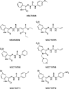
Structures of NSC10580, NSC168468, NSC292408, and NSC713048.
Figure 6
Figure 6
Isatin-β-thiosemicarbazones identified as MDR-selective compounds in the Developmental Therapeutics Program data set.
Figure 7
Figure 7
Summary of structure–activity relationships of thiosemicarbazone derivatives targeting MDR cells overexpressing P-gp.
Figure 8
Figure 8
Summary of structure–activity relationships derived from desmosdumotin derivatives targeting MDR cells overexpressing P-gp.
Figure 9
Figure 9
Pro-oxidant compounds targeting MRP1-overexpressing cancer cells. Chalcone derivatives, chrysin, and HZ08 increase the toxicity of pro-oxidants such as cisplatin, doxorubicin, and curcumin in MRP1-overexpressing cancer cells by triggering cellular GSH depletion through MRP1, which induces mitochondrial dysfunction (for 2′,5′-DHC and chrysin) or cell cycle arrest and apoptosis signaling (for HZ08).
Figure 10
Figure 10
Structure–activity relationships of verapamil derivatives targeting MRP1-overexpressing cells. Verapamil stimulates MRP1-mediated GSH export, which triggers selective apoptosis of MRP1-overexpressing cells. Iodinated derivatives were designed to increase verapamil potency.
Figure 11
Figure 11
Structure–activity relationships of substituted xanthones. Xanthone derivatives trigger selective death of MRP1-overexpressing cells through MRP1-mediated GSH export with a marked dependency on their structure.
Figure 12
Figure 12
Structure–activity relationships of substituted flavones. A number of flavone derivatives trigger selective death of MRP1-overexpressing cells through MRP1-mediated GSH export in relation to their structures. Substitution at position 3 appears to be critical.
Figure 13
Figure 13
Structures of additional compounds displaying a selective cytotoxicity in MRP1-overexpressing cells. HNE, BSO, indomethacin, and tiopronin selectively sensitize MRP1-overexpressing cells through either an induced GSH depletion (HNE, BSO, and indomethacin) or currently unknown mechanisms (tiopronin).
Figure 14
Figure 14
MDR-selective compounds identified by correlating toxicity profiles and ABCG2 function pattern in the NCI-60 cell panel.
Figure 15
Figure 15
Photosensitive imidazoacridinones produce reactive oxygen species (ROS) upon illumination, causing damage to extracellular vesicles overexpressing ABCG2.
Figure 16
Figure 16
Possible mechanism of action of MDR-selective agents. MDR transporters might change the intracellular milieu, unshielding MDR cells by exposing the molecular targets (squares) of the MDR-selective compounds (stars). Alternatively, MDR-selective compounds might initiate a yet unknown, transporter-mediated signaling pathway, or the transporters might simply increase their intracellular accumulation. It is also possible that MDR transporters efflux an endogenous molecule (X), thus increasing the activation of the compounds. Finally, MDR-selective compounds might modulate the transport/substrate specificity of the transporters, which would result in the export and/or cellular depletion of essential endogenous molecules (circles), such as glutathione.

References

    1. Willingham A. T.; Deveraux Q. L.; Hampton G. M.; Aza-Blanc P. Oncogene 2004, 23, 8392. - PubMed
    1. Szybalski W.; Bryson V. J. Bacteriol. 1952, 64, 489. - PMC - PubMed
    1. Borst P. Open Biol. 2012, 2, 120066. - PMC - PubMed
    1. Komarova N. L.; Wodarz D. Proc. Natl. Acad. Sci. U.S.A. 2005, 102, 9714. - PMC - PubMed
    1. Szakacs G.; Paterson J. K.; Ludwig J. A.; Booth-Genthe C.; Gottesman M. M. Nat. Rev. Drug. Discovery 2006, 5, 219. - PubMed

Publication types

MeSH terms

Substances