. 2014 Apr 24:2:47.
doi: 10.1186/2051-5960-2-47.

The instability of the BTB-KELCH protein Gigaxonin causes Giant Axonal Neuropathy and constitutes a new penetrant and specific diagnostic test

Affiliations

The instability of the BTB-KELCH protein Gigaxonin causes Giant Axonal Neuropathy and constitutes a new penetrant and specific diagnostic test

Alexia Boizot et al. Acta Neuropathol Commun. .

Abstract

Background: The BTB-KELCH protein Gigaxonin plays key roles in sustaining neuron survival and cytoskeleton architecture. Indeed, recessive mutations in the Gigaxonin-encoding gene cause Giant Axonal Neuropathy (GAN), a severe neurodegenerative disorder characterized by a wide disorganization of the Intermediate Filament network. Growing evidences suggest that GAN is a continuum with the peripheral neuropathy Charcot-Marie-Tooth diseases type 2 (CMT2). Sharing similar sensory-motor alterations and aggregation of Neurofilaments, few reports have revealed that GAN and some CMT2 forms can be misdiagnosed on clinical and histopathological examination. The goal of this study is to propose a new differential diagnostic test for GAN/CMT2. Moreover, we aim at identifying the mechanisms causing the loss-of-function of Gigaxonin, which has been proposed to bind CUL3 and substrates as part of an E3 ligase complex.

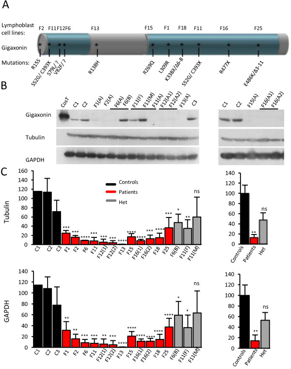
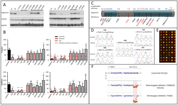
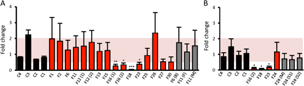
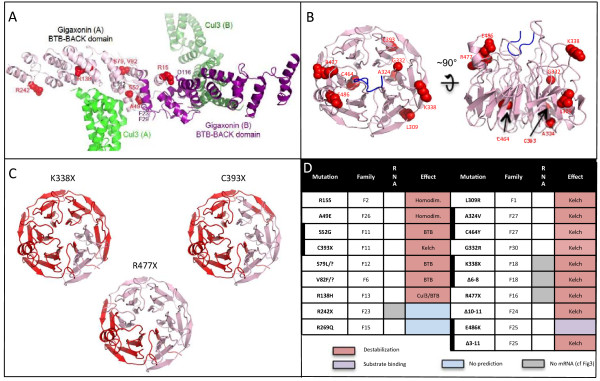
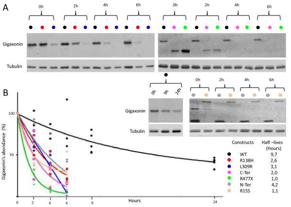
Results: We establish that determining Gigaxonin level constitutes a very valuable diagnostic test in discriminating new GAN cases from clinically related inherited neuropathies. Indeed, in a set of seven new families presenting a neuropathy resembling GAN/CMT2, only five exhibiting a reduced Gigaxonin abundance have been subsequently genetically linked to GAN. Generating the homology modeling of Gigaxonin, we suggest that disease mutations would lead to a range of defects in Gigaxonin stability, impairing its homodimerization, BTB or KELCH domain folding, or CUL3 and substrate binding. We further demonstrate that regardless of the mutations or the severity of the disease, Gigaxonin abundance is severely reduced in all GAN patients due to both mRNA and protein instability mechanisms.

Conclusions: In this study, we developed a new penetrant and specific test to diagnose GAN among a set of individuals exhibiting CMT2 of unknown etiology to suggest that the prevalence of GAN is probably under-evaluated among peripheral neuropathies. We propose to use this new test in concert with the clinical examination and prior to the systematic screening of GAN mutations that has shown strong limitations for large deletions. Combining the generation of the structural modeling of Gigaxonin to an analysis of Gigaxonin transcripts and proteins in patients, we provide the first evidences of the instability of this E3 ligase adaptor in disease.


Figures

Figure 1
Figure 1
Decreased abundance of disease-associated Gigaxonin. A Schematic representation of Gigaxonin and the corresponding known mutations in GAN patients. The N-terminal BTB and C-terminal KELCH domains are represented in blue. Lymphoblast cell lines derived from GAN patients are numbered F1-F25 and their respective mutations are mapped on Gigaxonin. All patients are severely affected by the disease with the exception of patients F2 and F13, who are mild cases reported previously. B Abundance of Gigaxonin, as revealed by immunoblotting using the GigA antibody [8]. Cost and c1-c3 correspond to ectopic Flag-tagged Gigaxonin expressed in COS cells and to unrelated control individuals, respectively. (A), (B), (F) and (M) stand for Affected, non-affected Brother, Father and Mother, respectively. A1 and A2 are two affected children from the same family. Please note that immunoblottings of patients F18 and F25 are shown in Figure 2A. C Quantification of Gigaxonin in GAN patients and their relatives. Left: Percentage of Gigaxonin for each individual in comparison to wild type Gigaxonin, as the average of 3–5 independent experiments, after normalization with tubulin and GAPDH. Right: Mean abundance of Gigaxonin in patients and heterozygous individuals, as measured by the percentage in comparison to wild type Gigaxonin. (T-test, *, p < 0,05; **, p < 0,01, ***, p < 0,001 and ***, p < 0,0001; error bars represent standard deviation).
Figure 2
Figure 2
Diminished levels of Gigaxonin corroborate with identification of mutations in the GAN locus. A Immunodetection of Gigaxonin in new patient’s lymphoblast cell lines. (S1) and (S2) are unaffected sisters of patient F24. B Quantification of Gigaxonin in patients and their relatives, using Tubulin or GAPDH as internal controls. Individual level of Gigaxonin is compared with the range of wild type Gigaxonin (left panel) and mutated Gigaxonin in known GAN patients (as presented in Figure 1, right panel). The red lines correspond to the maximum individual mean value from patients. Please note that Gigaxonin abundance was so low (undetectable) for F24 and F30 that it was detected as significantly different from mutated Gigaxonin. N = 3-5 experiments. (T-test, *, p < 0,05; **, p < 0,01, ***, p < 0,001 and ***, p < 0,0001; error bars represent standard deviation). C Schematic representation of Gigaxonin and the mutations identified in known patients (black) and new patients (red). D Electropherograms representing the point mutations identified by systematic screening of the 11 exons of the GAN gene. E Illustration and F results of the CGH data, that revealed homozygous genomic deletion encompassing exons 10 and 11 in patient F24 and heterozygosity for sister 1.
Figure 3
Figure 3
Nonsense mediated mRNA decay in some GAN patients. Gigaxonin mRNA levels of four control individuals (c1-4, in black), all GAN patients (in red) and their relatives (in grey) are measured using quantitative RT-PCR with GAN-exons9-11 (A) and GAN-exons4-5 (B), and normalized using HPRT mRNA levels. Each mRNA level is expressed as the fold change to the mean value of the four control mRNAs. (B), (F) and (M) (S1 or S2) stand for non-affected Brother, Father, Mother, and sister, respectively N = 3 experiments. (T-test, *, p < 0,05; **, p < 0,01, ***, p < 0,001; error bars represent standard deviation SD. A 2-fold change is statistically different).
Figure 4
Figure 4
Structural modeling of Gigaxonin and predicted destabilization due to mutations. A Structural model of the homodimer BTB-BACK domain of Gigaxonin (purple A, B), in complex with with Cul3 (green A, B). Patient mutations lying in this domain are represented in red. B Representation of the top and side views of a structural model for the 6-bladed β-propeller KELCH domain of Gigaxonin. Mutations found in patients are represented in red. C Top view of the structural model for the KELCH domain of Gigaxonin, with regions deleted by the indicated truncation mutants shown in red. D Summary of the effects predicted from the modelization of Gigaxonin for all patients included in the study. Heterozygous mutations are represented by a thick vertical bar. Most of the mutations are predicted to destabilize Gigaxonin (red), whereas one of them would impair substrate binding (purple). The effect of two mutations could not be determined by the 3D model (blue).
Figure 5
Figure 5
GAN-linked Gigaxonins exhibit shorter half-lives. A Representative autoradiograms and immunoblots of the pulse chase assay for wild type, mutant Gigaxonins, and the BTB (N-ter) and KELCH (C-ter) domains. At different times (2, 4, 6, 9, 24 h) after the beginning of the chase (0 h), Gigaxonin was immunoprecipitated and processed for autoradiography (top panel). The signals were normalized to the tubulin immunoblotting of the supernatent fractions of the IPs and plotted at 100% for the time point 0 h. B Half-lives of Gigaxonins. Each of the 3–4 experiments realized per construction is plotted on the graph, to define the corresponding curve.

References

    1. Bomont P, Cavalier L, Blondeau F, Ben Hamida C, Belal S, Tazir M, Demir E, Topaloglu H, Korinthenberg R, Tuysuz B, Landrieu P, Hentati F, Koenig M. The gene encoding gigaxonin, a new member of the cytoskeletal BTB/kelch repeat family, is mutated in giant axonal neuropathy. Nat Genet. 2000;26:370–374. doi: 10.1038/81701. - DOI - PubMed
    1. Asbury AK, Gale MK, Cox SC, Baringer JR, Berg BO. Giant axonal neuropathy: a unique case with segmental neurofilamentous masses. Acta Neuropathol. 1972;20:237–247. doi: 10.1007/BF00686905. - DOI - PubMed
    1. Berg BO, Rosenberg SH, Asbury AK. Giant axonal neuropathy. Pediatrics. 1972;49:894–899. - PubMed
    1. Azzedine H, Ravise N, Verny C, Gabreels-Festen A, Lammens M, Grid D, Vallat JM, Durosier G, Senderek J, Nouioua S, Hamadouche T, Bouhouche A, Guilbot A, Stendel C, Ruberg M, Brice A, Birouk N, Dubourg O, Tazir M, LeGuern E. Spine deformities in Charcot-Marie-Tooth 4C caused by SH3TC2 gene mutations. Neurology. 2006;67:602–606. doi: 10.1212/01.wnl.0000230225.19797.93. - DOI - PubMed
    1. Lus G, Nelis E, Jordanova A, Lofgren A, Cavallaro T, Ammendola A, Melone MA, Rizzuto N, Timmerman V, Cotrufo R, De Jonghe P. Charcot-Marie-Tooth disease with giant axons: a clinicopathological and genetic entity. Neurology. 2003;61:988–990. doi: 10.1212/WNL.61.7.988. - DOI - PubMed

Publication types