. 2014 Jun;3(6):745-59.
doi: 10.5966/sctm.2013-0200. Epub 2014 Apr 23.

Crosstalk between adrenergic and toll-like receptors in human mesenchymal stem cells and keratinocytes: a recipe for impaired wound healing

Affiliations

Crosstalk between adrenergic and toll-like receptors in human mesenchymal stem cells and keratinocytes: a recipe for impaired wound healing

Mohan R Dasu et al. Stem Cells Transl Med. 2014 Jun.

Abstract

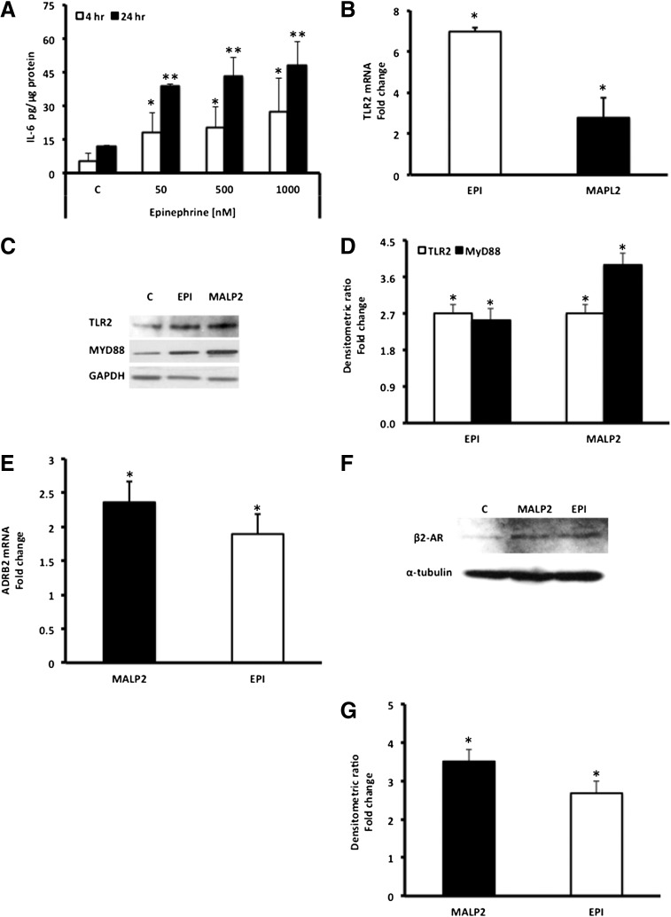
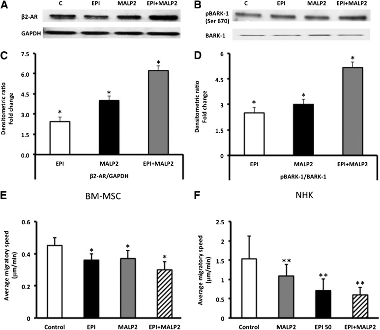
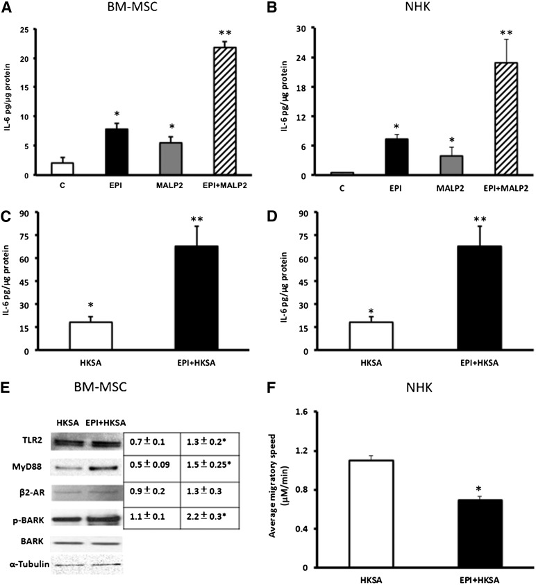
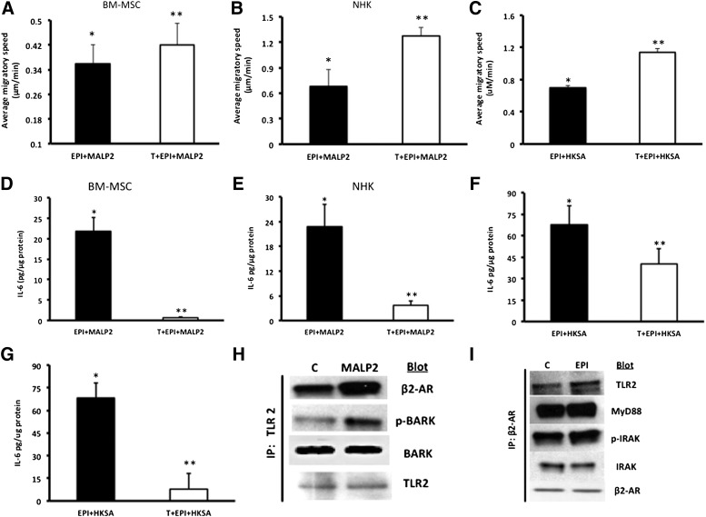
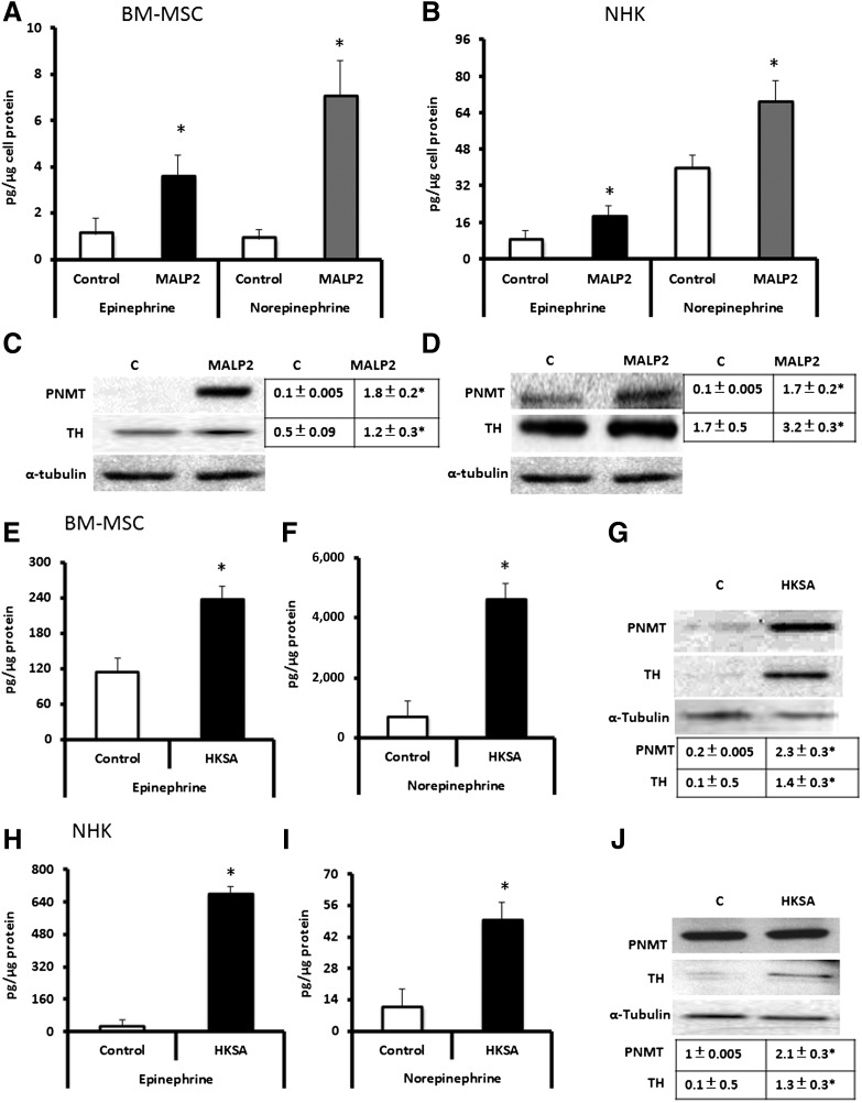
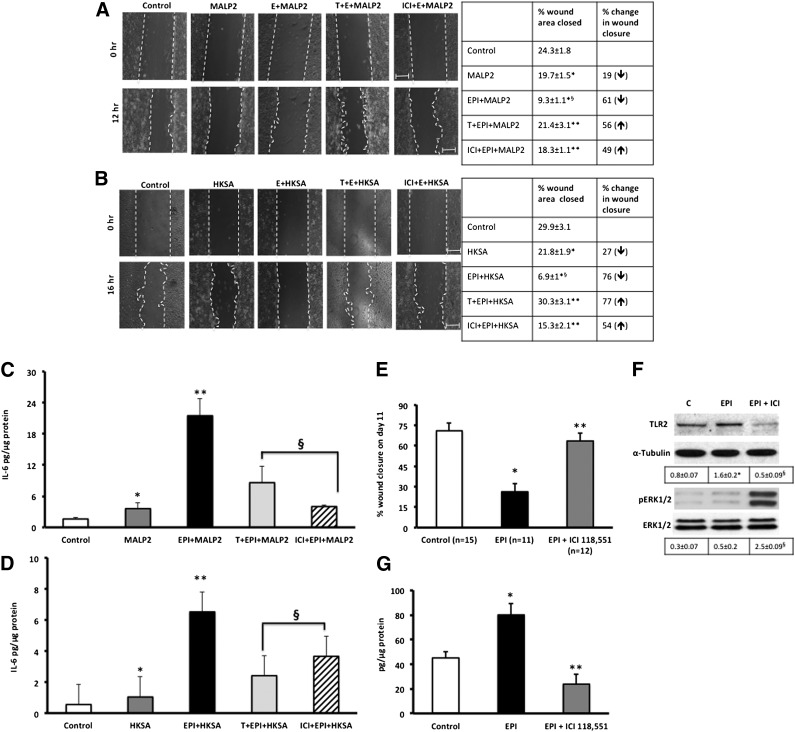
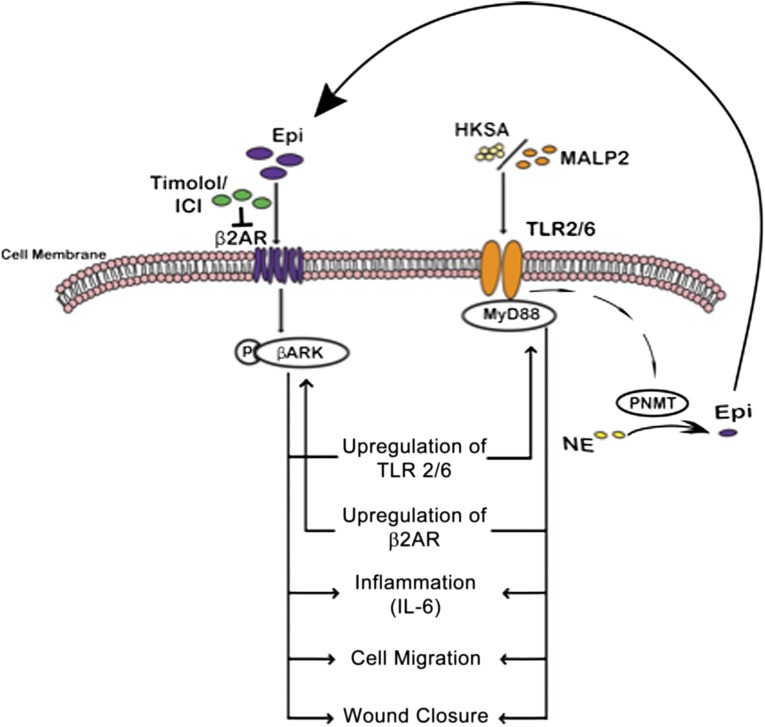
Previous studies demonstrate that skin wounds generate epinephrine (EPI) that can activate local adrenergic receptors (ARs), impairing healing. Bacterially derived activators of Toll-like receptors (TLRs) within the wound initiate inflammatory responses and can also impair healing. In this study, we examined the hypothesis that these two pathways crosstalk to one another, using EPI and macrophage-activating lipopeptide-2 (MALP2) to activate ARs and TLR2, respectively, in human bone marrow-derived mesenchymal stem cells (BM-MSCs) and neonatal keratinocytes (NHKs). BM-MSCs exposed to EPI significantly (p < .05) increased TLR2 message (sevenfold BM-MSCs), TLR2 protein (twofold), and myeloid differentiation factor 88 (MyD88) (fourfold). Conversely, activation of TLR2 by MALP2 in these cells increased β2-AR message (twofold in BM-MSCs, 2.7-fold in NHKs), β2-AR protein (2.5-fold), phosphorylation of β-AR-activated kinase (p-BARK, twofold), and induced release of EPI from both cell types (twofold). Treating cells with EPI and MALP2 together, as would be encountered in a wound, increased β2-AR and p-BARK protein expression (sixfold), impaired cell migration (BM-MSCs- 21%↓ and NHKs- 60%↓, p < .002), and resulted in a 10-fold (BM-MSCs) and 51-fold (NHKs) increase in release of IL-6 (p < .001) responses that were remarkably reduced by pretreatment with β2-AR antagonists. In vivo, EPI-stressed animals exhibited impaired healing, with elevated levels of TLR2, MyD88, and IL-6 in the wounds (p < .05) relative to nonstressed controls. Thus, our data describe a recipe for decreasing cell migration and exacerbating inflammation via novel crosstalk between the adrenergic and Toll-like receptor pathways in BM-MSCs and NHKs.

Keywords: Cell migration; Cell signaling; Mesenchymal stem cells; Stem cell-microenvironment interactions; Tissue regeneration.


Figures

Figure 1.
Figure 1.
EPI induces TLR2, MyD88, and IL-6 expression in BM-MSCs. (A): BM-MSCs (passages 3–5) were exposed to EPI (50–1,000 nM), and secreted IL-6 levels in the cell culture supernatants were determined by enzyme-linked immunosorbent assay, as described in Materials and Methods. Values are expressed as pg/μg protein (mean ± SD). ∗∗, p < .05 versus 4-hour control (C); ∗∗, p < .05 versus 24-hour control (n = 4). (B): TLR2 mRNA expression was measured in EPI (50 nM)-treated BM-MSCs using reverse transcription-polymerase chain reaction (RT-PCR), as described in Materials and Methods. Values are expressed as fold change versus control. MALP2 (100 ng/ml) was used as a positive control, as described in Materials and Methods. ∗, p < .05 versus control (n = 4). (C): TLR2 and MyD88 protein levels were measured in EPI (50 nM)-treated BM-MSC lysates using Western blot assay. GAPDH was used as the loading control, and MALP2 (100 ng/ml) as positive control. (D): Densitometric analysis of the Western blots. Protein/GAPDH ratio values are expressed as fold change versus control (mean ± SD). ∗, p < .05 versus control (n = 4). MALP2 induces β2-AR mRNA and protein expression in BM-MSCs. (E): β2-AR (ADRB2) mRNA expression was measured in MALP2 (100 ng/ml)-treated cells using RT-PCR. Values are expressed as fold change (in mRNA/housekeeping gene ratio) versus control (mean ± SD), as described in Materials and Methods. ∗, p < .001 versus control (n = 3). EPI (50 nM) was used as a positive control. (F): β2-AR protein expression was measured in MALP2 (100 ng/ml)-treated cells by Western blot. α-Tubulin was used as the loading control, and EPI (50 nM) as positive control. (G): Densitometric analysis of the Western blots. β2-AR/α-tubulin ratio values are expressed as fold change versus control (mean ± SD). ∗, p < .05 versus control (n = 4). Abbreviations: ADRB2, β2-adrenergic receptor; β2-ARs, β2-adrenergic receptors; C, control; EPI, epinephrine; GAPDH, glyceraldehyde-3-phosphate dehydrogenase; IL-6, interleukin-6; MALP2, macrophage-activating lipopeptide-2; MyD88, myeloid differentiation factor 88; TLRs, Toll-like receptors.
Figure 2.
Figure 2.
Synergistic effects of EPI and MALP2 on β2-AR protein expression and BARK-1 phosphorylation in BM-MSCs. (A): Western blot analysis of β2-AR protein expression in EPI (50 nM)-, MALP2 (100 ng/ml)-, and EPI + MALP2-treated cells, as described in Materials and Methods. GAPDH was used as the loading control. (B): Western blot analysis of BARK-1 phosphorylation in EPI (50 nM)-, MALP2 (100 ng/ml)-, and EPI + MALP2-treated cells, as described in Materials and Methods. Total BARK-1 was used as the internal control. (C): Densitometric analysis of the Western blots. β2-AR/GAPDH ratio values are expressed as fold change versus control (mean ± SD). ∗, p < .01 versus control (n = 4). (D): Densitometric analysis of the Western blots. pBARK-1/BARK-1 ratio values are expressed as fold change versus control (mean ± SD). ∗, p < .05 versus control (n = 4). EPI and MALP2 effects on BM-MSC and NHK single-cell migration (SCM). β2-AR and TLR2 activation reduced BM-MSC and NHK migration. (E, F): Migratory speeds of BM-MSCs (E) and NHKs (F) plated on collagen-coated glass-bottomed culture dishes and treated with serum-free growth medium (control), EPI (50 nM), MALP2 (100 ng/ml), or EPI + MALP2 were determined. SCM rates of at least 50 cells per treatment were measured, as described in Materials and Methods. Each panel represents the mean ± SD of at least four experiments (160– 200 cells per treatment) using four cell strains isolated from four different donors. ∗, p < .05 versus BM-MSC control; ∗∗, p < .001 versus NHK control. Abbreviations: β2-ARs, β2-adrenergic receptors; BARK, β-adrenergic receptor-activated kinase; BM-MSCs, bone marrow-derived mesenchymal stem cells; C, control; EPI, epinephrine; GAPDH, glyceraldehyde-3-phosphate dehydrogenase; MALP2, macrophage-activating lipopeptide-2; NHKs, neonatal keratinocytes; pBARK, phospho-BARK.
Figure 3.
Figure 3.
Synergistic effects of EPI and MALP2 or EPI and HKSA on IL-6 secretion in BM-MSCs and NHKs. (A, C): BM-MSCs were exposed to EPI (50 nM), MALP2 (100 ng/ml), HKSA (104 cells per milliliter), and EPI + MALP2 or EPI + HKSA for 4 hours, and secreted IL-6 levels in supernatants were determined by enzyme-linked immunosorbent assay, as described in Materials and Methods. Values are expressed as pg/μg protein (mean ± SD). ∗, p < .05 versus control; ∗∗, p < .001 versus EPI or MALP2/HKSA (n = 4 experiments). (B, D): NHKs were exposed to EPI (50 nM), MALP2 (100 ng/ml), HKSA (104 cells per milliliter), and EPI + MALP2 or EPI + HKSA for 4 hours, and secreted IL-6 levels in supernatants were determined by ELISA, as described in Materials and Methods. Values are expressed as pg/μg protein (mean ± SD). ∗, p < .05 versus control; ∗∗, p < .001 versus EPI or MALP2/HKSA (n = 4 experiments). (E): Western blot showing the combined effects of EPI + HKSA on TLR2-MyD88 and β2-AR-BARK1 expression in BM-MSCs. Cells were exposed to HKSA (104 cells per milliliter) and EPI + HKSA, and total cell protein was subjected to Western blot assay, as described in Materials and Methods. α-Tubulin was used as the loading control, and total BARK-1 was used as the internal control for phospho-BARK-1 (n = 4 experiments). Densitometric ratios (TLR2/α-tubulin, MyD88/α-tubulin, β2-AR/α-tubulin, or pBARK-1/BARK-1) are shown in the adjacent table. ∗, p < .05 versus HKSA. (F): EPI and HKSA effects on NHK single-cell migration. NHKs were plated on collagen-coated glass-bottomed culture dishes and treated with HKSA (104 cells per milliliter) and EPI + HKSA in serum-free growth medium, and single-cell migration rates of at least 50 cells per treatment were determined, as described in Materials and Methods. Each panel represents the mean values and standard deviations of at least four experiments (200 cells). ∗, p < .05 versus HKSA (n = 4 experiments). Abbreviations: β2-ARs, β2-adrenergic receptors; BARK, β-adrenergic receptor-activated kinase; BM-MSCs, bone marrow-derived mesenchymal stem cells; C, control; EPI, epinephrine; HKSA, heat-killed Staphylococcus aureus; IL-6, interleukin-6; MALP2, macrophage-activating lipopeptide-2; MyD88, myeloid differentiation factor 88; NHKs, neonatal keratinocytes; p-BARK, phospho-BARK; TLRs, Toll-like receptors.
Figure 4.
Figure 4.
Timolol (a nonselective β2-AR antagonist) reverses the combined effects of EPI + MALP2 or EPI + HKSA on single-cell migration (SCM) and IL-6 secretion in BM-MSCs and NHKs. (A): BM-MSCs were exposed to EPI (50 nM) + MALP2 (100 ng/ml) or pretreated with Timolol (10 μM, 30 minutes) and were further treated for 4 hours with EPI + MALP2. SCM rates of at least 50 cells per treatment were determined, as described in Materials and Methods. Values are expressed as average migratory speed (μm/min; mean ± SD). ∗, p < .05 versus EPI or MALP2; ∗∗, p < .005 versus EPI + MALP2 (n = 3 experiments; total of 150 cells). (B, C): NHKs were exposed to EPI (50 nM) + MALP2 (100 ng/ml), EPI + HKSA (104 cells per milliliter), or pretreated with Timolol (10 μM, 30 minutes) and were further treated for 4 hours with EPI + MALP2 or EPI + HKSA. SCM rates of at least 60 cells per treatment were determined, as described in Materials and Methods. Values are expressed as average migratory speed (μm/min; mean ± SD). ∗, p < .05 versus EPI or MALP2; ∗∗, p < .05 versus EPI + MALP2; ∗∗, p < .01 versus EPI + HKSA (n = 2 experiments; 120 cells). (D, F): BM-MSCs were exposed to EPI (50 nM) + MALP2 (100 ng/ml), EPI (50 nM) + HKSA (104 cells per milliliter), or pretreated with Timolol (10 μM, 30 minutes) and were further treated for 4 hours with EPI + MALP2 or EPI + HKSA. Secreted IL-6 in the cell culture supernatant was determined using enzyme-linked immunosorbent assay, as described in Materials and Methods. Values are expressed as pg/μg protein (mean ± SD). ∗, p < .05 versus EPI or MALP2; ∗∗, p < .001 versus EPI + MALP2; ∗∗, p < .05 versus EPI + HKSA (n = 4 experiments). (E, G): NHKs were exposed to EPI (50 nM) + MALP2 (100 ng/ml), EPI (50 nM) + HKSA (104 cells per milliliter), or pretreated with Timolol (10 μM, 30 minutes) and were further treated for 4 hours with EPI + MALP2 or EPI + HKSA. Secreted IL-6 levels were determined in the cell culture supernatants, as described in Materials and Methods. Values are expressed as pg/μg protein (mean ± SD). ∗, p < .05 versus EPI or MALP2; ∗∗, p < .005 versus EPI + MALP2; ∗∗, p < .01 versus EPI + HKSA (n = 4 experiments). Physical interaction between β2-AR and TLR2 signaling pathways in NHKs. (H): Western blot showing enhanced expression of β2-ARs and pBARK-1 in NHK cell lysates immunoprecipitated with TLR2 antibody after MALP2 (100 ng/ml) challenge, as detailed in Materials and Methods. TLR2 was used as internal/positive/negative controls (n = 4 experiments), as described in Materials and Methods. (I): Western blot showing enhanced expression of TLR2, MyD88, and pIRAK-1 in NHK cell lysates immunoprecipitated with β2-AR antibody after EPI (50 nm) challenge, as detailed in Materials and Methods. β2-ARs and total IRAK-1 were used as internal controls (n = 4 experiments) in addition to the negative controls, as described in Materials and Methods. Abbreviations: β2-ARs, β2-adrenergic receptors; BARK, β-adrenergic receptor-activated kinase; BM-MSCs, bone marrow-derived mesenchymal stem cells; C, control; EPI, epinephrine; HKSA, heat-killed Staphylococcus aureus; IL-6, interleukin-6; IP, immunoprecipitation; IRAK, interleukin receptor-activated kinase; MALP2, macrophage-activating lipopeptide-2; MyD88, myeloid differentiation factor 88; NHKs, neonatal keratinocytes; p-BARK, phospho-BARK; p-IRAK, phospho-interleukin receptor-activated kinase; T, Timolol; TLRs, Toll-like receptors.
Figure 5.
Figure 5.
MALP2 and HKSA induce catecholamine secretion and catecholamine-producing enzymes in BM-MSCs/NHKs. (A, B): BM-MSCs (A) and NHKs (B) were stimulated with 100 ng/ml MALP2 in vitro. Cell culture supernatants were collected and analyzed by high-performance liquid chromatography (HPLC) for EPI and norepinephrine, as described in Materials and Methods. All data are presented as pg/μg cell protein (mean ± SD). ∗, p < .05 versus control (n = 4 experiments). (C, D): BM-MSCs (C) and NHKs (D) were stimulated in vitro with 100 ng/ml MALP2. Total cell protein was isolated and subjected to Western blot analysis for PNMT or TH enzymes. α-Tubulin was used as a loading control. Densitometric ratios (PNMT/α-tubulin and TH/α-tubulin) are shown in the adjacent table. ∗, p < .05 versus control (C) (n = 4 experiments). (E, F): Epinephrine (E) and norepinephrine (F) levels in BM-MSCs stimulated with HKSA (104 cells per milliliter) in vitro. Cell culture supernatants were collected and analyzed by HPLC for EPI and norepinephrine, as described in Materials and Methods. All data are presented as pg/μg cell protein (mean ± SD). ∗, p < .05 versus control. (G): BM-MSCs were stimulated in vitro with 104 cells per milliliter HKSA in vitro. Total cell protein was isolated and subjected to Western blot analysis for PNMT or TH enzymes. α-Tubulin was used as a loading control. Densitometric ratios (PNMT/α-tubulin and TH/α-tubulin) are shown in the adjacent table. ∗, p < .05 versus control (C) (n = 4 experiments). (H, I) Epinephrine (H) and norepinephrine (I) levels in NHKs stimulated with HKSA (104 cells per milliliter) in vitro. Cell culture supernatants were collected and analyzed by HPLC for EPI and norepinephrine, as described in Materials and Methods. All data are presented as pg/μg cell protein (mean ± SD). ∗, p < .05 versus control (n = 3). (J): NHKs were stimulated in vitro with 104 cells per milliliter HKSA in vitro. Total cell protein was isolated and subjected to Western blot analysis for PNMT or TH enzymes. α-Tubulin was used as a loading control. Densitometric ratios (PNMT/α-tubulin and TH/α-tubulin) are shown in the adjacent table. ∗, p < .05 versus control (C) (n = 4 experiments). Abbreviations: BM-MSCs, bone marrow-derived mesenchymal stem cells; C, control; HKSA, heat-killed Staphylococcus aureus; MALP2, macrophage-activating lipopeptide-2; NHKs, neonatal keratinocytes; PNMT, phenylethanolamine N-methyltransferase; TH, tyrosine hydroxylase.
Figure 6.
Figure 6.
Blocking β2-ARs with ICI 118,551 or Timolol reverses EPI + MALP2- or EPI + HKSA-delayed NHK migration and increased IL-6 production in injured NHKs. (A): NHK monolayers were pretreated with Timolol or ICI 118,551 (10 μM, 30 minutes), followed by EPI (50 nM), MALP2 (100 ng/ml), or EPI + MALP2 treatment, and then wounded by scratches, as described in Materials and Methods. The defined areas were photographed at 0 and 12 hours after wounding. The percentage of wound area closed was calculated and presented in adjacent table along with percentage of change in wound closure (↓ = decreased wound closure; ↑ = increased wound closure). Values represent mean ± SD; ∗, p < .05 versus control; ∗∗, p < .05 versus EPI + MALP2; §, p < .01 versus MALP2 (n = 3 experiments). (B): NHK monolayers were pretreated with Timolol or ICI 118,551 (10 μM, 30 minutes), followed by EPI (50 nM), HKSA (104 cells per milliliter), or EPI + HKSA treatment, and then wounded by scratches, as described in Materials and Methods. The defined areas were photographed at 0 and 16 hours after wounding. The percentage of open and closed wound areas was calculated and presented in bar graph panel. The percentage of wound area closed was calculated and presented in adjacent table along with percentage of change in wound closure (↓ = decreased wound closure; ↑ = increased wound closure). Values represent mean ± SD, ∗, p < .05 versus control; ∗∗, p < .05 versus EPI + MALP2; §, p < .01 versus MALP2 (n = 3 experiments). (C): NHK monolayers were pretreated with ICI 118,551 or Timolol (10 μM, 30 minutes), followed by EPI (50 nM), MALP2 (100 ng/ml), or EPI + MALP2 treatment, and then wounded by scratches, as described in Materials and Methods. Cell supernatants were collected for IL-6 enzyme-linked immunosorbent assays. Values represent mean ± SD; ∗, p < .05 versus control; ∗∗, p < .05 versus MALP2; §, p < .05 versus E + MALP2 (n = 3 experiments). (D): NHK monolayers were pretreated with ICI 118,551 or Timolol (10 μM, 30 minutes), followed by EPI (50 nM), HKSA (104 cells per milliliter), or EPI + HKSA treatment, and then wounded by scratches, as described in Materials and Methods. Cell supernatants were collected for IL-6 ELISA analyses. Values represent mean ± SD; ∗, p < .05 versus control; ∗∗, p < .05 versus HKSA; §, p < .05 versus E + HKSA (n = 3 experiments). Full-thickness cutaneous wounds of EPI-stressed C57BL6J mice show decreased wound closure (E), increased TLR2 protein expression and decreased ERK1/2 phosphorylation (F), and increased local IL-6 secretion (G). Blocking β2-ARs with ICI 118,551 improves healing, decreases IL-6, TLR2 expression, and increases ERK1/2 phosphorylation in vivo. Densitometric ratios (TLR2/α-tubulin and pERK1/2/ERK1/2) are presented below the blots (F). Values represent mean ± SD; ∗, p < .05 versus control; ∗∗, p < .05 versus EPI; §, p < .05 versus EPI (n = 10–15 mice/group). Abbreviations: C, control; E, epinephrine; EPI, epinephrine; ERK, extracellular regulated kinase; HKSA, heat-killed Staphylococcus aureus; ICI, ICI-118,551; IL-6, interleukin-6; MALP2, macrophage-activating lipopeptide-2; T, Timolol; TLRs, Toll-like receptors.
Figure 7.
Figure 7.
Schema illustrating the crosstalk between β2-ARs and TLR2 in BM-MSCs and NHKs. Inflammatory effects of EPI and TLR2 ligands are mediated through phosphorylation of BARK-1 and engagement of MyD88, respectively, leading to decreased cell migration and increased IL-6 secretion. In addition, TLR2 ligands also induce catecholamine secretion by increasing TH and PNMT levels in BM-MSCs and NHKs, paving way for an autocrine inflammatory loop. Blocking β2-ARs with selective (ICI 118,551) or nonselective (Timolol) antagonists can reverse some effects. Abbreviations: β2AR, β2-adrenergic receptor; BARK, β-adrenergic receptor-activated kinase; Epi, epinephrine; HKSA, heat-killed Staphylococcus aureus; ICI, ICI-118,551; IL-6, interleukin-6; MALP2, macrophage-activating lipopeptide-2; NE, norepinephrine; PNMT, phenylethanolamine N-methyltransferase; TLRs, Toll-like receptors.

Similar articles

Cited by

References

    1. Sen CK, Gordillo GM, Roy S, et al. Human skin wounds: A major and snowballing threat to public health and the economy. Wound Repair Regen. 2009;17:763–771. - PMC - PubMed
    1. Falanga V. Wound healing and its impairment in the diabetic foot. Lancet. 2005;366:1736–1743. - PubMed
    1. Brem H, Sheehan P, Boulton AJ. Protocol for treatment of diabetic foot ulcers. Am J Surg. 2004;187:1S–10S. - PubMed
    1. Davis SC, Ricotti C, Cazzaniga A, et al. Microscopic and physiologic evidence for biofilm-associated wound colonization in vivo. Wound Repair Regen. 2008;16:23–29. - PubMed
    1. Dasu MR, Isseroff RR. Toll-like receptors in wound healing: Location, accessibility, and timing. J Invest Dermatol. 2012;132:1955–1958. - PubMed

Publication types

MeSH terms