. 2014 Apr 23;34(17):5754-64.
doi: 10.1523/JNEUROSCI.3667-13.2014.

Nerve injury-induced neuropathic pain causes disinhibition of the anterior cingulate cortex

Affiliations

Nerve injury-induced neuropathic pain causes disinhibition of the anterior cingulate cortex

Sigrid Marie Blom et al. J Neurosci. .

Abstract

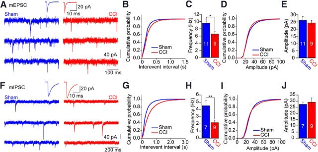
Neuropathic pain caused by peripheral nerve injury is a debilitating neurological condition of high clinical relevance. On the cellular level, the elevated pain sensitivity is induced by plasticity of neuronal function along the pain pathway. Changes in cortical areas involved in pain processing contribute to the development of neuropathic pain. Yet, it remains elusive which plasticity mechanisms occur in cortical circuits. We investigated the properties of neural networks in the anterior cingulate cortex (ACC), a brain region mediating affective responses to noxious stimuli. We performed multiple whole-cell recordings from neurons in layer 5 (L5) of the ACC of adult mice after chronic constriction injury of the sciatic nerve of the left hindpaw and observed a striking loss of connections between excitatory and inhibitory neurons in both directions. In contrast, no significant changes in synaptic efficacy in the remaining connected pairs were found. These changes were reflected on the network level by a decrease in the mEPSC and mIPSC frequency. Additionally, nerve injury resulted in a potentiation of the intrinsic excitability of pyramidal neurons, whereas the cellular properties of interneurons were unchanged. Our set of experimental parameters allowed constructing a neuronal network model of L5 in the ACC, revealing that the modification of inhibitory connectivity had the most profound effect on increased network activity. Thus, our combined experimental and modeling approach suggests that cortical disinhibition is a fundamental pathological modification associated with peripheral nerve damage. These changes at the cortical network level might therefore contribute to the neuropathic pain condition.

Keywords: anterior cingulate cortex; chronic pain; disinhibition; neuronal network; structural plasticity.


Figures

Figure 1.
Figure 1.
Average paw withdrawal thresholds in response to electronic von Frey filament testing. CCI animals developed mechanical hyperalgesia reflected in a decrease in the mechanical withdrawal threshold on the left (injured) side over time (red circles), whereas the withdrawal threshold on the right side did not change (red squares). Sham operated animals did not show any mechanical sensitization on either side (blue symbols). Error bars indicate SEM. n = 64 (sham) and n = 56 (CCI). ***p < 0.001.
Figure 2.
Figure 2.
Nerve injury increases the intrinsic excitability of L5 pyramidal neurons in the anterior cingulate cortex. A, Reconstruction of a biocytin-filled L5 pyramidal neuron. Examples of evoked responses elicited by 700 ms current injection of 90, 120, and 150 pA for the sham (B) and CCI (C) condition. Average values for AP threshold (D), input resistance (E), and membrane potential (F) and the frequency of the two first APs in a spike train evoked by a 150 pA current injection (G) for the two conditions. H, Average F-I curves for the two conditions. Statistical significance was determined by two-tailed Student's t test in D–G and by two-way ANOVA followed by Bonferroni's post hoc test in H. Numbers in bar graphs indicate number of cells. Error bars indicate SEM. *p < 0.05. **p < 0.01. ***p < 0.001.
Figure 3.
Figure 3.
Nerve injury does not change the intrinsic excitability of L5 fast-spiking interneurons. A, Reconstruction of a biocytin-filled L5 interneuron. B, Example of an evoked response elicited by 700 ms current injection of 150 pA for the CCI condition. Magnification of a single AP is shown in the inset. Average values for AP threshold (C) and membrane potential (D) for the two conditions. E, Average F-I curves for the two conditions. Statistical significance was determined by two-tailed Student's t test in C and D and by two-way ANOVA followed by Bonferroni's post hoc test in E. Numbers in bar graphs indicate number of cells. Error bars indicate SEM.
Figure 4.
Figure 4.
Nerve injury changes the neuronal connectivity pattern in L5 of the ACC. A, Biocytin staining of pyramidal neurons. B, Example voltage traces showing presynaptic and postsynaptic recordings for a pyramidal cell–pyramidal cell connection. C, Biocytin staining of a connected pyramidal cell and a FS interneuron. D, Example voltage traces showing presynaptic and postsynaptic recordings for a pyramidal cell–FS interneuron connection (left) and FS interneuron–pyramidal cell connection (right). E, Connectivity probability for pyramidal cell–pyramidal cell connections (left), pyramidal cell–FS interneuron connections (middle), and FS interneuron–pyramidal cell connections (right). *p < 0.05.
Figure 5.
Figure 5.
Properties of unitary synaptic connections are not changed after nerve injury. A, Summary of paired recordings from pyramidal to pyramidal neurons in L5 of ACC. A train of 8 APs (30 Hz) followed by one AP 500 ms later in a presynaptic pyramidal neuron resulted in corresponding EPSPs in the postsynaptic pyramidal neuron. The average postsynaptic response in the sham condition is shown in blue (middle traces) and in the CCI condition in red (lower traces). Traces from individual experiments are shown in gray. B, Summary of paired recordings from pyramidal cells to FS interneurons and the corresponding responses to the standard AP pattern. C, Same as in B for FS interneuron to pyramidal cell connections. D, Average peak EPSP amplitudes for the pyramidal cell to pyramidal cell connection as a function of EPSP number. Solid lines indicate fits to the average, and shaded areas represent fits to ±SEM according to the Tsodyks–Markram model. E, Average peak EPSP amplitudes for the pyramidal cell to FS interneuron connection as a function of EPSP number. F, Average peak IPSP amplitudes for the FS interneuron to pyramidal cell connection as a function of IPSP number. G–I, Averaged amplitude of the first postsynaptic potential and paired-pulse ratio (PPR) for the different conditions indicating that the unitary connection efficacy was not changed by CCI surgery. Error bars indicate SEM.
Figure 6.
Figure 6.
Frequency of mEPSCs and mIPSCs is reduced after nerve injury. A, Example traces showing mEPSCs recorded in FS interneurons in L5 of ACC for sham (left) and CCI (right). Average mEPSCs are shown above the traces. B, Cumulative probability plot for the interevent interval for mEPSCs. C, Average mEPSC frequency for the two conditions. D, Cumulative probability plot for mEPSC amplitudes. E, Average mEPSC amplitude for the two conditions. F, Example traces showing mIPSCs recorded in pyramidal neurons in L5 of ACC for sham (left) and CCI (right). Average mIPSCs are shown above the traces. G, Cumulative probability plot for the interevent interval for mIPSCs. H, Average mIPSC frequency for the two conditions. I, Cumulative probability plot for mIPSC amplitudes. J, Average mIPSC amplitude for the two conditions. Numbers in bar graphs indicate number of cells. Error bars indicate SEM. *p < 0.05. **p < 0.01.
Figure 7.
Figure 7.
Model of L5 ACC. A, Network architecture. B, C, Calibration of the model. B, F-I curve for pyramidal neurons. C, F-I curve for FS interneurons in sham (blue) and CCI (red) conditions. The average F-I curve from the data (large disks) is fitted with the model (solid line). ±SEM of the data (small disks) are also fitted with the model (shaded area). D, E, Response of the network (D, excitatory neurons; E, inhibitory neurons) to step current in the pool of excitatory neurons for the sham condition (blue) and for the CCI condition (red). F, G, Stationary network response (F, excitatory neurons; G, inhibitory neurons) to a given excitatory current for the sham condition (blue) and for the CCI condition (red). The dashed line indicates the effective transfer function for the mean parameters. The solid line indicates the median; the shaded area represents the interval between the first and third quartile when the parameters are sampled consistently with the uncertainty of the mean (see Materials and Methods). Inset, Distribution of responses at 300 pA.
Figure 8.
Figure 8.
Loss of connectivity is the most prominent factor for the transition from sham to CCI conditions. A, Relative effect of the model parameters (or group of parameters) on the firing rate of the excitatory neurons for a given input current (300 pA). All the parameters are taken from the sham dataset, except the parameter under investigation, which is taken from the CCI dataset (for a formal definition of this relative effect, see Materials and Methods). B, Same as in A for a given input current of I = 150 pA.

Similar articles

Cited by

References

    1. Apkarian AV, Baliki MN, Geha PY. Towards a theory of chronic pain. Prog Neurobiol. 2009;87:81–97. doi: 10.1016/j.pneurobio.2008.09.018. - DOI - PMC - PubMed
    1. Basbaum AI, Bautista DM, Scherrer G, Julius D. Cellular and molecular mechanisms of pain. Cell. 2009;139:267–284. doi: 10.1016/j.cell.2009.09.028. - DOI - PMC - PubMed
    1. Bennett GJ, Xie YK. A peripheral mononeuropathy in rat that produces disorders of pain sensation like those seen in man. Pain. 1988;33:87–107. doi: 10.1016/0304-3959(88)90209-6. - DOI - PubMed
    1. Breivik H, Collett B, Ventafridda V, Cohen R, Gallacher D. Survey of chronic pain in Europe: prevalence, impact on daily life, and treatment. Eur J Pain. 2006;10:287–333. doi: 10.1016/j.ejpain.2005.06.009. - DOI - PubMed
    1. Calejesan AA, Kim SJ, Zhuo M. Descending facilitatory modulation of a behavioral nociceptive response by stimulation in the adult rat anterior cingulate cortex. Eur J Pain. 2000;4:83–96. doi: 10.1053/eujp.1999.0158. - DOI - PubMed

Publication types