. 2014 Apr 23;34(17):5861-73.
doi: 10.1523/JNEUROSCI.0021-14.2014.

Activation of dopaminergic D2/D3 receptors modulates dorsoventral connectivity in the hippocampus and reverses the impairment of working memory after nerve injury

Affiliations

Activation of dopaminergic D2/D3 receptors modulates dorsoventral connectivity in the hippocampus and reverses the impairment of working memory after nerve injury

Helder Cardoso-Cruz et al. J Neurosci. .

Abstract

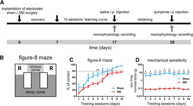
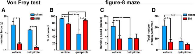
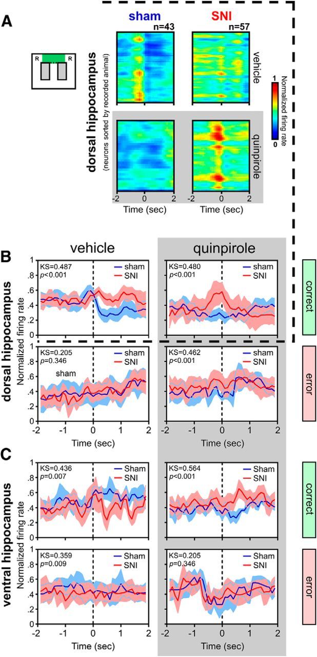
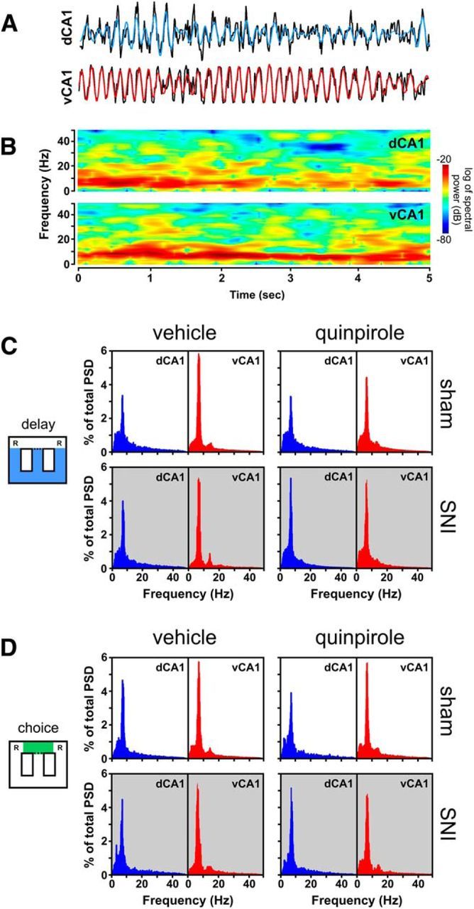
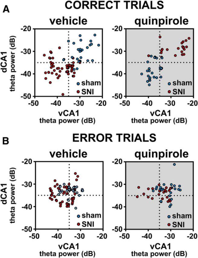
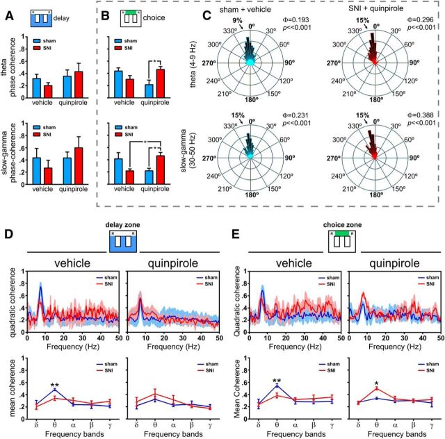
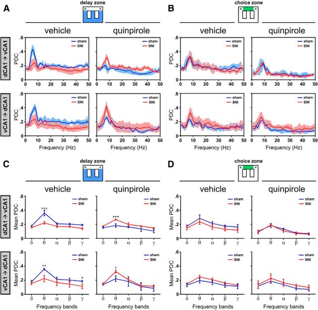
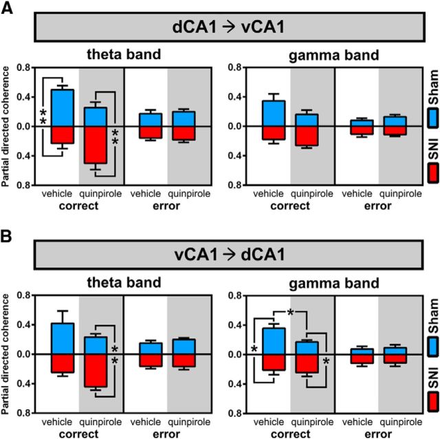
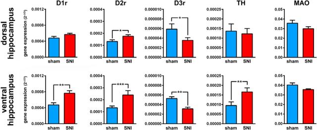
Dopamine plays an important role in several forms of synaptic plasticity in the hippocampus, a crucial brain structure for working memory (WM) functioning. In this study, we evaluated whether the working-memory impairment characteristic of animal models of chronic pain is dependent on hippocampal dopaminergic signaling. To address this issue, we implanted multichannel arrays of electrodes in the dorsal and ventral hippocampal CA1 region of rats and recorded the neuronal activity during a food-reinforced spatial WM task of trajectory alternation. Within-subject behavioral performance and patterns of dorsoventral neuronal activity were assessed before and after the onset of persistent neuropathic pain using the Spared Nerve Injury (SNI) model of neuropathic pain. Our results show that the peripheral nerve lesion caused a disruption in WM and in hippocampus spike activity and that this disruption was reversed by the systemic administration of the dopamine D2/D3 receptor agonist quinpirole (0.05 mg/kg). In SNI animals, the administration of quinpirole restored both the performance-related and the task-related spike activity to the normal range characteristic of naive animals, whereas quinpirole in sham animals caused the opposite effect. Quinpirole also reversed the abnormally low levels of hippocampus dorsoventral connectivity and phase coherence. Together with our finding of changes in gene expression of dopamine receptors and modulators after the onset of the nerve injury model, these results suggest that disruption of the dopaminergic balance in the hippocampus may be crucial for the clinical neurological and cognitive deficits observed in patients with painful syndromes.

Keywords: awake animal physiology; dopamine; hippocampus; neuropathy; pain; working memory.


Figures

Figure 1.
Figure 1.
Experimental timeline, arena, and behavioral performance. A, Timeline of experimental protocol. Briefly, each animal was implanted with multielectrode arrays in the dorsal and ventral hippocampus and subjected to a sham or SNI lesion surgery. After recovery, the animals were trained for 10 d and then had electrophysiological recording sessions before and after injection of quinpirole. B, Diagram of the figure-8 maze of spatial alternation used in the study. Starting from the center of the maze, the animal had to visit two reward sites (R) in alternation to obtain chocolate-flavored pellets. C, Gain in performance during the 10 training sessions. The nerve-lesioned animals (SNI: n = 7) showed a significantly lower performance compared with control animals (sham: n = 4). D, Level of mechanical sensitivity measured by withdrawal response to stimulation with Von Frey filaments. As expected, a large decrease was observed in the threshold required to induce a paw response in SNI group and the sensitivity level was stable throughout the training period. Values are presented as mean ± SEM. Comparisons between experimental groups are based on two-way ANOVA (group × time), followed by post hoc Bonferroni. *p < 0.05; **p < 0.01; ***p < 0.001.
Figure 2.
Figure 2.
Effect of quinpirole injection on neuropathic nociception and behavioral performance. A, Quinpirole did not affect the already significant difference in mechanical threshold between sham (n = 4) and SNI (n = 7) animals. B, Injection of quinpirole reversed the behavioral performance between sham and SNI animals. The percentage of completed correct alternations between reward sites was larger in the sham animals after vehicle administration, but it was larger in the SNI animals after quinpirole. The major effect of quinpirole was a decrease in the performance of the sham animals. C, Quinpirole decreased the running speed of the sham animals to the same range of the SNI animals, but did not affect the running speed of the SNI group. D, Quinpirole reduced the total number of trials (visits to the reward site after completing a full turn in the lateral corridors) executed by the sham animals. Note that despite the low number of completed trials by the SNI animals, their percentage of correct trials was larger (B). Values are presented as mean ± SEM. Comparisons between experimental groups are based on two-way ANOVA (group × treatment), followed by post hoc Bonferroni. *p < 0.05; **p < 0.01; ***p < 0.001.
Figure 3.
Figure 3.
Peridecision neuronal activity during correct and error trials. A, Color-coded raster plots of neuronal activity (bin resolution of 50 ms) for all dorsal hippocampus recorded neurons during the 4 s period centered in the moment of crossing from the delay to the choice area in the figure-8 maze during correct trials. dCA1 neurons of sham animals present a peak of neuronal firing rate immediately preceding the approach of the decision point that is followed by a sudden decrease in firing rate. In contrast, dCA1 neurons of SNI animals show a more stable firing rate during the peridecision period. Injection of quinpirole reverses this electrophysiological pattern, with the SNI animals now presenting a peak of activity and sham animals showing a stable firing rate. B, C, Detailed peridecision histograms of population neuronal activity for both dorsal (B) and ventral (C) hippocampus during either correct (top) or error (bottom) trials. Values are presented as mean ± SEM. Comparisons of firing distributions of experimental groups are based on the KS2 test; p < 0.05.
Figure 4.
Figure 4.
Spectral analysis of dorsal and ventral CA1 hippocampus LFP signals. A, Example trace of raw recording representing 5 s of ongoing LFP activity in a sham animal recorded during delay zone navigation. Blue trace represent the dorsal hippocampus θ filtered signal (4–9 Hz), whereas the red trace represent the ventral hippocampus θ filtered signal (4–9 Hz); the black traces undern each colored trace are the unfiltered LFP signal of each area. B, Full spectrograms for the same example of 5 s recording used in A show the predominance of θ oscillations in both areas of the hippocampus during active navigation. C, D, Power spectra density plots of dorsal and ventral LFP activity during navigation in the delay zone (C) and choice zone (D) for both sham (n = 4) and SNI (n = 7) animals. Data were calculated for each entire recording session independently of correct or error trials. PSD traces show that quinpirole does not affect the overall pattern of θ predominance.
Figure 5.
Figure 5.
Dissociation between dorsal and ventral CA1 field θ power activity during correct and error trials. Scatter plots of the distribution of dorsoventral θ ratios per trials of sham (blue dots) and SNI animals (red dots). A, During correct trials, sham animals presented higher θ power in dorsal hippocampus, whereas SNI animals presented higher θ power in ventral hippocampus; this pattern of dorsoventral ratio was reversed by quinpirole (shaded scatter plot in A). B, In contrast, the during error trials, there was no preferential dorsoventral θ ratio in either groups of animals before or after the administration of quinpirole.
Figure 6.
Figure 6.
Spectral analysis of θ and γ LFP activity in dorsal and ventral hippocampus during maze navigation. A, B, dCA1-vCA1 phase coherence (Φ) activity for θ (4–9 Hz) and γ (30–50 Hz) frequency bands during delay and choice zones navigation. No significant differences were observed in delay zone between experimental groups or quinpirole administration (A). In contrast, during choice zone navigation (B), quinpirole reversed the phase coherence between sham and SNI animals in both the θ and γ bands of frequency. C, Examples of rose plots of LFP phase distributions after vehicle (in a sham animal) and quinpirole (in an SNI animal). The number in the upper right corner of each rose plot represents the value of Φ. All circular concentration distributions are significantly nonuniform (Rayleigh test, p < 0.01). D, E, Spectral quadratic coherence between CA1 and vCA1 LFP signals show in all cases a strong coherence in the θ band of frequency (top). Detailed analysis of the quadratic coherence per band of frequency (bottom) shows that sham animals in both areas of maze have the largest θ quadratic coherence, whereas after quinpirole, the maximal θ coherence occurs in the SNI animals. Frequency bands: δ, 1–4 Hz; θ, 4–9 Hz; α, 9–15 Hz; β, 15–30 Hz; γ, 30–50 Hz. Values are presented as mean ± SEM. Comparisons between experimental groups are based on two-way ANOVA (group × treatment) or (group × frequency band), followed by post hoc Bonferroni. *p < 0.05; **p < 0.01.
Figure 7.
Figure 7.
Dorsoventral functional connectivity during maze navigation. A, B, Bidirectional traces of PDC across the spectral range of frequencies for both the sham and SNI groups before and after quinpirole administration. In all cases, there is a peak of PDC values in the θ range, reflecting that hippocampus dorsoventral functional connectivity is particularly important in this band of frequency. C, D, Bidirectional analysis of PDC per band of frequencies in both sham and SNI groups before and after quinpirole administration. The functional connectivity is only different between experimental groups during navigation in the delay zone, not in the choice zone. During navigation in the delay zone, quinpirole reversed the functional connectivity between experimental groups: before quinpirole, the sham animals presented the largest dorsoventral connectivity, whereas after quinpirole, the largest connectivity occurs in the SNI animals. Frequency bands: δ, 1–4 Hz; θ, 4–9 Hz; α, 9–15 Hz; β, 15–30 Hz; γ, 30–50 Hz. Values are presented as mean ± SEM. Comparisons between experimental groups are based on two-way ANOVA (group × frequency band), followed by post hoc Bonferroni. *p < 0.05; **p < 0.01; ***p < 0.001.
Figure 8.
Figure 8.
Differences in dorsoventral functional connectivity between correct and error trials. A, B, Values of bidirectional PDC separated by group, treatment, and correct versus error trials. The separation of correct from error trials reveal distinct patterns of connectivity: whereas during correct trials, there are high connectivity PDC values in θ and γ bands and a quinpirole-induced reversal of connectivity between sham and SNI animals (as described previously in Fig. 7), the error trials are characterized by low dorsoventral connectivity, no difference between sham or SNI animals, and no change after quinpirole administration. Frequency bands: θ, 4–9 Hz; γ, 30–50 Hz. Values are presented as mean ± SEM. Comparisons between experimental groups are based on nonparametric Kruskal–Wallis test, followed by Dunn's post hoc test for multiple comparisons. *p < 0.05; **p < 0.01.
Figure 9.
Figure 9.
Nerve injury associated changes in dopamine-related gene expression in dorsal and ventral hippocampus. Gene expression was assessed by real-time PCR using GAPDH as a housekeeping gene. Each bar represents the average of individual tissue samples run separately (10 animals per group). Comparisons between experimental groups are based on nonparametric Mann–Whitney test for unpaired samples. TH, tyrosine hydroxylase; MAO, monoamine oxidase A. *p < 0.05; **p < 0.01; ***p < 0.001.

Similar articles

Cited by

References

    1. Aguiar P, Mendonça L, Galhardo V. OpenControl: a free opensource software for video tracking and automated control of behavioral mazes. J Neurosci Methods. 2007;166:66–72. doi: 10.1016/j.jneumeth.2007.06.020. - DOI - PubMed
    1. Altier N, Stewart J. The role of dopamine in the nucleus accumbens in analgesia. Life Sci. 1999;65:2269–2287. doi: 10.1016/S0024-3205(99)00298-2. - DOI - PubMed
    1. Amano T, Wada E, Yamada D, Zushida K, Maeno H, Noda M, Wada K, Sekiguchi M. Heightened amygdala long-term potentiation in neurotensin receptor type-1 knockout mice. Neuropsychopharmacology. 2008;33:3135–3145. doi: 10.1038/npp.2008.38. - DOI - PubMed
    1. Arnsten AF, Cai JX, Steere JC, Goldman-Rakic PS. Dopamine D2 receptor mechanisms contribute to age-related cognitive decline: the effects of quinpirole on memory and motor performance in monkeys. J Neurosci. 1995;15:3429–3439. - PMC - PubMed
    1. Baccalá LA, Sameshima K. Partial directed coherence: a new concept in neural structure determination. Biol Cybern. 2001;84:463–474. doi: 10.1007/PL00007990. - DOI - PubMed

Publication types

MeSH terms