. 2014 Apr 23;34(17):5895-908.
doi: 10.1523/JNEUROSCI.0672-14.2014.

The role of Arp2/3 in growth cone actin dynamics and guidance is substrate dependent

Affiliations

The role of Arp2/3 in growth cone actin dynamics and guidance is substrate dependent

José E San Miguel-Ruiz et al. J Neurosci. .

Abstract

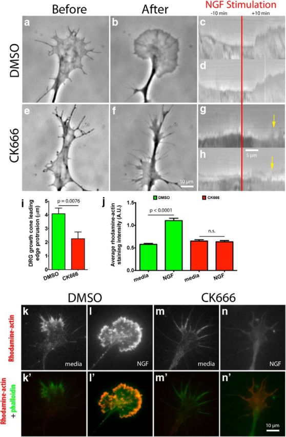
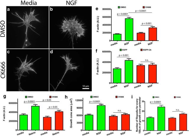
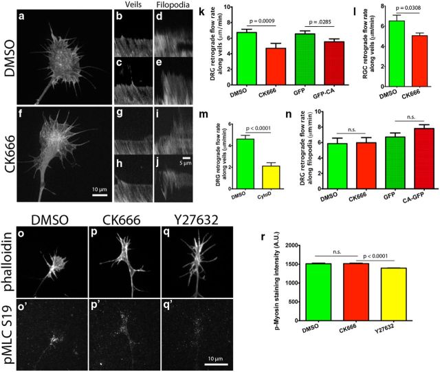
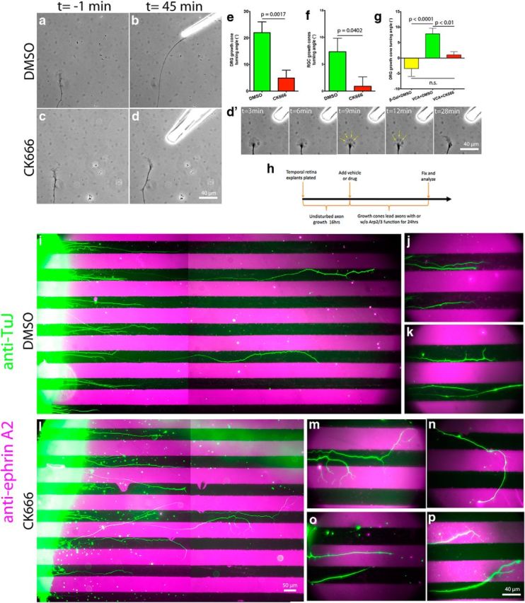
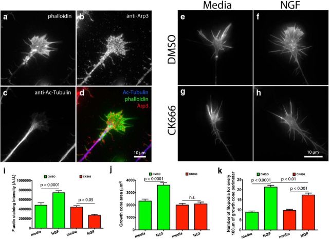
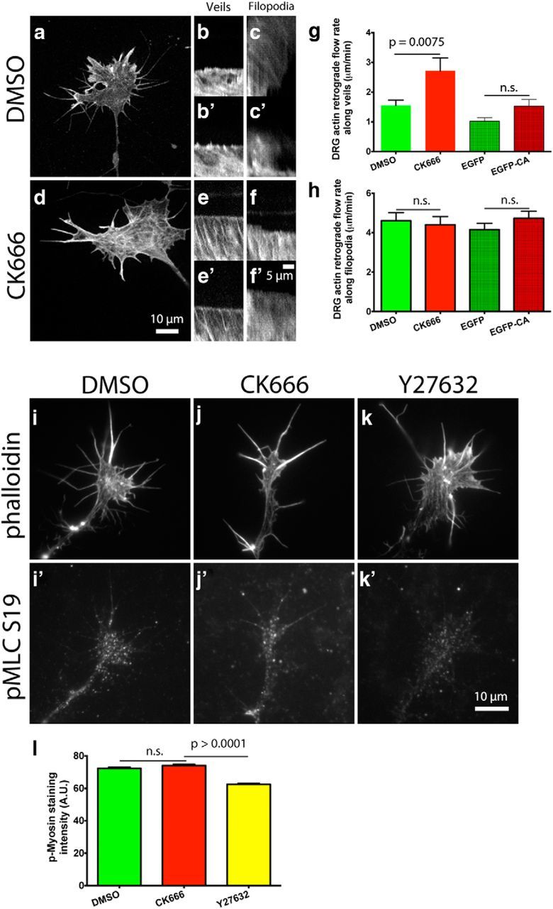
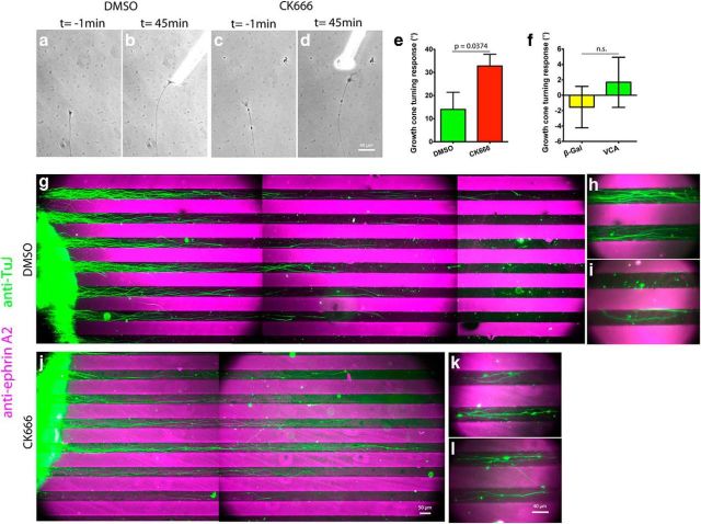
During development extrinsic guidance cues modulate the peripheral actin network in growth cones to direct axons to their targets. We wanted to understand the role of the actin nucleator Arp2/3 in growth cone actin dynamics and guidance. Since growth cones migrate in association with diverse adhesive substrates during development, we probed the hypothesis that the functional significance of Arp2/3 is substrate dependent. We report that Arp2/3 inhibition led to a reduction in the number of filopodia and growth cone F-actin content on laminin and L1. However, we found substrate-dependent differences in growth cone motility, actin retrograde flow, and guidance after Arp2/3 inhibition, suggesting that its role, and perhaps that of other actin binding proteins, in growth cone motility is substrate dependent.

Keywords: Arp2/3; L1 CAM; actin; growth cone; guidance; laminin.


Figures

Figure 1.
Figure 1.
Arp2/3 is present in the leading edge of growth cones on L1. a–j, E7 DRG explants were grown overnight on L1, treated globally with 50 ng/ml NGF for 15 min, fixed, and stained with phalloidin and antibodies against acetylated α-tubulin (Ac-Tubulin), p34, Arp3, and L1 (8D9). Images were acquired with standard confocal microscopy; (c) p34 staining was also imaged with the TIRF modality to show its localization near the substrate.
Figure 2.
Figure 2.
Arp2/3 mediates leading edge protrusion and barbed-end creation after global NGF stimulation on L1. a, b, e, f, E7 DRG explants were grown overnight on L1 and treated with DMSO or 50 μm CK666 (Arp2/3 inhibitor) for 4 h; growth cones were then imaged with phase contrast microscopy for 10 min, before and after, global application of 50 ng/ml NGF. Representative kymographs depicting leading edge behavior during NGF stimulation (red bar) for (c, d) DMSO-treated and (g, h) CK666-treated growth cones. Yellow arrows point to leading edge collapse. i, Quantification of leading edge protrusion 10 min after global NGF application. k–n, E7 DRG explants were grown on L1, treated with DMSO or CK666 for 4 h, then with 50 ng/ml NGF globally for 10 min, permeabilized with buffer containing phalloidin and Rhodamine-actin for 4 min, then fixed. k′–n′, Rhodamine-actin (red) and phalloidin (green) labeling were overlaid to show barbed-end distribution in growth cones on L1. j, Quantification of the average Rhodamine-actin incorporated into growth cones. Graphs show mean ± SEM.
Figure 3.
Figure 3.
Arp2/3 mediates actin polymerization, surface area expansion, and increase in filopodial numbers in growth cones on L1. a–d, E7 DRG explants were grown overnight on L1, treated with DMSO or 50 μm CK666 for 4 h, stimulated with 50 ng/ml NGF or control media for 15 min, fixed, stained with Alexa Fluor 488 phalloidin, (e) and its fluorescent intensity quantified. f, Quantification of Alexa Fluor 488 phalloidin fluorescent intensity bound to DRG growth cones transfected with EGFP, or the EGFP-CA (Arp2/3 dominant-negative construct). g, Quantification of Alexa Fluor 488 phalloidin fluorescent intensity bound to RGC growth cones treated with DMSO or 50 μm CK666 for 4 h, stimulated with 500 ng/ml Netrin or media as a control for 15 min, fixed, and stained with phalloidin. h, Measured DRG growth cone area. i, Average number of filopodia for every 100 μm of growth cone perimeter. Graphs show mean ± SEM.
Figure 4.
Figure 4.
Arp2/3 inhibition reduces actin retrograde flow at the leading edge of growth cones on L1, independent of myosin II activity. a, f, E7 DRG or RGC neurons were transfected with mCherry-actin and grown overnight on L1. Arp2/3 was inhibited by coexpression of EGFP-CA with mCherry-actin, or by 50 μm CK666 treatment 4 h before imaging. Growth cones were imaged in a spinning disc confocal microscope and kymographs made from these time-lapse videos. b, c, g, h, Representative kymographs along veils and (d, e, i, j) filopodia of DMSO- and CK666-treated growth cones. k, l, Quantification of the actin retrograde flow along veils of DRG and RGC growth cones and (m) the effect that 2 μm cytochalasin D has on it. Note that in this experiment DMSO and cytochalasin D were added 3 min before imaging, instead of the normal 4 h, hence the different DMSO control rates. n, Quantification of the actin retrograde flow along filopodia of DRG growth cones. o–q′, As a control, DRG growth cones on L1 were treated with DMSO, 50 μm CK666, or 10 μm Y27632 for 4 h, fixed, and stained with phalloidin and an antibody against phospho myosin light chain (Ser19), (r) and anti-pMLC average fluorescent intensity quantified. Graphs show mean ± SEM.
Figure 5.
Figure 5.
Arp2/3 activity is necessary for efficient guidance and sufficient to trigger growth cone turning on L1. E7 DRG or RGC explants were grown on L1 overnight and treated with DMSO or 50 μm CK666 4 h before the turning assay. NGF (DRG) or Netrin (RGC) gradients were established with a micropipette located at 45° and ∼80 μm from the growth cone leading edge. a–d, Representative images of DRG growth cones 1 min before and 45 min after positioning the micropipette. d′, Montage of Arp2/3-inhibited growth cone response to the NGF gradient showing multiple points of veil collapse (yellow arrows). e, f, DRG and RGC growth cone turning angle quantification. g, Turning angle quantification for DRG growth cones on L1 in response to a gradient of Chariot complexed to VCA-GST or β-galactosidase as a control, delivered as above. h, Diagram depicting the experimental design for the substrate-bound ephrin A2 chemorepulsive assay. i, l, Boundary assay for control (DMSO) and Arp2/3-inhibited (CK666) retina explants (left) with alternating lanes of laminin (black) and ephrin A2 + laminin (magenta) with axons stained with antibodies against the neuronal β3-tubulin (green). j, k, Distal tips of control and (m–p) Arp2/3-inhibited axons shown at higher magnification. Note the guidance deficits and the multiple processes sprouted by Arp2/3-inhibited axons over the ephrin A2 lanes. Graphs show mean ± SEM.
Figure 6.
Figure 6.
Arp2/3 mediates actin polymerization, surface area expansion, and increase in number of filopodia in growth cones after NGF stimulation on laminin. a–d, E7 DRG growth cones on laminin, treated with NGF, and stained with phalloidin and antibodies against Arp3 and acetylated α-tubulin (Ac-Tubulin). e–h, E7 DRG growth cones on laminin treated with DMSO or CK666 for 4 h, stimulated with NGF or media as control, fixed, stained with Alexa Fluor 488 phalloidin, (i) and phalloidin fluorescent intensity quantified. j, Quantification of growth cone area. k, Average number of filopodia for every 100 μm of growth cone perimeter. Graphs show mean ± SEM.
Figure 7.
Figure 7.
Arp2/3 inhibition increases the actin retrograde flow rate at the leading edge of growth cones on laminin in a myosin II-independent manner. a, d, E7 DRG neurons were transfected with mCherry-actin and grown overnight on laminin. Arp2/3 was inhibited by coexpression of EGFP-CA with mCherry-actin, or by 50 μm CK666 treatment 4 h before imaging. Growth cones were imaged in a spinning disc confocal and kymographs made from these time-lapse videos. b, b′,e, e′, Representative kymographs along veils and (c, c′,f, f′) filopodia of DMSO- and CK666-treated growth cones. g, h, Quantification of the actin retrograde flow rate along veils and filopodia. i–k′, As a control, DRG growth cones on L1 were treated with DMSO, 50 μm CK666, or 10 μm Y27632 for 4 h, fixed, and stained with phalloidin and an antibody against phospho myosin light chain (Ser19), (l) and anti-pMLC average fluorescent intensity quantified. Graphs show mean ± SEM.
Figure 8.
Figure 8.
Arp2/3 inhibition reduces the area and number of paxillin-GFP puncta in growth cones on laminin. a–d, E7 DRG neurons were transfected with paxillin-GFP and mCherry-CA, grown overnight on laminin, and imaged through wide-field and TIRF microscopy. e, f, Thresholded TIRF images were used to measure the area and number of paxillin-GFP puncta.
Figure 9.
Figure 9.
Arp2/3 activity is not necessary for efficient guidance and insufficient to trigger growth cone turning on laminin. E7 DRG explants were grown on laminin and treated with DMSO or 50 μm CK666 4 h before the turning assay. An NGF gradient was established with a micropipette located at 45° and ∼80 μm from the growth cone leading edge. a–d, Representative images of DRG growth cones 1 min before and 45 min after positioning the micropipette. e. f, Growth cone turning angle quantification. g, Growth cone turning angle quantification in response to a gradient of Chariot complexed to VCA-GST or β-galactosidase as a control, delivered as above. h, k, Boundary assay for control (DMSO) and Arp2/3-inhibited (CK666) retina explants (left) with alternating lanes of laminin (black) and ephrin A2 + laminin (magenta) with axons stained with antibodies against the neuronal β3-tubulin (green). i, j, l, Show axons in close apposition to the ephrin A2 + laminin lanes at higher magnification.

Similar articles

Cited by

References

    1. Bentley D, Toroian-Raymond A. Disoriented pathfinding by pioneer neurone growth cones deprived of filopodia by cytochalasin treatment. Nature. 1986;323:712–715. doi: 10.1038/323712a0. - DOI - PubMed
    1. Burden-Gulley SM, Payne HR, Lemmon V. Growth cones are actively influenced by substrate-bound adhesion molecules. J Neurosci. 1995;15:4370–4381. - PMC - PubMed
    1. Butler B, Cooper JA. Distinct roles for the actin nucleators Arp2/3 and hDia1 during NK-mediated cytotoxicity. Curr Biol. 2009;19:1886–1896. doi: 10.1016/j.cub.2009.10.029. - DOI - PMC - PubMed
    1. Carlstrom LP, Hines JH, Henle SJ, Henley JR. Bidirectional remodeling of beta1-integrin adhesions during chemotropic regulation of nerve growth. BMC Biol. 2011;9:82. doi: 10.1186/1741-7007-9-82. - DOI - PMC - PubMed
    1. Chan CE, Odde DJ. Traction dynamics of filopodia on compliant substrates. Science. 2008;322:1687–1691. doi: 10.1126/science.1163595. - DOI - PubMed

Publication types

LinkOut - more resources