. 2014 Apr 23;34(17):5971-84.
doi: 10.1523/JNEUROSCI.5109-13.2014.

Decoding ventromedial hypothalamic neural activity during male mouse aggression

Affiliations

Decoding ventromedial hypothalamic neural activity during male mouse aggression

Annegret L Falkner et al. J Neurosci. .

Abstract

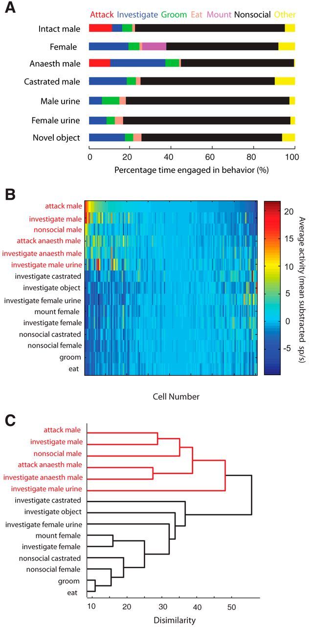
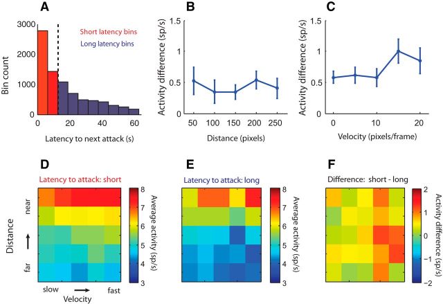
The ventromedial hypothalamus, ventrolateral area (VMHvl) was identified recently as a critical locus for inter-male aggression. Optogenetic stimulation of VMHvl in male mice evokes attack toward conspecifics and inactivation of the region inhibits natural aggression, yet very little is known about its underlying neural activity. To understand its role in promoting aggression, we recorded and analyzed neural activity in the VMHvl in response to a wide range of social and nonsocial stimuli. Although response profiles of VMHvl neurons are complex and heterogeneous, we identified a subpopulation of neurons that respond maximally during investigation and attack of male conspecific mice and during investigation of a source of male mouse urine. These "male responsive" neurons in the VMHvl are tuned to both the inter-male distance and the animal's velocity during attack. Additionally, VMHvl activity predicts several parameters of future aggressive action, including the latency and duration of the next attack. Linear regression analysis further demonstrates that aggression-specific parameters, such as distance, movement velocity, and attack latency, can model ongoing VMHvl activity fluctuation during inter-male encounters. These results represent the first effort to understand the hypothalamic neural activity during social behaviors using quantitative tools and suggest an important role for the VMHvl in encoding movement, sensory, and motivation-related signals.

Keywords: aggression; hypothalamus; motivation; physiology.


Figures

Figure 1.
Figure 1.
VMHvl response during social and nonsocial interactions. A, Schematic of time spent in social and nonsocial behaviors in the presence of each stimulus class (percentage of total time). B, Response profiles of VMHvl neurons shown across all stimulus conditions. Heat map shows average mean subtracted firing rate of each VMHvl neuron (x-axis) during each behavior (y-axis). Neurons are sorted left to right in descending order according to their average response during attack male (n = 161 neurons tested across all stimulus conditions in 5 animals). C, Dendrogram of social and nonsocial behaviors according to hierarchical clustering of activity matrix shown in B exhibits two primary clusters. Male-specific behaviors are shown in red, and non-male behaviors are shown in black.
Figure 2.
Figure 2.
VMHvl neurons transiently increase activity during attack and investigation of male mice. A, B, PSTH of an example neuron (A) and population average (B) of activity of male responsive neurons during attack (red) and investigation (blue) shown aligned to the onset (left) and offset (right) of behavior. (n = 105 neurons in 5 animals). Error bar is SEM, and bin width for all PSTHs is 50 ms with Gaussian smoothing. C, Average VMHvl cell activity during attack male is not significantly different from that during investigating male. Red circle indicates example cell shown in A. D, Distribution of the difference in activity between attack/investigation and interleaved nonsocial periods of male responsive neurons. Red, blue, and green dots show attack, investigation, and dual responsive cells, respectively (n = 105).
Figure 3.
Figure 3.
VMHvl neurons respond to male-derived olfactory cues. A, Test stimuli to control for sensory modality included intact male (red), anesthetized male (blue), castrated male (black), female (purple), male mouse urine (orange), and object (green). Color conventions are consistent across the panels. B, Population PSTH showing activity of male responsive neurons during investigation of various stimuli listed in A. Responses are aligned to investigation onset (left) and investigation offset (right). Bin size is 50 ms. C–F, Comparison of activity change in male responsive neurons during investigation of various tested stimuli. Activity change during investigation of an intact male is significantly greater than activity change during investigation of either an anesthetized male (C, n = 82) or a castrated male (D, n = 72) but is not significantly different from activity change during investigation of a source of male mouse urine (E, n = 75). Activity change during investigation of male urine is significantly greater than that during novel object investigation (F, n = 75). G, Comparison of averaged activity during pre-interaction, during-interaction, and post-interaction epochs. Activity of male responsive neurons increases during investigation relative to pre-interaction epoch and remains elevated after removal of male-related stimuli but not other stimuli (*p < 0.05, **p < 0.01, ***p < 0.001, one-sample t test using a subpopulation of male responsive neurons tested in each condition; intact male, n = 105; anesthetized male, n = 79; castrated male, n = 65; female, n = 102; male urine, n = 75; novel object, n = 92; all male responsive neurons from n = 5 animals).
Figure 4.
Figure 4.
VMHvl activity of male responsive neurons is modulated by inter-male distance and movement velocity during attack. A, B, Mean subtracted firing rate as a function of distance (A) and velocity (B) for all periods (red) and periods excluding investigation and attack (black). Cell activity is significantly tuned to both parameters when all periods are included, although only distance remains tuned after removing attack and investigation epochs. C, D, Average firing rates of male responsive cells plotted as a function of the distance (50–400 pixels using 50 pixel spacing) between the resident and the male intruder and the instantaneous velocity (0–30 pixels/frame using 5 pixels/frame spacing) of the resident mouse for all time bins during inter-male encounters (C) or all time bins excluding investigating and attacking male (D). E–H, Male responsive neurons are also tuned to the distance from a source of male mouse urine even when investigation episodes are excluded, but activity is not modulated by movement velocity during male urine trials. Male responsive neurons are from all five animals (**p ≪ 0.01, n = 95 neurons in AD, n = 75 neurons in EH).
Figure 5.
Figure 5.
VMHvl activity during investigation correlates with the likelihood of a future attack. A, Population average PSTH of activity during investigating male that was followed either by an attack (red) or a nonsocial behavior (blue). Activity is normalized for each neuron by the average activity across both conditions (investigate to attack and investigate to nonsocial) and includes only investigative episodes that lasted a minimum duration of 250 ms. B, Comparison of average neural activity in 250 ms bin at the onset of investigation between investigate to attack and investigate to nonsocial trials (red dots, gray bar in A, left) and neural activity in 250 ms bin at the offset of behavior transition to attack or nonsocial (black dots, gray bar in A, right; n = 75 investigative responsive neurons in 5 animals). One outlier point was removed for visualization. C, Scatter plot showing significantly longer median investigation time preceding attack males than those preceding nonsocial behaviors (n = 36 sessions). Black lines in B and C indicate the line of equality.
Figure 6.
Figure 6.
VMHvl activity at attack onset correlates with attack duration and time elapsed from last attack. A, Population average PSTHs of the mean firing rate of male responsive neurons aligned to attack onset (left) and offset (right) for attack trials separated into quartiles within each neuron based on attack duration. Red, green, blue, and black lines indicate PSTHs composed of attack trials lasting the shortest to the longest. Gray bar shows pre-attack activity epoch used in B and C. B, Trial-to-trial correlation of pre-attack activity (1 s before attack) and attack duration for an example neuron. Line shows the least squares regression. C, Histogram of distribution of correlation coefficients (rduration) between attack duration and pre-attack response (mean firing rate during −1 to 0 s) for all male responsive neurons. Blue overlay shows cells with significant correlation. D, Population average PSTHs of the mean firing rate of male responsive neurons aligned to attack offset (left) and onset (right) for attack trials separated into quartiles based on inter-attack interval. Gray bar shows time window used for analysis in E and F. Color conventions as in A. E, Scatter plot showing trial-to-trial correlation between attack onset activity (500 ms before attack to 500 ms after attack onset) and the ln(inter-attack interval) of an example cell. F, Histogram showing the distribution of rinter-attack interval between ln(inter-attack interval) and onset response of the next attack. Blue overlay shows cells with significant correlation. For all population analyses, n = 105 male responsive neurons from 5 animals; PSTH bin width for A and D is 100 ms.
Figure 7.
Figure 7.
VMHvl activity correlates with latency to next attack independent of changes in inter-male distance and movement velocity. Activity during each inter-male interaction was separated into short- and long-latency attack time bins. A, Example separation of time bins (1 s) during single inter-male sessions by median attack latency (dotted line) into short latency to attack (red) and long latency (blue) to attack. B and C, Across the population, VMHvl activity increases for short-latency attack time bins relative to long-latency attack bins regardless of inter-male distance (B) or movement velocity (C). Error bars show ±SEM. D–F, Heat maps showing the average activity of male responsive cells as a function of distance and velocity during short-latency attack time bins (D), long-latency attack time bins (E), and the difference between these maps (F). Activity differences between short- and long-latency maps were computed for each neuron and then averaged across neurons. (n = 93 male responsive neurons in 5 animals for B–F).
Figure 8.
Figure 8.
Model of VMHvl activity during inter-male interactions with aggression-specific parameters using regularized linear regression. A, Number of neurons including each parameter in the sparse model. Neurons were regressed individually, and the y-axis indicates the number of neurons that include each parameter as a significant regressor to the model. Darker bars indicate parameters most frequently contributing to the model and used for subsequent model. B, Distribution of regression coefficients for parameters identified as contributing to the sparse model (*p < 0.05, **p < 0.001, t test; n = 93 neurons in 5 animals). C, Variance explained (r2) by regularized model for each neuron for the full model (with minimized mean squared error, open bars), overlaid with r2 values for the sparse model (filled bars). Neurons with all zero coefficients were eliminated from this analysis. D, One example showing the fit between the model and cell activity during male stimulus presentation. The example model includes only two parameters: inter-male distance and latency to attack.

Similar articles

Cited by

References

    1. Archer J. The effect of strange male odor on aggressive behavior in male mice. J Mammal. 1968;49:572–575. doi: 10.2307/1378238. - DOI - PubMed
    1. Bard P. A diencephalic mechanism for the expression of rage with special reference to the sympathetic nervous system. Am J Physiol. 1928;84:490–515.
    1. Bean NJ. Modulation of agonistic behavior by the dual olfactory system in male mice. Physiol Behav. 1982;29:433–437. doi: 10.1016/0031-9384(82)90262-1. - DOI - PubMed
    1. Bruce HM. Effect of castration on the reproductive pheromones of male mice. J Reprod Fertil. 1965;10:141–143. doi: 10.1530/jrf.0.0100141. - DOI - PubMed
    1. Canteras NS, Simerly RB, Swanson LW. Connections of the posterior nucleus of the amygdala. J Comp Neurol. 1992;324:143–179. doi: 10.1002/cne.903240203. - DOI - PubMed

Publication types

LinkOut - more resources