Residue 41 of the Eurasian avian-like swine influenza a virus matrix protein modulates virion filament length and efficiency of contact transmission
- PMID: 24760887
- PMCID: PMC4054412
- DOI: 10.1128/JVI.00119-14
Residue 41 of the Eurasian avian-like swine influenza a virus matrix protein modulates virion filament length and efficiency of contact transmission
Abstract
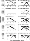
Position 41 of the influenza A virus matrix protein encodes a highly conserved alanine in human and avian lineages. Nonetheless, strains of the Eurasian avian-like swine (Easw) lineage contain a change at this position: position 41 of A/swine/Spain/53207/04 (H1N1) (SPN04) encodes a proline. To assess the impact of this naturally occurring polymorphism on viral fitness, we utilized reverse genetics to produce recombinant viruses encoding wild-type M1 41P (rSPN04-P) and consensus 41A (rSPN04-A) residues. Relative to rSPN04-A, rSPN04-P virus displayed reduced growth in vitro. In the guinea pig model, rSPN04-P was transmitted to fewer contact animals than rSPN04-A and failed to infect guinea pigs that received a low-dose inoculum. Moreover, the P41A change altered virion morphology, reducing the number and length of filamentous virions, as well as reducing the neuraminidase activity of virions. The lab-adapted human isolate, A/PR/8/34 (H1N1) (PR8), is nontransmissible in the guinea pig model, making it a useful background in which to identify certain viral factors that enhance transmissibility. We assessed transmission in the context of single-, double-, and triple-reassortant viruses between PR8 and SPN04; PR8/SPN04 M, PR8/SPN04 M+NA, and PR8/SPN04 M+NA+HA, encoding either matrix 41 A or P, were generated. In each case, the virus possessing 41P transmitted less well than the corresponding 41A-encoding virus. In summary, we have identified a naturally occurring mutation in the influenza A virus matrix protein that impacts transmission efficiency and can alter virion morphology and neuraminidase activity.
Importance: We have developed a practical model for examining the genetics underlying transmissibility of the Eurasian avian-like swine lineage viruses, which contributed M and NA segments to the 2009 pandemic strain. Here, we use our system to investigate the impact on viral fitness of a naturally occurring polymorphism at matrix (M1) position 41 in an Easw isolate. Position 41 has been implicated previously in adaptation to laboratory substrates and to mice. Here we show that the polymorphism at M1 41 has a limited effect on growth in vitro but changes the morphology of the virus and impacts growth and transmission in the guinea pig model.
Copyright © 2014, American Society for Microbiology. All Rights Reserved.
Figures


Similar articles
-
The M segment of the 2009 pandemic influenza virus confers increased neuraminidase activity, filamentous morphology, and efficient contact transmissibility to A/Puerto Rico/8/1934-based reassortant viruses.J Virol. 2014 Apr;88(7):3802-14. doi: 10.1128/JVI.03607-13. Epub 2014 Jan 15. J Virol. 2014. PMID: 24429367 Free PMC article.
-
The neuraminidase and matrix genes of the 2009 pandemic influenza H1N1 virus cooperate functionally to facilitate efficient replication and transmissibility in pigs.J Gen Virol. 2012 Jun;93(Pt 6):1261-1268. doi: 10.1099/vir.0.040535-0. Epub 2012 Feb 15. J Gen Virol. 2012. PMID: 22337640 Free PMC article.
-
Identification of Key Amino Acids in the PB2 and M1 Proteins of H7N9 Influenza Virus That Affect Its Transmission in Guinea Pigs.J Virol. 2019 Dec 12;94(1):e01180-19. doi: 10.1128/JVI.01180-19. Print 2019 Dec 12. J Virol. 2019. PMID: 31597771 Free PMC article.
-
Enhancement of influenza virus transmission by gene reassortment.Curr Top Microbiol Immunol. 2014;385:185-204. doi: 10.1007/82_2014_389. Curr Top Microbiol Immunol. 2014. PMID: 25048543 Review.
-
Influenza virus assembly and budding.Virology. 2011 Mar 15;411(2):229-36. doi: 10.1016/j.virol.2010.12.003. Epub 2011 Jan 14. Virology. 2011. PMID: 21237476 Free PMC article. Review.
Cited by
-
Swine influenza A virus isolates containing the pandemic H1N1 origin matrix gene elicit greater disease in the murine model.Microbiol Spectr. 2024 Mar 5;12(3):e0338623. doi: 10.1128/spectrum.03386-23. Epub 2024 Feb 1. Microbiol Spectr. 2024. PMID: 38299860 Free PMC article.
-
Multiple Gene Segments Are Associated with Enhanced Virulence of Clade 2.3.4.4 H5N8 Highly Pathogenic Avian Influenza Virus in Mallards.J Virol. 2021 Aug 25;95(18):e0095521. doi: 10.1128/JVI.00955-21. Epub 2021 Aug 25. J Virol. 2021. PMID: 34232725 Free PMC article.
-
A Virus Is a Community: Diversity within Negative-Sense RNA Virus Populations.Microbiol Mol Biol Rev. 2022 Sep 21;86(3):e0008621. doi: 10.1128/mmbr.00086-21. Epub 2022 Jun 23. Microbiol Mol Biol Rev. 2022. PMID: 35658541 Free PMC article. Review.
-
Antigenicity and genetic properties of an Eurasian avian-like H1N1 swine influenza virus in Jiangsu Province, China.Biosaf Health. 2024 Nov 20;6(6):319-326. doi: 10.1016/j.bsheal.2024.11.007. eCollection 2024 Dec. Biosaf Health. 2024. PMID: 40078985 Free PMC article.
-
Molecular patterns of matrix protein 1 (M1): A strong predictor of adaptive evolution in H9N2 avian influenza viruses.Proc Natl Acad Sci U S A. 2025 Mar 4;122(9):e2423983122. doi: 10.1073/pnas.2423983122. Epub 2025 Feb 28. Proc Natl Acad Sci U S A. 2025. PMID: 40020189 Free PMC article.
References
-
- Kyriakis CS, Brown IH, Foni E, Kuntz-Simon G, Maldonado J, Madec F, Essen SC, Chiapponi C, Van Reeth K. 2011. Virological surveillance and preliminary antigenic characterization of influenza viruses in pigs in five European countries from 2006 to 2008. Zoonoses Public Health 58:93–101. 10.1111/j.1863-2378.2009.01301.x - DOI - PubMed
Publication types
MeSH terms
Substances
Grants and funding
LinkOut - more resources
Full Text Sources
Other Literature Sources
Research Materials
Miscellaneous

