. 2014 Jul;88(14):7738-52.
doi: 10.1128/JVI.00214-14. Epub 2014 Apr 23.

Host and viral determinants of Mx2 antiretroviral activity

Affiliations

Host and viral determinants of Mx2 antiretroviral activity

Idoia Busnadiego et al. J Virol. 2014 Jul.

Abstract

Myxovirus resistance 2 (Mx2/MxB) has recently been uncovered as an effector of the anti-HIV-1 activity of type I interferons (IFNs) that inhibits HIV-1 at an early stage postinfection, after reverse transcription but prior to proviral integration into host DNA. The mechanistic details of Mx2 antiviral activity are not yet understood, but a few substitutions in the HIV-1 capsid have been shown to confer resistance to Mx2. Through a combination of in vitro evolution and unbiased mutagenesis, we further map the determinants of sensitivity to Mx2 and reveal that multiple capsid (CA) surfaces define sensitivity to Mx2. Intriguingly, we reveal an unanticipated sensitivity determinant within the C-terminal domain of capsid. We also report that Mx2s derived from multiple primate species share the capacity to potently inhibit HIV-1, whereas selected nonprimate orthologs have no such activity. Like TRIM5α, another CA targeting antiretroviral protein, primate Mx2s exhibit species-dependent variation in antiviral specificity against at least one extant virus and multiple HIV-1 capsid mutants. Using a combination of chimeric Mx2 proteins and evolution-guided approaches, we reveal that a single residue close to the N terminus that has evolved under positive selection can determine antiviral specificity. Thus, the variable N-terminal region can define the spectrum of viruses inhibited by Mx2. Importance: Type I interferons (IFNs) inhibit the replication of most mammalian viruses. IFN stimulation upregulates hundreds of different IFN-stimulated genes (ISGs), but it is often unclear which ISGs are responsible for inhibition of a given virus. Recently, Mx2 was identified as an ISG that contributes to the inhibition of HIV-1 replication by type I IFN. Thus, Mx2 might inhibit HIV-1 replication in patients, and this inhibitory action might have therapeutic potential. The mechanistic details of how Mx2 inhibits HIV-1 are currently unclear, but the HIV-1 capsid protein is the likely viral target. Here, we determine the regions of capsid that specify sensitivity to Mx2. We demonstrate that Mx2 from multiple primates can inhibit HIV-1, whereas Mx2 from other mammals (dogs and sheep) cannot. We also show that primate variants of Mx2 differ in the spectrum of lentiviruses they inhibit and that a single residue in Mx2 can determine this antiviral specificity.


Figures

FIG 1
FIG 1
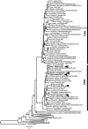
The Mx2 variants under investigation are orthologous. Maximum-likelihood phylogeny was calculated using the TIM2+I+G substitution model (log likelihood = −84,923.10). Bootstrap values below 70% are not shown for clarity, and asterisks (*) indicate 100% bootstrap support. The arrows indicate sequences functionally characterized in the present study.
FIG 2
FIG 2
Multiple primate Mx2s inhibit HIV-1 infection, and group O is inhibited in a species-specific fashion. (A) Western blot analysis (α-c-Myc and α-actin) of HsMx2, MacMx2, AGMMx2, OvMx2, and CanMx2 (in addition to TagRFP and Macaca mulatta TRIM5α controls) expression in the MT-4 cells used throughout the manuscript (with the exception of Fig. 3 and 6), in the presence or absence of pretreatment with doxycycline. The level of infection of these MT-4 CD4+ T cells expressing doxycycline-inducible variants of Mx2 (or TagRFP and Macaca mulatta TRIM5α controls) with (B) HIV-1 (pCCGW) (81), (C) FIV (pGiNSin) (82), (D) EIAV (pONY system), (E) MLV (pCNCG), (F) HERV-K (pCCGBX) (51), or (G) MPMV (pSARM-EGFP) (49) in the presence (gray bars) or absence (black bars) of pretreatment with doxycycline is shown. (H) Western blot analysis (α-c-Myc and α-actin) of HsMx2, MacMx2, AGMMx2, OvMx2, and CanMx2 expression (in addition to TagRFP and Macaca mulatta TRIM5α controls) in MT-4 TMZ R5 cells in the presence or absence of pretreatment with doxycycline. (I and J) Infection of MT-4 TMZ R5-expressing doxycycline-inducible variants of Mx2 (or TagRFP and Macaca mulatta TRIM5α controls) with (I) group M HIV-1 (NL4-3) and (J) group O HIV-1 (CMO2.5) in the presence (gray bars) or absence (black bars) of pretreatment with doxycycline.
FIG 3
FIG 3
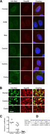
Variation in subcellular localization among Mx2 proteins. (A and B) Deconvolution microscopic images of HOS cells stably expressing HA-tagged Mx2 proteins from the indicated species and stained with antibodies against the HA tag (green) and Nup98 (red). An overlay image, also displaying DAPI-stained nuclei, is also shown. In panel A, optical sections approximately at the center of the vertical dimension of the nucleus are shown, and the scale bar represents 5 μm. Two examples of cells expressing canine Mx2 are shown to reflect the diversity in localization that was observed for this particular Mx2 protein. In panel B, an expanded segment of an optical section coincident with the dorsal surface of the nucleus is shown, and the scale bar represents 1 μm. (C) Pearson correlation coefficient for colocalization of Nup98 and HA-Mx2 proteins from the indicated species. Each symbol represents the Pearson correlation coefficient for an individual cell. (D) Alignment of the N-terminal 25 amino acids of Mx2 from primates and nonprimates. Arginine and lysine residues (predicted to be important for nuclear localization) are highlighted in blue.
FIG 4
FIG 4
In vitro evolution reveals an Mx2 sensitivity determinant in the C-terminal domain of CA. (A) The level of infection with HIV-1 (NHG) or SIVagmTAN in cells expressing doxycycline-inducible HsMx2 or macaque TRIMcyp in the presence (gray bars) or absence (black bars) of pretreatment with doxycycline and the presence or absence of 5 μM CsA or D025 is shown. (B) The level of HIV-1 (NHG) infection in cells expressing doxycycline-inducible HsMx2 treated (filled circles, dashed line) or not treated (unfilled circles, solid line) with doxycycline is shown over time. Cell-free passages are indicated with arrows. (C) The positions of P207S, G208R, and T210K point mutants are highlighted as solid gray spheres on the CA hexamer structure (3GV2). (D) The level of infection of MT-4 CD4+ T cells expressing doxycycline-inducible Mx2 variants (or TagRFP control) with HIV-1 (NHGcapNM) or P207S, G208R, or T210K point mutants in the presence (gray bars) or absence (black bars) of pretreatment with doxycycline is shown.
FIG 5
FIG 5
CA library screening identifies multiple substitutions conferring resistance to Mx2. (A) The level of infection (relative to non-doxycycline-treated controls) of 46 HIV-1 CA mutants in doxycycline-treated MT-4 CD4+ T cells expressing doxycycline-inducible Mx2. The dashed line indicates the threshold used to select resistant mutants for further consideration. (B) The positions of the seven point mutants most resistant to Mx2 identified in panel A are highlighted as yellow spheres on the CA hexamer structure (3GV2). Residues identified through in vitro evolution are also highlighted as green spheres. (C) The level of infection of MT-4 CD4+ T cells expressing doxycycline-inducible Mx2 variants (or TagRFP control) with HIV-1 (NHGcapNM) or the indicated CA point mutants in the presence (gray bars) or absence (black bars) of pretreatment with doxycycline is shown.
FIG 6
FIG 6
Chimeric Mx2s reveal an N-terminal antiviral specificity determinant. (A) Diagrammatic representation of the chimeric human and canine Mx2 proteins used in panels B and C. (B) The level of infection of MT-4 CD4+ T cells expressing doxycycline-inducible HsMx2, CanMx2, or human-canine chimeric Mx2s with HIV-1 (NHG) or MLV (CNCG) in the presence (gray bars) or absence (black bars) of doxycycline pretreatment is shown. (C) Western blot analysis (α-c-Myc and α-actin) of HsMx2, CanMx2, and chimeric Mx2s in CD4+ MT-4 cells in the presence or absence of doxycycline pretreatment (D) Deconvolution microscopic images of HOS cells stably expressing HA-tagged Mx2 proteins from the indicated chimera and stained with antibodies against the HA tag (green) and Nup98 (red). An overlay image, also displaying DAPI-stained nuclei, is also shown. In panel D, optical sections approximately at the center of the vertical dimension of the nucleus are shown, and the scale bar represents 5 μm. Two examples of cells expressing Can29Hs are shown to reflect the diversity in localization that was observed for this particular Mx2 chimera. In panel E, an expanded segment of an optical section coincident with the dorsal surface of the nucleus is shown, and the scale bar represents 1 μm. The Pearson correlation coefficient for the colocalization of Nup98 and HA-Mx2 proteins from the indicated chimera is also shown. Each symbol represents the Pearson correlation coefficient for an individual cell. (F) Diagrammatic representation of the chimeric macaque and AGM Mx2s used in panels G and H. (G) The level of infection of MT-4 CD4+ T cells expressing doxycycline-inducible HsMx2, AGMMx2, or chimeric Mx2s with HIV-1 (NHGcapNM), HIV-1 P207S, or MLV (CNCG) in the presence (gray bars) or absence (black bars) of pretreatment with doxycycline is shown. (H) Western blot analysis (α-c-Myc and α-actin) of MacMx2, AGMMx2, and chimeric Mx2s in CD4+ MT-4 cells in the presence or absence of pretreatment with doxycycline. (I) Diagrammatic representation of macaque and AGM Mx2 highlighting the regions of divergence between these orthologs. (J) Alignment of a short section of human (Hs) and AGM Mx2 highlighting divergent residues in this region. (K) The level of infection of MT-4 CD4+ T cells expressing doxycycline-inducible HsMx2, AGMMx2, or chimeric Mx2s with group M HIV-1 (NL4-3) or group O (CMO2.5) HIV-1 in the presence (gray bars) or absence (black bars) of pretreatment with doxycycline is shown. (L) Western blot analysis (α-c-Myc and α-actin) of HsMx2, AGMMx2, and chimeric Mx2s in CD4+ MT-4 cells in the presence or absence of pretreatment with doxycycline.
FIG 7
FIG 7
A single residue determines species-dependent inhibition of the HIV-1 P207S CA mutant. (A) A diagram representing the various domains of Mx2 is shown (BSE indicates the bundle signaling element), above plots of the dN/dS ratio (upper plot) and the Bayes factor for dN>dS (lower plot) at each codon in an alignment of primate Mx2 coding sequences. Ten residues identified by REL and M3 methods (from Table 1) are highlighted with arrows. The analogous loop 4 (L4) of Mx1 (62) is also highlighted. (B) Alignment of a short section of MacMx2 and AGMMx2 highlighting divergent residues in this region. (C) Western blot analysis (α-c-Myc and α-actin) of HsMx2, AGMMx2, and chimeric Mx2s in CD4+ MT-4 cells in the presence or absence of pretreatment with doxycycline. (D) The level of infection of MT-4 CD4+ T cells expressing doxycycline-inducible MacMx2, AGMMx2 or point mutants thereof with HIV-1 (NHGcapNM) or HIV-1 P207S in the presence (gray bars) or absence (black bars) of pretreatment with doxycycline is shown.

Similar articles

Cited by

References

    1. Ho DD, Hartshorn KL, Rota TR, Andrews CA, Kaplan JC, Schooley RT, Hirsch MS. 1985. Recombinant human interferon alfa-A suppresses HTLV-III replication in vitro. Lancet i:602–604 - PubMed
    1. Asmuth DM, Murphy RL, Rosenkranz SL, Lertora JJ, Kottilil S, Cramer Y, Chan ES, Schooley RT, Rinaldo CR, Thielman N, Li XD, Wahl SM, Shore J, Janik J, Lempicki RA, Simpson Y, Pollard RB. 2010. Safety, tolerability, and mechanisms of antiretroviral activity of pegylated interferon alfa-2a in HIV-1-monoinfected participants: a phase II clinical trial. J. Infect. Dis. 201:1686–1696. 10.1086/652420 - DOI - PMC - PubMed
    1. Kirmaier A, Wu F, Newman RM, Hall LR, Morgan JS, O'Connor S, Marx PA, Meythaler M, Goldstein S, Buckler-White A, Kaur A, Hirsch VM, Johnson WE. 2010. TRIM5 suppresses cross-species transmission of a primate immunodeficiency virus and selects for emergence of resistant variants in the new species. PLoS Biol. 8:1–12. 10.1371/journal.pbio.1000462 - DOI - PMC - PubMed
    1. Liberatore RA, Bieniasz PD. 2011. Tetherin is a key effector of the antiretroviral activity of type I interferon in vitro and in vivo. Proc. Natl. Acad. Sci. U. S. A. 108:18097–18101. 10.1073/pnas.1113694108 - DOI - PMC - PubMed
    1. Okeoma CM, Lovsin N, Peterlin BM, Ross SR. 2007. APOBEC3 inhibits mouse mammary tumour virus replication in vivo. Nature 445:927–930. 10.1038/nature05540 - DOI - PubMed

Publication types

LinkOut - more resources