. 2014 Jul 1;307(1):C55-65.
doi: 10.1152/ajpcell.00045.2014. Epub 2014 Apr 23.

Lysophosphatidic acid stimulation of NHE3 exocytosis in polarized epithelial cells occurs with release from NHERF2 via ERK-PLC-PKCδ signaling

Affiliations

Lysophosphatidic acid stimulation of NHE3 exocytosis in polarized epithelial cells occurs with release from NHERF2 via ERK-PLC-PKCδ signaling

Boyoung Cha et al. Am J Physiol Cell Physiol. .

Abstract

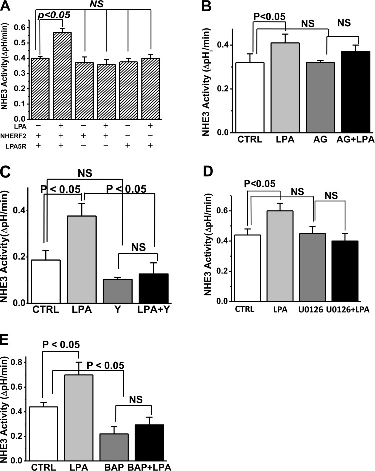
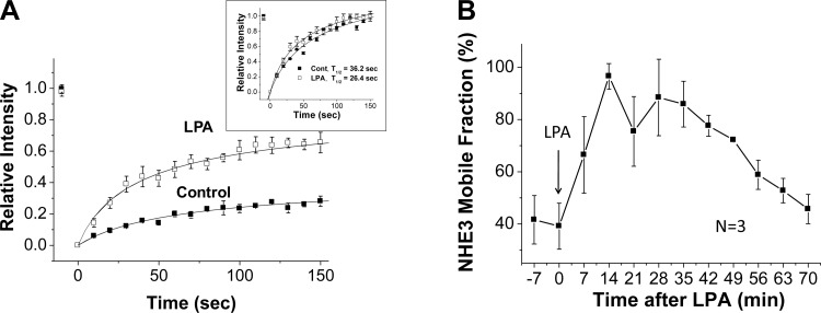
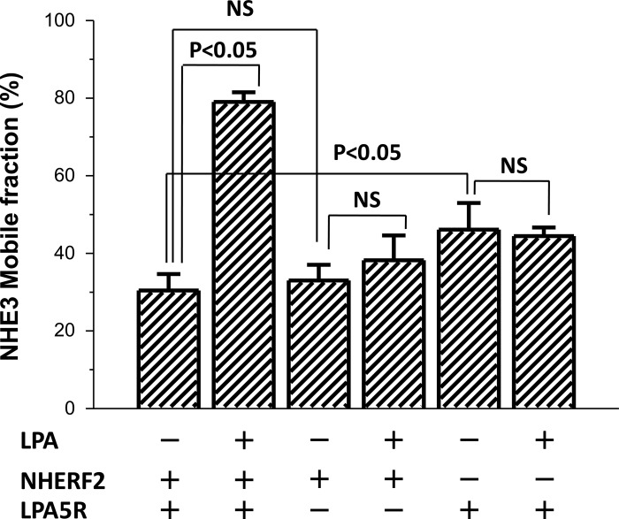
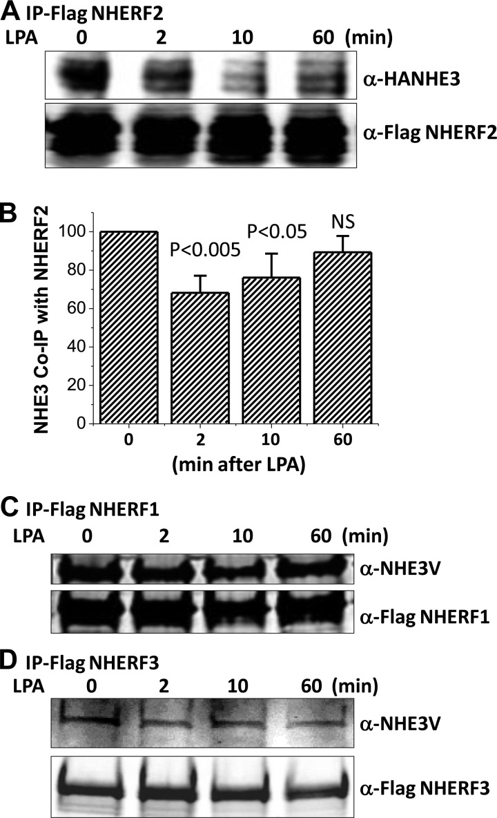
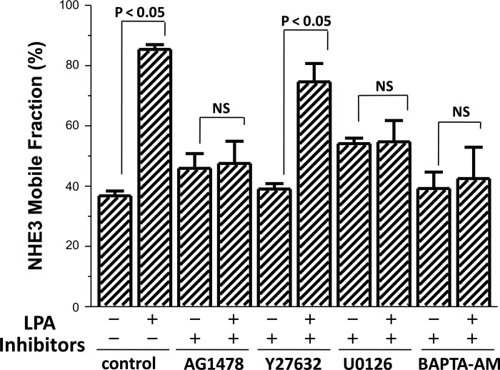
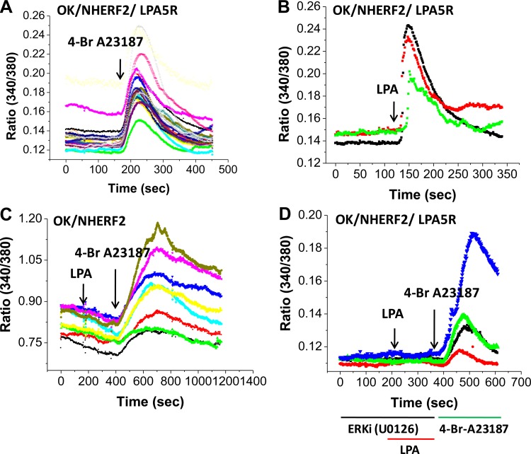
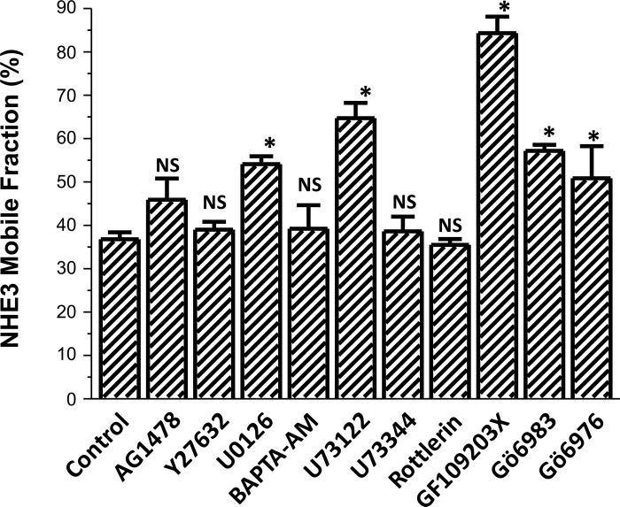
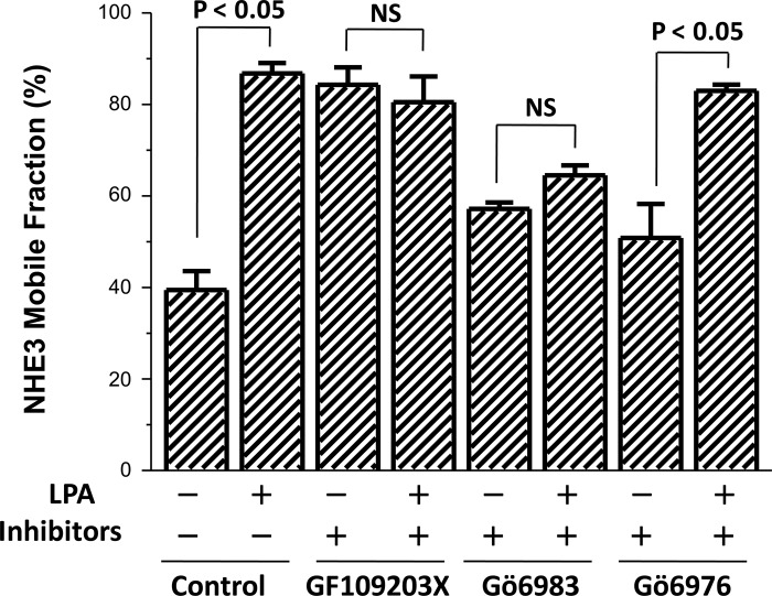
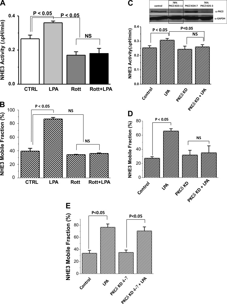
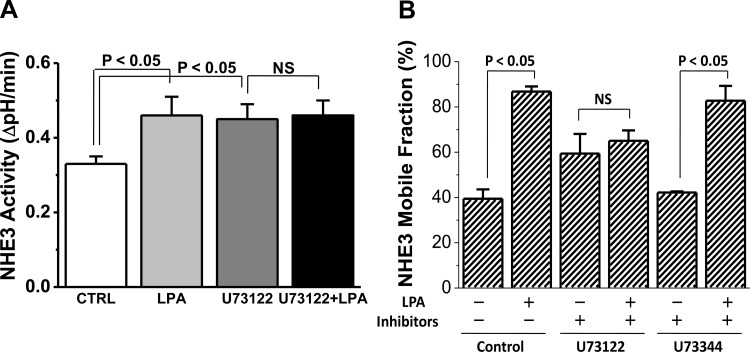
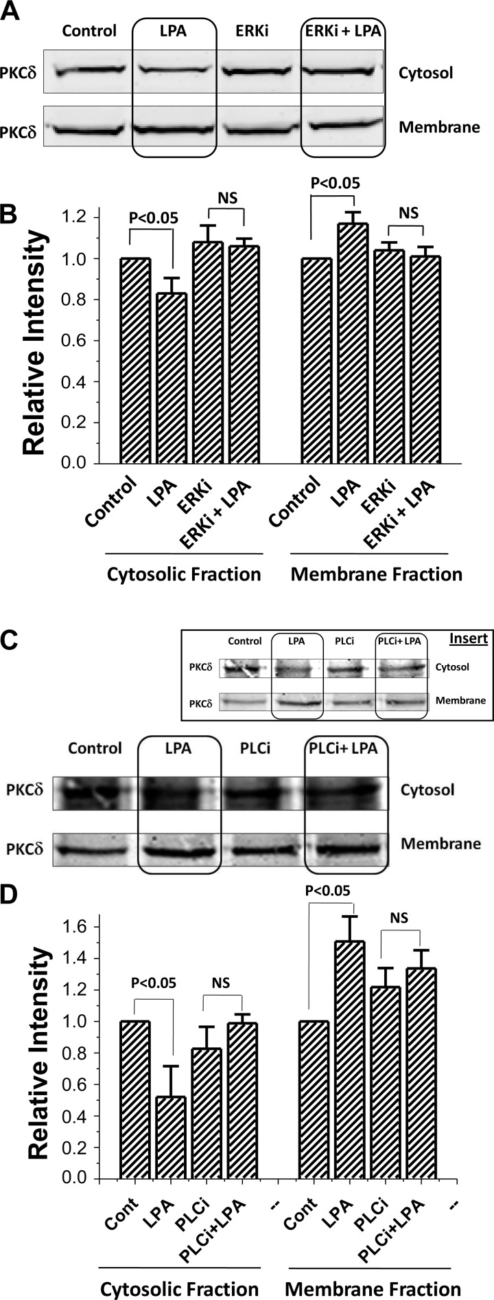
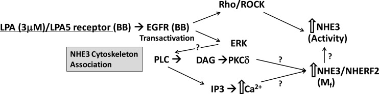
The Na(+)/H(+) exchanger 3 (NHE3) is a brush border (BB) Na(+)/H(+) antiporter that accounts for the majority of physiologic small intestinal and renal Na(+) absorption. It is regulated physiologically and in disease via changes in endocytosis/exocytosis. Paradoxically, NHE3 is fixed to the microvillar (MV) actin cytoskeleton and has little basal mobility. This fixation requires NHE3 binding to the multi-PDZ domain scaffold proteins Na(+)/H(+) exchanger regulatory factor (NHERF)1 and NHERF2 and to ezrin. Coordinated release of NHE3 from the MV cytoskeleton has been demonstrated during both stimulation and inhibition of NHE3. However, the signaling molecules involved in coordinating NHE3 trafficking and cytoskeletal association have not been identified. This question was addressed by studying lysophosphatidic acid (LPA) stimulation of NHE3 in polarized renal proximal tubule opossum kidney (OK) cells that occurs via apical LPA5 receptors and is NHERF2 dependent and mediated by epidermal growth factor receptor (EGFR), Rho/Rho-associated kinase (ROCK), and ERK. NHE3 activity was determined by BCECF/fluorometry and NHE3 microvillar mobility by FRAP/confocal microscopy using NHE3-EGFP. Apical LPA (3 μM)/LPA5R stimulated NHE3 activity, increased NHE3 mobility, and decreased the NHE3/NHERF2 association. The LPA stimulation of NHE3 was also PKCδ dependent. PKCδ was necessary for LPA stimulation of NHE3 mobility and NHE3/NHERF2 association. Moreover, the LPA-induced translocation to the membrane of PKCδ was both ERK and phospholipase C dependent with ERK acting upstream of PLC. We conclude that LPA stimulation of NHE3 exocytosis includes a signaling pathway that regulates fixation of NHE3 to the MV cytoskeleton. This involves a signaling module consisting of ERK-PLC-PKCδ, which dynamically and reversibly releases NHE3 from NHERF2 to contribute to the changes in NHE3 MV mobility.

Keywords: FRAP; NHE3; PDZ domain; microvillus; trafficking.


Figures

Fig. 1.
Fig. 1.
Lysophosphatidic acid (LPA)/LPA5R signaling in polarized epithelial cells stimulates Na+/H+ exchanger 3 (NHE3) activity. LPA stimulates NHE3 activity, but this only occurs in the presence of both Na+/H+ exchanger regulatory factor 2 (NHERF2) and LPA5R (A). The LPA/LPA5R stimulation of NHE3 requires the epidermal growth factor receptor (EGFR; B), Rho/Rho-associated kinase (ROCK; C), ERK (D), and elevated intracellular Ca2+ (E). Results are means ± SE of initial rates of NHE3 activity in polarized opossum kidney (OK) cells with inhibitors preincubated for 30 min. LPA stimulation and effects of inhibitors were similar after 2, 30, and 60 min of LPA exposure; results shown are for 2 and 30 min (combined) of LPA exposure. Concentration and number of experiments for each condition were as follows: LPA in A (n > 4 for all conditions); the EGFR inhibitor AG1478 (25 nM; n = 6); the Rho/ROCK inhibitor Y27632 (50 μM; n = 6); the ERK inhibitor U0126 (10 μM; n = 5); and BAPTA-AM (10 μM; n = 4). CTRL, control.
Fig. 2.
Fig. 2.
LPA/LPA5R acutely increases the mobile fraction (Mf) of apical NHE3. A: kinetic analysis of the change in polarized OK cell apical NHE3 Mf 30 min after LPA exposure. Results are means ± SE for 3 different regions of interest (ROI) in a single experiment. Inset: results rescaled from 0–1 for each control and LPA ROI to compare time to reach a new constant fluorescent signal (half-time). The y-axis sets fluorescence prebleach for each ROI as 1.0 and immediately postbleach as 0.0. B: NHE3 Mf was determined in the apical domain of OK cells starting various times after LPA addition. All results are means ± SE of n = 3 separate experiments with each experiment made up of multiple cells which were averaged to create a single number which was used for the calculation. All results were significantly increased compared with 0 time except 70 min. The first time point was obtained as quickly as possible using the experimental setup.
Fig. 3.
Fig. 3.
LPA/LPA5R acutely increase the Mf of apical NHE3 by a NHERF2- and LPA5R-dependent process. Transient expression of NHERF2 or LPA5R separately or together were examined for effects on NHE3 mobility. LPA (3 μM) fails to increase the NHE3 Mf in the apical domain of OK cells in the absence of either NHERF2 or LPA5R. Results are means ± SE of n > 4 replicates per condition. NHERF2/LPA5R-containing cells (2 bars at left) were used as a positive control for cells lacking either NHERF2 (2 bars at right) or LPA5R (2 bars at middle). P values compare LPA effects to control conditions lacking LPA.
Fig. 4.
Fig. 4.
LPA dynamically decreases the association between NHE3 and NHERF2 but not between NHE3 and NHERF1 or NHERF3 (A and B). OK/FLAG-NHERF2/HA-NHE3 cells were exposed to LPA (3 μM) for 0, 2, 10, and 60 min. FLAG-NHERF2 was immunoprecipitation (IP) using anti-FLAG M2 magnetic beads and the coprecipitated HA-NHE3 and FLAG-NHERF2 were detected by immunoblotting with anti-HA antibody and anti-FLAG antibody. A: immunoblot (IB) of single experiment. B: densitometry of immunoblots from single experiments were used to calculate means ± SE. Coprecipitated NHE3 values are means ± SE of 5 different experiments each of which was normalized to FLAG-NHERF2 intensity in the same experiment set as 100. p values compared with zero time (untreated) control. OK/FLAG-NHERF1/NHE3V (C) or OK/FLAG-NHERF3/NHE3V (D) cells were exposed to LPA (3 μM) for 0, 2, 10, and 60 min, washed with cold PBS (4°C) and then kept at 4°C for 30 min. Total cell lysates were then prepared for immunoprecipitation. FLAG-NHERF1 was immunoprecipitated using anti-FLAG magnetic beads and the coprecipitated NHE3V was detected by immunoblotting with anti-VSV-G antibody. Coprecipitated NHE3V with NHERF1 and NHERF3 was not changed by LPA (3 μM) treatment comparing 0, 2, 10, and 60 min. Single experiment is shown and was repeated 3 times with similar results.
Fig. 5.
Fig. 5.
LPA/LPA5R acutely increases the Mf of apical NHE3 by an ERK-dependent process. NHE3 Mf in OK/NHERF2 cells transiently transfected with LPA5R were exposed for 30 min to the signaling inhibitors at the concentrations used in Fig. 1 in the absence and 30 min after LPA exposure (3 μM). P values represent LPA effect compared with the control for each inhibitor; n > 3 for all conditions.
Fig. 6.
Fig. 6.
LPA/LPA5R (3 μM) increases intracellular Ca2+ in polarized OK/NHERF2 cells, an LPA5R- and ERK-dependent effect. A: intracellular Ca2+ concentration ([Ca2+]i) was determined with fura-2 AM using a microscopy based system with 4-Br-A23187 as a positive control. Arrow indicates 4-Br-A23187 (1 μM) addition. B: LPA caused a rapid onset increase in [Ca2+]i that returned towards baseline in ∼4 min. Arrow indicates LPA (3 μM) addition. C: LPA failed to increase [Ca2+]i in the absence of LPA5R even in the presence of NHERF2 indicating dependence on the LPA5 receptor. D: the LPA/LPA5R increase in [Ca2+]i was prevented by 30 min preincubation with the ERK inhibitor, U0126 (10 μM), which did not alter the A23187 (1 μM) induced increase (2nd arrow). Results of a single experiment are shown and was repeated 3 times with similar results.
Fig. 7.
Fig. 7.
Effect of the pharmacologic inhibitors on basal Mf of NHE3. Effects on the apical NHE3 Mf in OK cells as above of the inhibitors used to determine the signaling through which LPA/LPA5R (3 μM) stimulates NHE3 activity. All experiments were compared with untreated or solvent controls done on the same day with P values representing comparison with experimental controls; n ≥ 3 for all agents. The agents that significantly increased the NHE3 Mf under basal conditions included the ERK inhibitor U0126; the PLC inhibitor U73122; the general PKC inhibitor GF109293X; the conventional and novel PKC inhibitor Gö6983; and the conventional PKC inhibitor Gö6976. *P < 0.05.
Fig. 8.
Fig. 8.
LPA/LPA5R increase in Mf is dependent on a novel isoform of PKC. LPA effects on the apical NHE3 Mf in OK cells under otherwise untreated control conditions and in the presence of several classes of PKC inhibitors are shown. The general PKC inhibitor GF109203X (1 μM); novel + conventional PKC inhibitor Gö6983 (30 nM); and conventional PKC isoforms only inhibitor, Gö6976 (20 nM) were tested. Each PKC inhibitor was preincubated for 30 min before exposure to LPA for 30 min followed by determination of mobility using FRAP. Results are means ± SE of n ≥ 3 separate studies. P values represent comparison of the LPA effect on Mf in the presence and absence of the same PKC inhibitor (paired t-test).
Fig. 9.
Fig. 9.
PKCδ Inhibition and knockdown (KD) Prevents the LPA stimulation of NHE3 Activity and Mf. Pretreatment with the PKCδ inhibitor rottlerin (10 μM, 30 min) prevented the LPA stimulation of NHE3 activity, while lowering basal NHE3 activity (n = 5; A) and prevented the LPA-induced increase in NHE3 Mf (n = 3; B). P values compare effect of LPA on basal NHE3 activity and Mf in otherwise untreated and rotterlin pretreated cells and also rotterlin alone compared with control conditions. PKCδ KD prevents LPA stimulation of NHE3 activity (C) and Mf (D). Inset: 3 siRNA oligomers were transfected into OK/LPA5R/NHERF2/NHE3 cells and 48 h later lysates prepared for immunoblotting for PKCδ expression. Two of the constructs KD >75% of the PKCδ, while one had no effect (PKCδ KD 4-7). This study was repeated 3 times. PKCδ KD 3-11 (outlined in inset) prevented the LPA/LPA5R increase in NHE3 activity (C) and apical NHE3 Mf (D) compared with the ineffective KD construct (KD 4-7; E) called control (n = 4).
Fig. 10.
Fig. 10.
LPA/LPA5R stimulation of NHE3 activity and Mf are PLC dependent. The role of PLC in LPA stimulation of NHE3 activity was determined by pretreating OK cells with the PLC inhibitor U73122 (5–10 μM, 30 min). A: U73122 pretreatment increased basal NHE3 activity and totally prevented the LPA stimulation of NHE3 activity (n = 5). B: similar pretreatment with U73122 prevented the LPA-induced increase in apical NHE3 Mf, while the PLC inhibitor control U73344 (5 μM, 30 min) did not affect the LPA stimulation of NHE3 Mf (n = 3).
Fig. 11.
Fig. 11.
LPA-dependent translocation of PKCδ was prevented by ERK inhibitor U0126 and by the PLC inhibitor U73122 in OK/NHERF2/LPA5R cells. A: LPA-dependent translocation of PKCδ increasing the cell membrane and reducing cytosolic PKCδ in OK/NHERF2/LPA5R cells was prevented by pretreatment with the ERK inhibitor U0126. A representative Western blot analysis of PKCδ is shown above. OK cells were incubated with vehicle (control; lane 1), 3 μM LPA for 30 min (lane 2), 10 μM U0126 for 60 min (lane 3), and preincubation of 10 μM U0126 for 30 min then 3 μM LPA added for the last 30 min (lane 4). Cytosol and membrane fractions of PKCδ were then collected as described in materials and methods. B: statistical analysis (means ± SE) of 3 different experiments each of which was normalized to total intensity of PKCδ (cytosol + membrane) under each condition with densitometry used to quantitate the Western blot band intensity using the LI-COR software of the Odyssey system. P values compare LPA effects in membrane and cytosolic fractions in otherwise untreated and in U0126 treated conditions (paired t-tests). C: LPA-dependent translocation of PKCδ increasing the cell membrane and reducing cytosolic PKCδ in OK/NHERF2/LPA5R cells was prevented by pretreatment with the PLC inhibitor, U73122. Studies were done as those in A and B with the PLC inhibitor U73122 used instead of the ERK inhibitor. Pretreatment was performed with the PLC inhibitor U73122 (5–10 μM; 30-min pretreatment; total treatment 60 min with LPA added for the last 30 min). A representative Western blot analysis of PKCδ is shown in C and the means ± SE of 3 different experiments normalized as determined in B above is shown in D. P values compare LPA effects in membrane and cytosolic fractions in otherwise untreated and in U73122 treated conditions (paired t-tests). In C, the original IB is shown, while in the inset this IB with contrast altered to show the changes in band density is included. Quantitation was done on the original.
Fig. 12.
Fig. 12.
Current understanding of LPA/LPA5R stimulation of NHE3 activity and mobility based on current studies and the previous reports from the laboratory of Yun and colleagues (17, 24). Please note that NHERF2 is involved in the pathway in at least three steps, anchoring the LPA5R (17), binding PLC (8), and binding and releasing NHE3;? identify steps at which detailed understanding remain to be identified. BB, brush border; IP3, inositol 1,4,5-trisphosphate; DAG, diacylglycerol.

Similar articles

Cited by

References

    1. Alexander RT, Furuya W, Azaszi K, Orlowski J, Grinstein S. Rhos GTPases dictate the mobility of the Na/H exchanger NHE3 in epithelia: role in apical retention and targeting. Proc Natl Acad Sci USA 102: 12253–12258, 2005 - PMC - PubMed
    1. Cha B, Kenworthy A, Murtazina R, Donowitz M. The lateral mobility of NHE3 on the apical membrane of renal epithelial OK cells is limited by the PDZ domain proteins NHERF1/2, but is dependent on an intact actin cytoskeleton as determined by FRAP. J Cell Sci 117: 3353–3365, 2004 - PubMed
    1. Cha B, Kim JH, Hut H, Hogema BM, Nadarja J, Zizak M, Cavet M, Lee-Kwon W, Lohmann SM, Smolenski A, Tse CM, Yun C, de Jonge HR, Donowitz M. cGMP inhibition of Na+/H+ antiporter 3 (NHE3) requires PDZ domain adapter NHERF2, a broad specificity protein kinase G-anchoring protein. J Biol Chem 280: 16642–16650, 2005 - PubMed
    1. Cha B, Zhu XC, Chen W, Jones M, Ryoo S, Zachos NC, Chen TE, Lin R, Sarke R, Kenworthy AK, Tse CM, Kovbasnjuk O, Donowitz M. NHE3 mobility in brush borders increases upon NHERF2-dependent stimulation by lyophosphatidic acid. J Cell Sci 123: 2434–2443, 2010 - PMC - PubMed
    1. Choi JW, Lim S, Oh YS, Kim EK, Kim SH, Kim YH, Heo K, Kim J, Kim JK, Yang YR, Ryu SH, Suh PG. Subtype-specific role of phospholipase C-beta in bradykinin and LPA signaling through differential binding of different PDZ scaffold proteins. Cell Signal 22: 1153–1161, 2010 - PubMed

Publication types

MeSH terms

LinkOut - more resources