. 2014 Jun 15;306(12):E1378-87.
doi: 10.1152/ajpendo.00656.2013. Epub 2014 Apr 22.

Acylcarnitines activate proinflammatory signaling pathways

Affiliations

Acylcarnitines activate proinflammatory signaling pathways

Jennifer M Rutkowsky et al. Am J Physiol Endocrinol Metab. .

Abstract

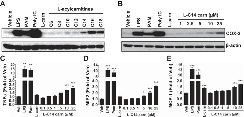
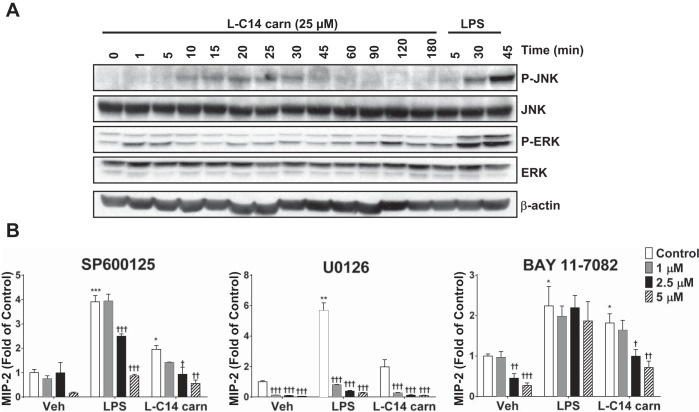
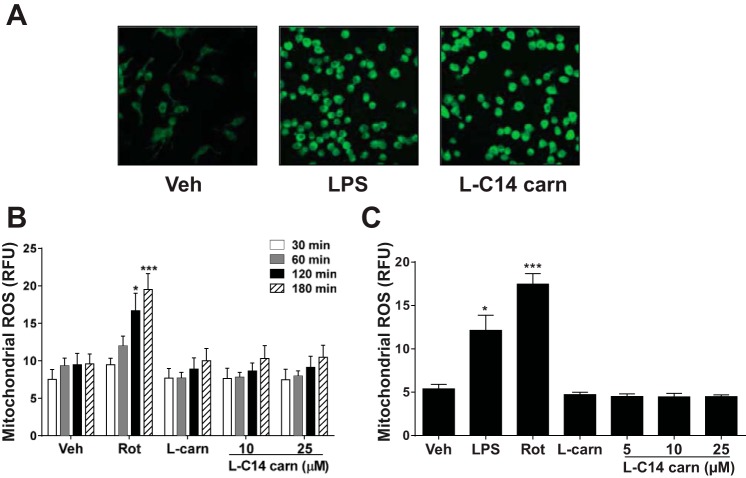
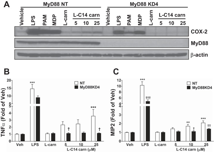
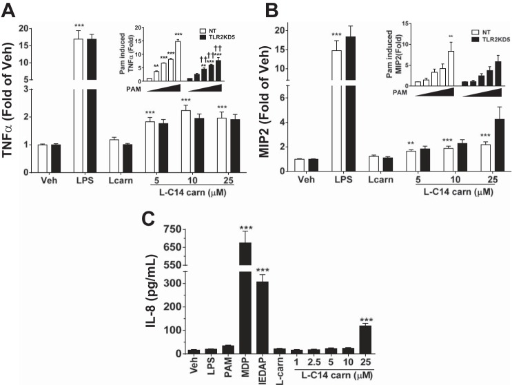
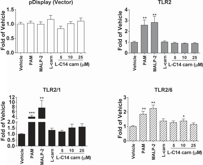
Incomplete β-oxidation of fatty acids in mitochondria is a feature of insulin resistance and type 2 diabetes mellitus (T2DM). Previous studies revealed that plasma concentrations of medium- and long-chain acylcarnitines (by-products of incomplete β-oxidation) are elevated in T2DM and insulin resistance. In a previous study, we reported that mixed D,L isomers of C12- or C14-carnitine induced an NF-κB-luciferase reporter gene in RAW 264.7 cells, suggesting potential activation of proinflammatory pathways. Here, we determined whether the physiologically relevant L-acylcarnitines activate classical proinflammatory signaling pathways and if these outcomes involve pattern recognition receptor (PRR)-associated pathways. Acylcarnitines induced the expression of cyclooxygenase-2 in a chain length-dependent manner in RAW 264.7 cells. L-C14 carnitine (5-25 μM), used as a representative acylcarnitine, stimulated the expression and secretion of proinflammatory cytokines in a dose-dependent manner. Furthermore, L-C14 carnitine induced phosphorylation of JNK and ERK, common downstream components of many proinflammatory signaling pathways including PRRs. Knockdown of MyD88, a key cofactor in PRR signaling and inflammation, blunted the proinflammatory effects of acylcarnitine. While these results point to potential involvement of PRRs, L-C14 carnitine promoted IL-8 secretion from human epithelial cells (HCT-116) lacking Toll-like receptors (TLR)2 and -4, and did not activate reporter constructs in TLR overexpression cell models. Thus, acylcarnitines have the potential to activate inflammation, but the specific molecular and tissue target(s) involved remain to be identified.

Keywords: TLR; acylcarnitine; inflammation; pattern recognition receptors; β-oxidation.


Figures

Fig. 1.
Fig. 1.
Induction of the proinflammatory gene product COX-2 by l-acylcarnitines in a chain length manner and l-C14 carnitine induction of COX-2 and secretion of TNFα, MIP2, and MCP1 in a dose-dependent manner in murine monocyte/macrophages. RAW 264.7 cells were serum-starved for 6 h (0.25% FBS/DMEM) then treated with LPS (TLR4 ligand, 0.2 ng/mL), PAM3CSK4 (TLR2 ligand, 2 ng/mL), Poly (I:C) (TLR 3 ligand, 0.25 μg/mL), 25 μM l-carnitine (l-carn) or (A) 25 μM of various chain lengths of l-acylcarnitine or (B) various doses of l-C14 carnitine (l-C14 carn, 0–25 μM) for 18 h. Whole cell lysates were analyzed by immunoblotting using COX-2 or β-actin antibodies. (Representative blot from 3 separate experiments). Media concentrations of proinflammatory cytokines TNFα, MIP2, and MCP1 (C, D, & E respectively) were measured by multiplex assay (Milliplex MAP). Controls: LPS (0.2 ng/mL), PAM (2 ng/mL), 25 μM l-carnitine. n = 6 over 3 experiments. One way ANOVAs with Dunnett's posttest for positive controls and for l-C14 carnitine dose response: *P < 0.05 **P < 0.01, ***P < 0.001 vs. vehicle-treated control (Veh); mean ± SE. Data are expressed as fold of vehicle control.
Fig. 2.
Fig. 2.
l-C14 carnitine increases phosphorylation of ERK and JNK in a time-dependent manner: MAP kinases and NF-κB contribution to l-C14 carnitine induced cytokine production. RAW 264.7 cells were serum-starved for 6 h (0.25% FBS/DMEM) then treated with LPS (100 ng/mL) or 25 μM l-C14 carn for indicated times (A). Whole cell lysates were prepared and analyzed for P-JNK, total JNK, P-ERK, total ERK. (Representative blot from 3 separate experiments). (B) RAW 264.7 cells were serum-starved for 3 h (0.25% FBS/DMEM) and then pretreated with JNK (Panel 1), ERK (Panel 2) or NF-κB (Panel 3) inhibitors for 1 h prior to treatment with LPS (100 ng/mL), or 25 μM l-C14 acylcarnitine (l-C14 carn) for 24 h. Media concentrations of proinflammatory cytokine MIP2 were measured by multiplex assay (Milliplex MAP); n = 4–6/treatment over 2–3 experiments. t-Tests for effect of treatment: *P < 0.05 **P < 0.01, ***P < 0.001. One way ANOVA with Dunnett's posttest for effect of inhibitor within treatment group: †P < 0.05 ††P < 0.01, †††P < 0.001 vs. vehicle-treated control (Veh); mean ± SE. Data are expressed as fold of vehicle control.
Fig. 3.
Fig. 3.
l-C14 carnitine induced reactive oxygen species (ROS) but not mitochondrial ROS in murine monocyte/macrophages. RAW 264.7 cells were serum-starved for 6 h (0.25% FBS/DMEM) then treated with LPS (100 ng/mL), 25 μM l-C14 acylcarnitine (l-C14 carn), or 25 μM l-carnitine for 45 min. (A) ROS production was determined from living cells by confocal microscopy as described in the Methods section. (B) RAW 264.7 cells were loaded with MitoSOX dye and then treated with rotenone (500 nM), l-carnitine (25 μM) or various doses of l-C14 carnitine (10 or 25 μM) for 30, 60, 120, or 180 min and then analyzed by flow cytometry. (C) RAW 264.7 cells were treated with LPS (1 μg/ml), rotenone (100 nM), l-carnitine (25 μM), or various doses of l-C14 carnitine (5, 10, or 25 μM) for 16 h. Cells were loaded with MitoSOX dye and analyzed by flow cytometry. One-way ANOVA with Dunnett's posttest *P < 0.05 **P < 0.01, ***P < 0.001 vs. vehicle-treated control (Veh). Data are expressed as RFU ± SE and n = 3.
Fig. 4.
Fig. 4.
Induction of COX-2 and proinflammatory cytokines are reduced in MyD88 knock-down RAW 264.7 cells. Stable MyD88 KD cells were generated by lentiviral mediated shRNA in RAW 264.7 cells. Cells were then serum-starved for 4–6 h (0.25% FBS/DMEM) then treated with LPS (0.2 ng/mL), PAM3CSK4 (2 ng/mL), MDP (25 μg/mL), 25 μM l-carnitine (l-carn) or l-C14 carnitine (l-C14 carn, 5–25 μM) for 18 h. Whole cell lysates were analyzed by immunoblotting using COX-2 or β-actin antibodies (A) (Representative blot from 3 separate experiments). Media concentrations of proinflammatory cytokines TNFα or MIP2 (B & C respectively) were measured by multiplex assay (Milliplex MAP). n = 6 over 3 experiments. One way ANOVAs with Dunnett's posttest for positive controls and for l-C14 carnitine dose response: *P < 0.05 **P < 0.01, ***P < 0.001 vs. vehicle-treated control (Veh); mean ± SE. Multi-comparison t-tests with Holm-Sidak correction for effect of KD †P < 0.05 ††P < 0.01, †††P < 0.001 vs. non-targeting (NT); mean ± SE. Data are expressed as fold of KD vehicle control.
Fig. 5.
Fig. 5.
Induction of proinflammatory cytokines is unaltered in TLR2 knock-down RAW cells and human colonic epithelial cells lacking TLR2 and TLR4. Stable non-targeting (NT) and TLR2 knockdown cells were generated by lentiviral mediated shRNA in RAW 264.7 cells. Cells were then serum-starved for 4–6 h (0.25% FBS/DMEM) then treated with LPS (0.2 ng/mL), 25 μM l-carnitine (l-carn) or l-C14 carnitine (l-C14 carn, 5–25 μM) for 18 h. Media concentrations of proinflammatory cytokines TNFα and MIP2 (A & B respectively) were measured by multiplex assay (Milliplex MAP). n = 6 over 3 experiments. Additionally, NT and TLR2KD cells were treated with PAM3CSK4 of various doses (0, 0.25, 0.5, 1 & 2 ng/mL) and media concentrations of TNFα and MIP2 (respective insets of A and B) were measured. One way ANOVAs with Dunnett's posttest for positive controls and for l-C14 carnitine dose response, or PAM3CSK4 dose response (inset): *P < 0.05 **P < 0.01, ***P < 0.001 vs. vehicle-treated control (Veh); mean ± SE. Multi-comparison t-tests with Holm-Sidak correction for effect of KD: †P < 0.05 ††P < 0.01, †††P < 0.001 vs. non-targeting (NT); mean ± SE. Data are expressed as fold of KD vehicle control. HCT116 cells were serum-starved for 6 h (0.25% FBS/DMEM) then treated with various doses of l-C14 carnitine (l-C14 carn, 0–25 μM) for 18 h (C). Media concentrations of proinflammatory cytokine IL-8 were measured by ELISA assay (BD Bioscience). Controls: LPS (1 μg/mL), PAM3CSK4 (1 μg/mL), MDP (25 μg/mL), C12-IEDAP (20 μg/mL), 25 μM l-carnitine. Data are expressed as pg/mL; mean ± SEM. One way ANOVAs with Dunnett's posttest for positive controls and for l-C14 carnitine dose response: *P < 0.05 **P < 0.01, ***P < 0.001 vs. vehicle (Veh); mean ± SE (n = 6 over 3 experiments).
Fig. 6.
Fig. 6.
l-C14 carnitine does not induce NFκB in TLR 2/1 or 2/6 in transfected HEK-293T cells. HEK-293T cells were cultured in 10% FBS/DMEM medium and cotransfected with TLR2 and TLR1, TLR2 and TLR6 in addition to NF-κB-luciferase reporter and β-galactosidase expression vectors. After 24 h, the cells were serum-starved in 0.25% FBS/DMEM for 6 h and then treated with l-C14 carnitine for 12 h. The cell lysates were assayed for luciferase and β-galactosidase activities. Values are expressed as RLA (relative luciferase activity). Controls: Pam (PamCSK4, TLR2/1 agonist, 10 ng/mL), MALP-2 (TLR2/6 agonist, 10 ng/mL), LPS (TLR4 agonist, 50 ng/mL). One way ANOVAs with Dunnett's posttest for positive controls and for l-C14 carnitine dose response: *P < 0.05 **P < 0.01, ***P < 0.001 vs. vehicle-treated control (Veh); mean ± SE. Data are expressed as fold of vehicle control.
Fig. 7.
Fig. 7.
DHA inhibits l-C14 carnitine-induced COX-2 and secretion of TNFα and MIP2. RAW 264.7 cells were pretreated with various doses of DHA for 1 h. Cells were co-treated with DHA and LPS (0.2 ng/mL) or 25 μM l-C14 carnitine for 24 h in a low serum media (0.25% FBS). Cells were serum-starved (0.25% FBS) for 6 h prior to l-C14 carnitine treatment. Whole cell lysates were analyzed by immunoblotting using COX-2 or β-actin antibodies (A). Representative blot at either 5 min or 60 min exposure from 3 independent experiments. Media concentrations of proinflammatory cytokines TNFα (B) and MIP2 (C) were measured by multiplex assay (Milliplex MAP). n = 6 over 3 experiments and values are mean ± SE (n = 4–8). T-tests for effect of treatment: *P < 0.05 **P < 0.01, ***P < 0.001. Multi-comparison t-tests with Holm-Sidak correction for effect of DHA within treatment group: †P < 0.05 ††P < 0.01, †††P < 0.001; mean ± SE. Data are expressed as fold of vehicle control.

References

    1. Adams SH, Hoppel CL, Lok KH, Zhao L, Wong SW, Minkler PE, Hwang DH, Newman JW, Garvey WT. Plasma acylcarnitine profiles suggest incomplete long-chain fatty acid beta-oxidation and altered tricarboxylic acid cycle activity in type 2 diabetic African-American women. J Nutr 139: 1073–1081, 2009 - PMC - PubMed
    1. Bastard JP, Maachi M, Lagathu C, Kim MJ, Caron M, Vidal H, Capeau J, Feve B. Recent advances in the relationship between obesity, inflammation, and insulin resistance. Eur Cytokine Netw 17: 4–12, 2006 - PubMed
    1. De Vogel-van den Bosch J, Hoeks J, Timmers S, Houten SM, van Dijk PJ, Boon W, Van Beurden D, Schaart G, Kersten S, Voshol PJ, Wanders RJ, Hesselink MK, Schrauwen P. The effects of long- or medium-chain fat diets on glucose tolerance and myocellular content of lipid intermediates in rats. Obesity (Silver Spring) 19: 792–799, 2011 - PubMed
    1. Ho JK, Duclos RI, Jr, Hamilton JA. Interactions of acyl carnitines with model membranes: a (13)C-NMR study. J Lipid Res 43: 1429–1439, 2002 - PubMed
    1. Huang S, Rutkowsky JM, Snodgrass RG, Ono-Moore KD, Schneider DA, Newman JW, Adams SH, Hwang DH. Saturated fatty acids activate TLR-mediated proinflammatory signaling pathways. J Lipid Res 53: 2002–2013, 2012 - PMC - PubMed

Publication types

MeSH terms