. 2014 Jun 15;306(12):E1460-7.
doi: 10.1152/ajpendo.00591.2013. Epub 2014 Apr 22.

Automated quantification of pancreatic β-cell mass

Affiliations

Automated quantification of pancreatic β-cell mass

Maria L Golson et al. Am J Physiol Endocrinol Metab. .

Abstract

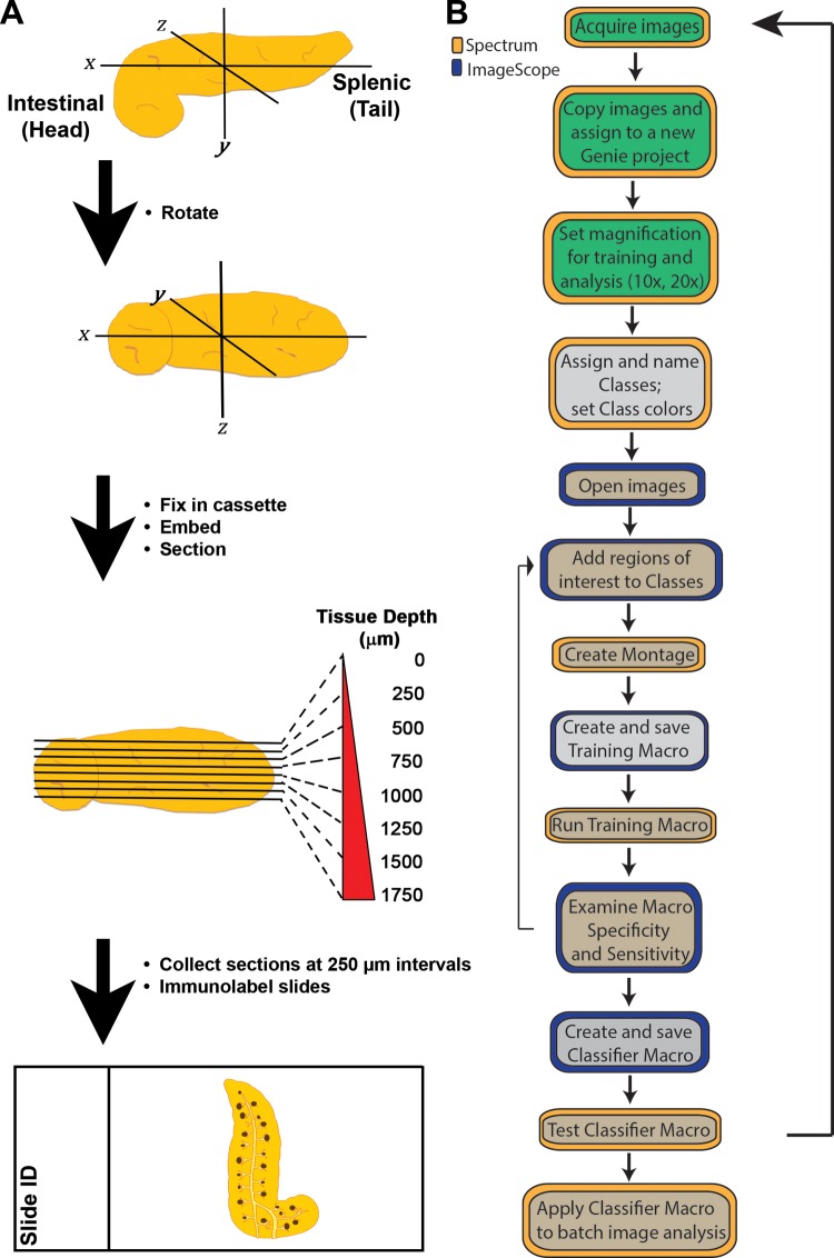
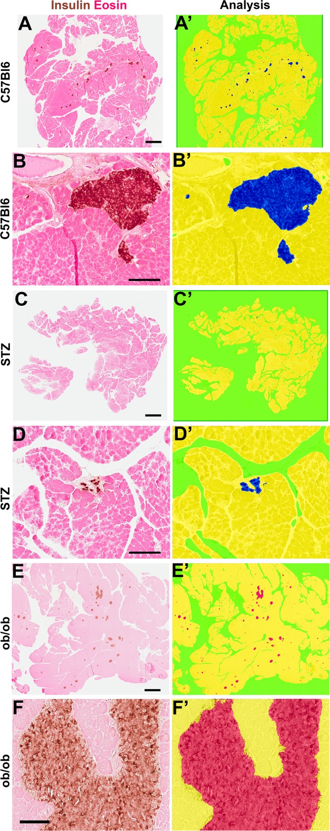
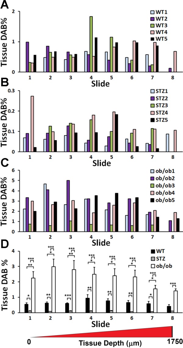
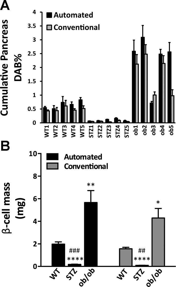
β-Cell mass is a parameter commonly measured in studies of islet biology and diabetes. However, the rigorous quantification of pancreatic β-cell mass using conventional histological methods is a time-consuming process. Rapidly evolving virtual slide technology with high-resolution slide scanners and newly developed image analysis tools has the potential to transform β-cell mass measurement. To test the effectiveness and accuracy of this new approach, we assessed pancreata from normal C57Bl/6J mice and from mouse models of β-cell ablation (streptozotocin-treated mice) and β-cell hyperplasia (leptin-deficient mice), using a standardized systematic sampling of pancreatic specimens. Our data indicate that automated analysis of virtual pancreatic slides is highly reliable and yields results consistent with those obtained by conventional morphometric analysis. This new methodology will allow investigators to dramatically reduce the time required for β-cell mass measurement by automating high-resolution image capture and analysis of entire pancreatic sections.

Keywords: automated analysis; diabetes; pancreatic islets; β-cell mass.


Figures

Fig. 1.
Fig. 1.
Workflow for automated β-cell mass analysis. A: schematic of pancreas processing and sectioning. B: schematic of image analysis. Building a macro for accurate image segmentation involves repeated rounds of tissue selection, training, and testing. Steps that should be omitted when tuning a macro are depicted in gray; optional steps are shown in green; required steps are shown in beige.
Fig. 2.
Fig. 2.
Selected tissue features for training montages with assigned Class color markup. A: clean glass. B: lightly stained acinar tissue. C: sinuous β-cell cluster adjacent to eosin-stained connective tissue in D. E: dirty glass. F: connective tissue with background staining. G: connective tissue. H: folded, intensely-stained acinar tissue. I: light DAB labeling within islet. J: folded, intensely-labeled DAB within an islet. Color markup for analyzed images: Glass (green), Eosin (yellow), and DAB (red or blue). All images are displayed at ×10 magnification; C and D are from the Macro 1 Montage; A and B and E–J are from the Macro 2 Montage. Glass, greeen class markup; Eeosin, yellow color markup; DAB, blue in C or red color markup in I and J. Yellow boxes in A–J denote a slide/tissue elements added to a montage.
Fig. 3.
Fig. 3.
Pattern recognition algorithm allows for automated detection of islets in pancreatic sections. Pancreatic sections from wild-type (A and B′), STZ-treated (C and D′), and Lepob//ob mice (E and F′) on a C57Bl6/J background were labeled for insulin and counterstained with eosin. Slides were scanned at ×20 magnification. A–F: original images; insulin (brown), eosin (pink). A′–F′: color markup of tissue Classes identified in A–F by image analysis with Macro 1 (A′–D′) or Macro 2 (E′ and F′). Glass (green), Eosin (yellow), DAB (blue in A′–D′ or red in E′ and F′). Scale bar in A, C, and E is 1 mm; scale bar in B, D, and F is 100 μm.
Fig. 4.
Fig. 4.
Percentage of tissue DAB representing pancreatic section area labeled for insulin varies in the pancreas with section depth. Percentage of tissue DAB was plotted in relationship to the tissue section location within the pancreas. A: Control mice, n = 5. B: STZ-treated mice, n = 5. C: Lepob/ob mice, n = 5. Note different scale on y-axis in A, B, and C. D: percentage of tissue DAB plotted as a function of section depth was averaged for all mice per each group. *P < 0.05, **P < 0.01, ***P < 0.001.
Fig. 5.
Fig. 5.
Validation of automated analysis for β-cell mass measurement. A: pancreas DAB% was not significantly different when analyzed by automated or conventional method across all mice (P = 0.6044). B: automated measurements of β-cell mass in control, STZ-treated, and Lepob/ob mice were compared with results generated by conventional morphometry. As expected, β-cell mass was significantly reduced in STZ-treated mice and increased in Lepob/ob mice compared with WT mice (P < 0.0001), but no significant difference was detected between automated and conventional analysis method (P = 0.1863). Automated analysis: ****P < 0.0001, STZ-treated vs. WT mice; ###P < 0.001, STZ-treated vs. Lepob/ob mice; **P < 0.01, Lepob/ob vs. WT mice. Conventional analysis: ****P < 0.0001, STZ-treated vs. WT mice; ##P < 0.01, STZ-treated vs. Lepob/ob mice; *P < 0.05 Lepob/ob vs. WT mice.

References

    1. Bock T, Pakkenberg B, Buschard K. Increased islet volume but unchanged islet number in ob/ob mice. Diabetes 52: 1716–1722, 2003 - PubMed
    1. Brissova M, Shostak A, Shiota M, Wiebe PO, Poffenberger G, Kantz J, Chen Z, Carr C, Jerome WG, Chen J, Baldwin HS, Nicholson W, Bader DM, Jetton T, Gannon M, Powers AC. Pancreatic islet production of vascular endothelial growth factor-A is essential for islet vascularization, revascularization, and function. Diabetes 55: 2974–2985, 2006 - PubMed
    1. Campbell-Thompson ML, Heiple T, Montgomery E, Zhang L, Schneider L. Staining protocols for human pancreatic islets. J Vis Exp: e4068, 2012 - PMC - PubMed
    1. Campbell-Thompson ML, Montgomery EL, Foss RM, Kolheffer KM, Phipps G, Schneider L, Atkinson MA. Collection protocol for human pancreas. J Vis Exp: e4039, 2012 - PMC - PubMed
    1. Chintinne M, Stangé G, Denys B, In't Veld P, Hellemans K, Pipeleers-Marichal M, Ling Z, Pipeleers D. Contribution of postnatally formed small beta cell aggregates to functional beta cell mass in adult rat pancreas. Diabetologia 53: 2380–2388, 2010 - PubMed

Publication types

MeSH terms

LinkOut - more resources