. 2013 Dec:31:86-100.
doi: 10.3767/003158513X669698. Epub 2013 Jun 17.

Phylogeny of chrysosporia infecting reptiles: proposal of the new family Nannizziopsiaceae and five new species

Affiliations

Phylogeny of chrysosporia infecting reptiles: proposal of the new family Nannizziopsiaceae and five new species

A M Stchigel et al. Persoonia. 2013 Dec.

Abstract

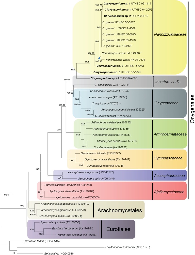
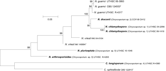
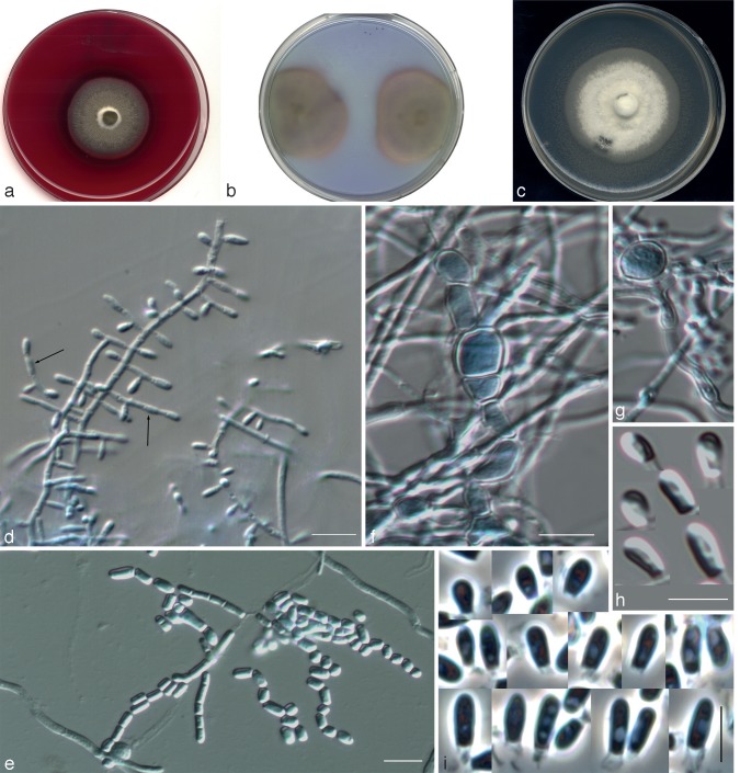
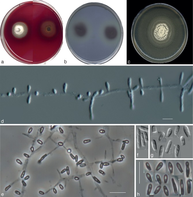
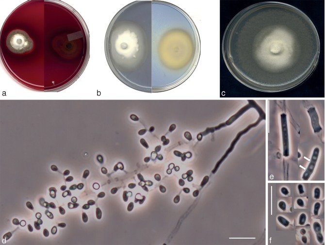
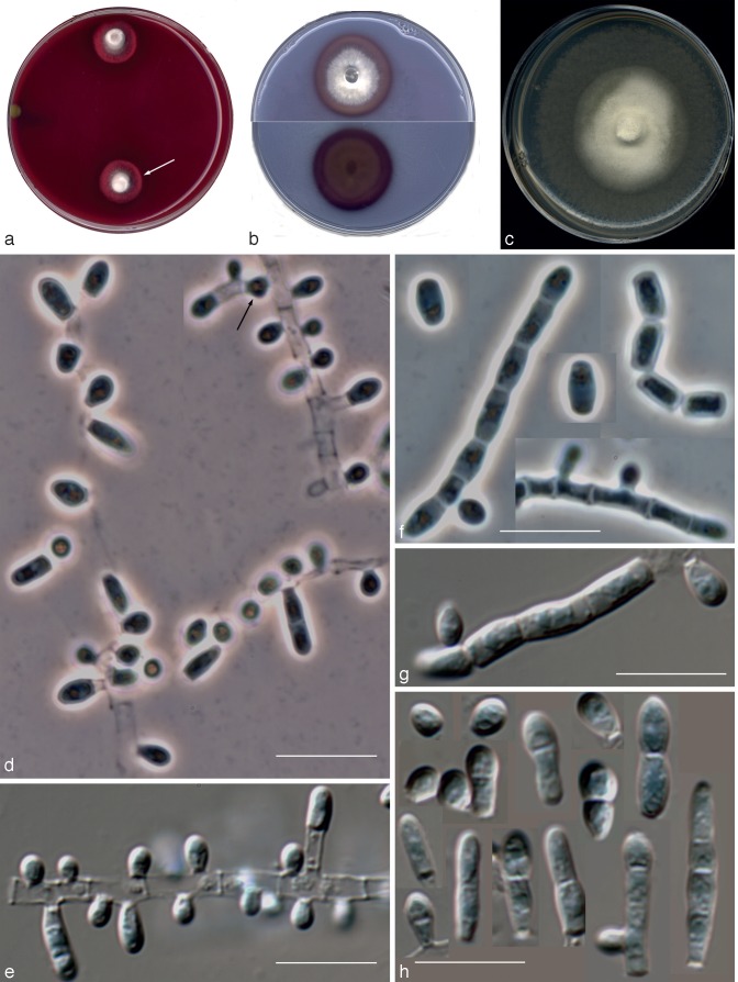
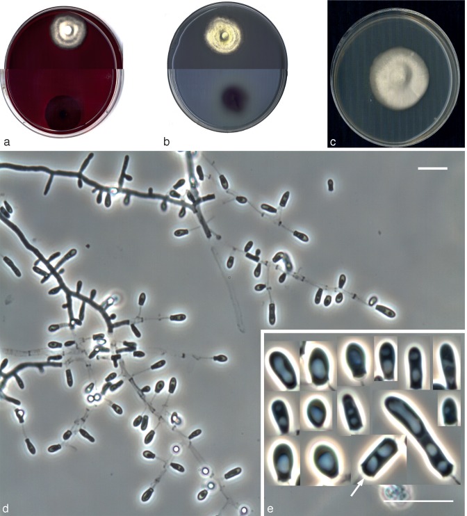
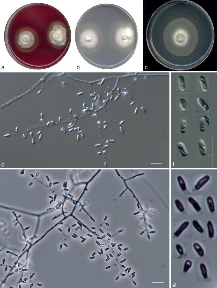
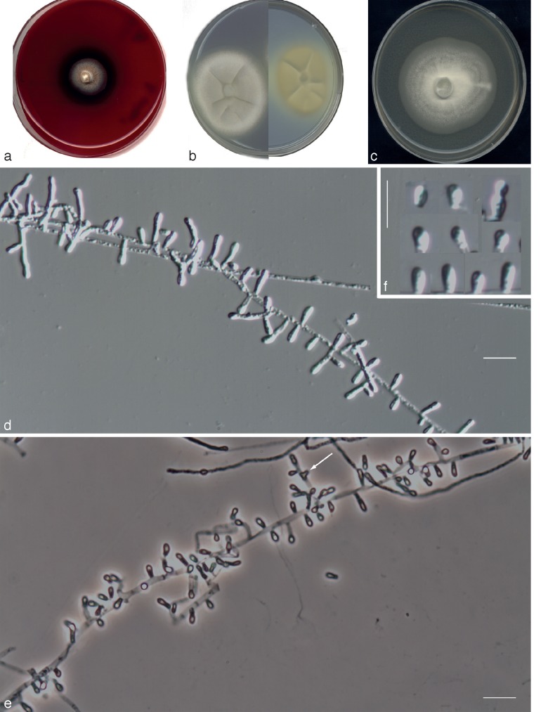
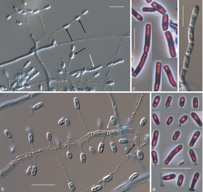
We have performed a phenotypic and phylogenetic study of a set of fungi, mostly of veterinary origin, morphologically similar to the Chrysosporium asexual morph of Nannizziopsis vriesii (Onygenales, Eurotiomycetidae, Eurotiomycetes, Ascomycota). The analysis of sequences of the D1-D2 domains of the 28S rDNA, including representatives of the different families of the Onygenales, revealed that N. vriesii and relatives form a distinct lineage within that order, which is proposed as the new family Nannizziopsiaceae. The members of this family show the particular characteristic of causing skin infections in reptiles and producing hyaline, thin- and smooth-walled, small, mostly sessile 1-celled conidia and colonies with a pungent skunk-like odour. The phenotypic and multigene study results, based on ribosomal ITS region, actin and β-tubulin sequences, demonstrated that some of the fungi included in this study were different from the known species of Nannizziopsis and Chrysosporium and are described here as new. They are N. chlamydospora, N. draconii, N. arthrosporioides, N. pluriseptata and Chrysosporium longisporum. Nannizziopsis chlamydospora is distinguished by producing chlamydospores and by its ability to grow at 5 °C. Nannizziopsis draconii is able to grow on bromocresol purple-milk solids-glucose (BCP-MS-G) agar alkalinizing the medium, is resistant to 0.2 % cycloheximide but does not grow on Sabouraud dextrose agar (SDA) with 3 % NaCl. Nannizziopsis arthrosporioides is characterised by the production of very long arthroconidia. Nannizziopsis pluriseptata produces 1- to 5-celled sessile conidia, alkalinizes the BCP-MS-G agar and grows on SDA supplemented with 5 % NaCl. Chrysosporium longisporum shows long sessile conidia (up to 13 μm) and does not produce lipase.

Keywords: Chrysosporium; Nannizziopsiaceae; Nannizziopsis; Onygenales; animal infections; ascomycetes; mycoses; reptiles.


Figures

Fig. 1
Fig. 1
Maximum-likelihood (ML) tree based on Tamura three-parameter corrected nucleotide distances among the D1 and D2 domains of the 28S rRNA gene sequences of taxa included in Table 1. Numbers on the branches are bootstrap ML values above 55 %, followed by Bayesian posterior probabilities (Bpp) above 0.6. Branch lengths are proportional to distance. Sequences not generated in this study and obtained from the GenBank database are indicated in parentheses. Ex-type strains of the different species are indicated with T. New species proposed in this study are indicated in bold. N. = Nannizziopsis. C. = Chrysosporium.
Fig. 2
Fig. 2
Maximum-likelihood (ML) tree obtained from the combined DNA sequence data from three loci (ITS, actin and β-tubulin). Bootstrap support values above 70 % are indicated at the nodes.
Fig. 3
Fig. 3
Nannizziopsis chlamydospora UTHSC 04-2056 (= Chrysosporium sp. 1). a. Colony on blood agar; b. colonies on BCP-MS-G (reverse); c. colony on TOTM; d. conidiophores bearing sessile and intercalary conidia (black arrow), and conidia on side branches; e. long chains of lateral and terminal arthroconidia; f. chlamydospores in chains; g. a solitary chlamydospore and thick-walled hyphae; h, i. conidia. — Scale bars: d–g = 10 μm; h, i = 5 μm (d–h, differential interference contrast; i, phase contrast).
Fig. 4
Fig. 4
Nannizziopsis draconii CCFVB CH12 (= Chrysosporium sp. 2). a. Colony on blood agar (surface and reverse); b. colonies on BCP-MS-G (reverse); c. colony on TOTM; d, e. conidiophores bearing sessile and terminal conidia; f. arthroconidia; g, h. sessile and terminal conidia. — Scale bars = 10 μm (d, f, g, differential interference contrast; e, h, phase contrast).
Fig. 5
Fig. 5
Nannizziopsis arthrosporioides UTHSC R-4263 (= Chrysosporium sp. 3). a. Colony on blood agar (surface and reverse); b. colony on BCP-MS-G (surface and reverse); c. colony on TOTM; d. fertile hyphae bearing sessile conidia; e. two singly intercalary conidia and a terminal chain of arthroconidia (white arrows show the septa); f. sessile (some 2-celled) conidia. — Scale bars = 10 μm (d–f, differential interference contrast).
Fig. 6
Fig. 6
Nannizziopsis pluriseptata UTHSC 10-1045 (= Chrysosporium sp. 5). a. Colonies on blood agar (the arrow shows the b-hemolysis halus); b. colony on BCP-MS-G (surface and reverse); c. colony on TOTM; d, e. fertile hyphae bearing mostly sessile conidia (arrow showing a intercalary conidium); f, g. arthroconidia; h. sessile conidia (observe the presence of up to 5-celled propagules). — Scale bars = 10 μm (d, f, differential interference contrast; e, g, h, phase contrast).
Fig. 7
Fig. 7
Chrysosporium longisporum UTHSC R-4380 (= Chrysosporium sp. 4). a. Colony on blood agar (surface and reverse); b. colony on BCP-MS-G (surface and reverse); c. colony on TOTM; d. fertile hyphae bearing mostly sessile conidia; e. sessile and intercalary (arrow) conidia. — Scale bars = 10 μm (d, e, phase contrast).
Fig. 8
Fig. 8
Nannizziopsis guarroi CBS 124553. a. Colonies on blood agar; b. colonies on BCP-MS-G; c. colony on TOTM; d, e. fertile hyphae bearing sessile conidia; f, g. sessile conidia. — Scale bars = 10 μm (d, f, differential interference contrast; e, g, phase contrast).
Fig. 9
Fig. 9
Chrysosporium anamorph of Nannizziopsis vriesii IMI 149994. a. Colony on blood agar; b. colony on BCP-MS-G (surface and reverse); c. colony on TOTM; d, e. fertile hyphae bearing mostly sessile conidia (arrow shows an intercalary conidium); f. sessile conidia. — Scale bars = 10 μm (d, f, differential interference contrast; e, phase contrast).
Fig. 10
Fig. 10
Chrysosporium ophiodiicola CBS 122913. a, b. Fertile hyphae bearing sessile conidia, intercalary conidia (black arrows) and intercalary chains of arthroconidia (white arrows); c, d. arthroconidia; e. sessile conidia. — Scale bars = 10 μm (a, b, d, differential interference contrast; c, e, phase contrast).

References

    1. Abarca ML, Castellá G, Martorell J, Cabañes FJ. 2010. Chrysosporium guarroi sp. nov. a new emerging pathogen of pet green iguanas (Iguana iguana). Medical Mycology 48: 365–372 - PubMed
    1. Abarca ML, Martorell J, Castellá G, Ramis A, Cabañes FJ. 2008. Cutaneous hyalohyphomycosis caused by a Chrysosporium species related to Nannizziopsis vriesii in two green iguanas (Iguana iguana). Medical Mycology 46: 349–354 - PubMed
    1. Abarca ML, Martorell J, Castellá G, Ramis A, Cabañes FJ. 2009. Dermatomycosis in a pet inland bearded dragon (Pogona vitticeps) caused by a Chrysosporium species related to Nannizziopsis vriesii. Veterinary Dermatology 20: 295–299 - PubMed
    1. Abdel-Razik M, Zaki SM. 2008. Experimental pathogenicity and molecular characterization of an environmental isolate of Chrysosporium zonatum Al-Musallam and Tan (Family: Onygenaceae, Order: Onygenales). International Journal of Agriculture and Biology 10: 273–277
    1. Allender MC, Dreslik M, Wylie S, Phillips C, Wylie DB, et al. 2011. Chrysosporium sp. infection in eastern massasauga rattlesnakes. Emerging Infectious Diseases 17: 2383–2384 - PMC - PubMed

LinkOut - more resources