. 2013 Sep 1;1(3):267-281.
doi: 10.4248/BR201303005.

Inhibition of Rgs10 Expression Prevents Immune Cell Infiltration in Bacteria-induced Inflammatory Lesions and Osteoclast-mediated Bone Destruction

Affiliations

Inhibition of Rgs10 Expression Prevents Immune Cell Infiltration in Bacteria-induced Inflammatory Lesions and Osteoclast-mediated Bone Destruction

Sen Yang et al. Bone Res. .

Abstract

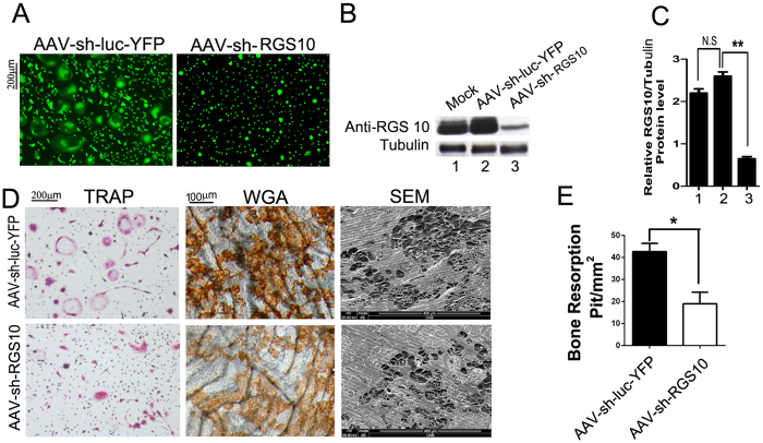
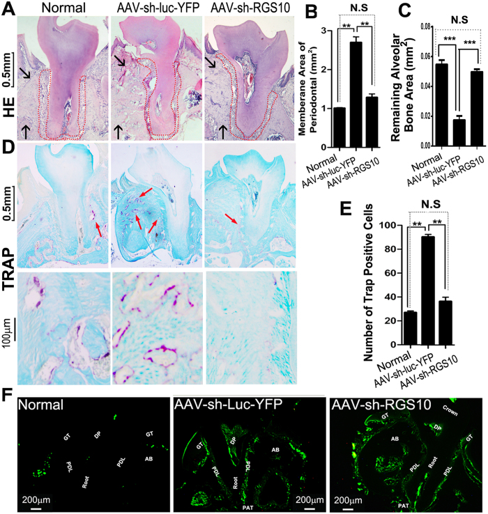
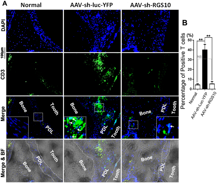
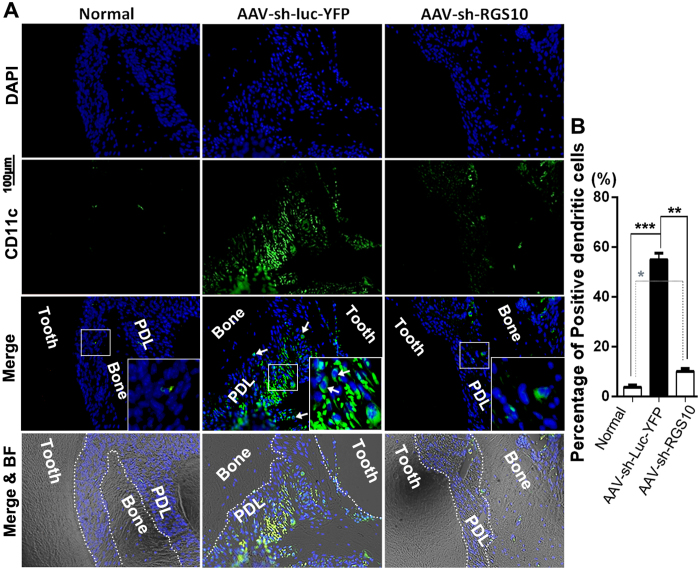
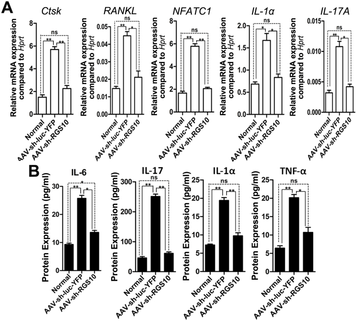
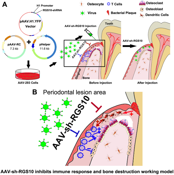
Regulator of G-protein Signaling 10 (Rgs10) plays an important function in osteoclast differentiation. However, the role of Rgs10 in immune cells and inflammatory responses, which activate osteoclasts in inflammatory lesions, such as bacteria-induced periodontal disease lesions, remains largely unknown. In this study, we used an adeno-associated virus (AAV-) mediated RNAi (AAV-shRNA-Rgs10) knockdown approach to study Rgs10's function in immune cells and osteoclasts in bacteria-induced inflammatory lesions in a mouse model of periodontal disease. We found that AAV-shRNA-Rgs10 mediated Rgs10 knockdown impaired osteoclastogenesis and osteoclast-mediated bone resorption, in vitro and in vivo. Interestingly, local injection of AAV-shRNA-Rgs10 into the periodontal tissues in the bacteria-induced inflammatory lesion greatly decreased the number of dendritic cells, T-cells and osteoclasts, and protected the periodontal tissues from local inflammatory damage and bone destruction. Importantly, AAV-mediated Rgs10 knockdown also reduced local expression of osteoclast markers and pro-inflammatory cytokines. Our results demonstrate that AAV-shRNA-Rgs10 knockdown in periodontal disease tissues can prevent bone resorption and inflammation simultaneously. Our data indicate that Rgs10 may regulate dendritic cell proliferation and maturation, as well as the subsequent stimulation of T-cell proliferation and maturation, and osteoclast differentiation and activation. Our study suggests that AAV-shRNA-Rgs10 can be useful as a therapeutic treatment of periodontal disease.

Keywords: AAV-mediated RNAi knockdown; Rgs10; bone resorption; gene therapy; gingival inflammation; immune cell; periodontal disease.


Figures

Figure 1
Figure 1
AAV-sh-Rgs10 targets Rgs10 mRNA and efficiently knocks down the expression of Rgs10. (A-E) MBM was stimulated with M-CSF/RANKL for 3 days to allow for differentiation of osteoclasts, which were then transduced with AAV-sh-luc-YFP (control vector) or AAV-sh-Rgs10 or left untreated (mock). (A) Fluorescence indicates effective transduction of pre-osteoclasts and osteoclasts. (B, C) Western blot and quantification of Rgs10 protein level demonstrate that AAV-sh-Rgs10 treated osteoclasts have significantly reduced expression of Rgs10 (P<0.01). (D) TRAP stain reveals that the number of osteoclast is decreased in the AAV-sh-Rgs10 treatment group. Resorption lacunae were visualized by wheat germ agglutinin (WGA) and scanning electron microscopy (SEM), which demonstrates that Rgs10 knockdown inhibits osteoclast-mediated bone resorption. (E) Quantification of resorption pits on bone slices visualized by SEM showed a significant decrease in the degree of bone resorption in the AAV-sh-Rgs10 treatment group (P<0.01). N.S: no significant; *P<0.05; **P<0.01.
Figure 2
Figure 2
AAV effectively transduced periodontal tissue and Rgs10 knockdown efficiently protected mice from bone loss in W50-stimulated periodontitis. (A) Mouse maxillary molars showing AAV injection sites (black arrows). Fluorescence microscope image reveals eGFP expression after local injection with AAV-sh-Rgs10 compared with normal mouse tissues that did not receive AAV treatment. (B) Representative figures of periodontal bone resorption and root exposure from different groups. (C) There was no significant difference in bone loss between the normal group and the AAV-sh-Rgs10 treatment group (P>0.05). In addition, the AAV-sh-Rgs10 treatment group had significantly less bone loss compared with the AAV-sh-luc-YFP treatment group (P<0.01) (N=9, n=3 per group, repeated 3 times). N.S: no significant; **P<0.01.
Figure 3
Figure 3
AAV-mediated Rgs10 knockdown decreased bone resorption and the number of TRAP-positive osteoclasts in the periodontal area. (A) Representative figures of H&E staining from uninfected mice (Normal), W50-infected mice treated with AAV-sh-Rgs10 or AAV-sh-luc-YFP. Black arrows indicate significant change of alveolar bone in different groups, which demonstrate that the alveolar bone resorption was severe in AAV-sh-luc-YFP treatment group. And the periodontal ligament area in AAV-sh-luc-YFP treated disease group showed irregular shape compared to the normal and AAV-sh-Rgs10 treated group (red dot area). (B, C) The area of periodontal ligament increased in the AAV-sh-luc-YFP treatment group, compared to the normal and AAV-sh-Rgs10 treatment groups (P<0.01) and remaining alveolar bone area was also decreased significantly in AAV-sh-luc-YFP treated disease group (P<0.001) (N=9, n=3 per group, repeated 3 times). (D) Representative figures of TRAP staining (counter stained with fast green) from uninfected mice (Normal), W50-infected mice treated with AAV-sh-Rgs10 or AAV-sh-luc-YFP (Red Arrows). (E) Compared with normal control group, the number of TRAP positive osteoclasts from AAV-sh-luc-YFP groups increased significantly. The AAV-sh-Rgs10 treatment group has obviously therapeutic effect, and the number of osteoclasts were close to normal control group (P<0.01). (F) Fluorescence microscope image of sections reveal YFP expression after local injection with AAV-sh-luc-YFP and eGFP expression of AAV-sh-Rgs10 compared with normal mouse tissues that did not receive AAV treatment (GT: Gingival Tissue, AB: Alveolar Bone, DP: Dental Pulp, PDL: Periodontal Ligament, PAT: Periapical tissue) (N=9, n=3 per group, repeated 3 times). N.S: no significant; **P<0.01; ***P<0.001.
Figure 4
Figure 4
AAV-mediated Rgs10 knockdown decreased the number of T-cells in periodontal lesions. (A) Representative figures from immunofluorescence staining of alveolar sections indicated that uninfected mice (Normal) and W50-infected mice treated with AAV-sh-Rgs10 had less CD3 positive (green) T-cells (white arrows), compared to W50-infected mice treated with AAV-sh-luc-YFP. Cell nuclei were labeled using DAPI stain (blue). (B) Quantification of CD3 positive T-cells. (N=9, n=3 per group, repeated 3 times). BF: Bright Field, PDL: periodontal ligament. N.S: no significant; *P<0.05; **P<0.01; ***P<0.001.
Figure 5
Figure 5
AAV-mediated Rgs10 knockdown decreased the number of DCs cells in periodontal lesions. (A) Representative figures from immunofluorescence staining of alveolar sections indicated that uninfected mice (Normal) and W50-infected mice treated with AAV-sh-Rgs10 had less CD11c (green) positive dendritic cells (white arrows), compared to W50-infected mice treated with AAV-sh-luc-YFP. Cell nuclei were labeled using DAPI staining (blue). (B) Quantification of CD11c positive dendritic cells (N=9, n=3 per group, repeated 3 times). BF: Bright Field, PDL: periodontal ligament. *P<0.05; **P<0.01; ***P<0.001.
Figure 6
Figure 6
AAV-sh-Rgs10 reduced the expression of osteoclast marker genes and cytokines in periodontal lesions. (A) qPCR of osteoclast marker genes (i.e., Cathepsin K), genes important for osteoclast differentiation (i.e., RANKL and NFATC1) and cytokines (i.e., IL-1α, and IL-17A) in the periodontal lesion from uninfected mice (Normal) or infected mice treated with AAV-sh-Luc-YFP or with AAV-sh-Rgs10. Hprt was used as an endogenous control. AAV-sh-Rgs10 reduced the expression of inflammatory cytokines in the periodontal tissues (pooled 3 samples each time in each group on three independent experiments). (B) ELISA experiment for inflammatory cytokine expression from osteoclast or T-cells in the periodontal tissues of uninfected mice (Normal) or W50-infected mice treated with AAV-sh-luc-YFP or with AAV-sh-Rgs10. The data of ELISA indicates that uninfected mice (Normal) and W50-infected mice treated with AAV-sh-Rgs10 have less inflammatory cytokines expression of IL-6, IL-1α, IL-17 and TNF-α, compared to W50-infected mice treated with AAV-sh-luc-YFP (pooled 3 samples each time in each group on three independent experiments). N.S: no significant; *P<0.05; **P<0.01; ***P <0.001.
Figure 7
Figure 7
Diagram model for the AAV-shRNA-Rgs10 inhibits immune response in periodontal lesion. (A) The whole experiment was starting from the design of shRNA sequences, annealing shRNA stands together, linearizing pAAV.H1 vector, inserting shRNA fragment into pAAV.H1 vector, verifying clones have inserted, and finally transfecting into AAV particles to create virus. Then the virus was injected into the periodontal lesion, in which the AAV-sh-Rgs10 inhibited the immune DCs and the subsequent decrease of T-cells. And the decreased pro-inflammatory cytokines resulted in less osteoclast differentiation and activation. (B) The following immune response mediated by immune cells (i.e. DCs and T-cells) and bone resorption or formation mediated by osteoclasts and osteoblasts respectively in periodontal lesion. After contact with bacterial (red arrow), the antigen presenting cells (APCs) (i.e. DCs) presented the bacterial pathogen (dark red arrow) to T-cells and activated T-cells. Then T-cells can activate the osteoclasts and inhibit osteoblasts (blue arrow) while AAV-sh-Rgs10 can inhibit this process, which will prevent further bone loss by over-activated osteoclasts.

Similar articles

Cited by

References

    1. Chambrone L, Guglielmetti MR, Pannuti CM, Chambrone LA. Evidence grade associating periodontitis to preterm birth and/or low birth weight: I. A systematic review of prospective cohort studies. J Clin Periodontol. 2011;38:795–808. - PubMed
    1. Kuula H, Salo T, Pirilä E, Tuomainen AM, Jauhiainen M, Uitto VJ, Tjäderhane L, Pussinen PJ, Sorsa T. Local and systemic responses in matrix metalloproteinase 8-deficient mice during Porphyromonas gingivalis-induced periodontitis. Infect Immun. 2009;77:850–859. - PMC - PubMed
    1. Van Dyke TE, Serhan CN. Resolution of inflammation: a new paradigm for the pathogenesis of periodontal diseases. J Dent Res. 2003;82:82–90. - PubMed
    1. D'Aiuto F, Parkar M, Andreou G, Suvan J, Brett PM, Ready D, Tonetti MS. Periodontitis and systemic inflammation: control of the local infection is associated with a reduction in serum inflammatory markers. J Dent Res. 2004;83:156–160. - PubMed
    1. Loe H. Periodontal disease. The sixth complication of diabetes mellitus. Diabetes Care. 1993;16:329–334. - PubMed

LinkOut - more resources