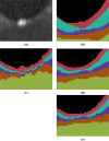
Multiple-object geometric deformable model for segmentation of macular OCT
- PMID: 24761289
- PMCID: PMC3986003
- DOI: 10.1364/BOE.5.001062
Multiple-object geometric deformable model for segmentation of macular OCT
Abstract
Optical coherence tomography (OCT) is the de facto standard imaging modality for ophthalmological assessment of retinal eye disease, and is of increasing importance in the study of neurological disorders. Quantification of the thicknesses of various retinal layers within the macular cube provides unique diagnostic insights for many diseases, but the capability for automatic segmentation and quantification remains quite limited. While manual segmentation has been used for many scientific studies, it is extremely time consuming and is subject to intra- and inter-rater variation. This paper presents a new computational domain, referred to as flat space, and a segmentation method for specific retinal layers in the macular cube using a recently developed deformable model approach for multiple objects. The framework maintains object relationships and topology while preventing overlaps and gaps. The algorithm segments eight retinal layers over the whole macular cube, where each boundary is defined with subvoxel precision. Evaluation of the method on single-eye OCT scans from 37 subjects, each with manual ground truth, shows improvement over a state-of-the-art method.
Keywords: (100.0100) Image processing; (170.4470) Ophthalmology; (170.4500) Optical coherence tomography.
Figures


References
-
- Gordon-Lipkin E., Chodkowski B., Reich D. S., Smith S. A., Pulicken M., Balcer L. J., Frohman E. M., Cutter G., Calabresi P. A., “Retinal nerve fiber layer is associated with brain atrophy in multiple sclerosis,” Neurology 69, 1603–1609 (2007). 10.1212/01.wnl.0000295995.46586.ae - DOI - PubMed
-
- Saidha S., Syc S. B., Ibrahim M. A., Eckstein C., Warner C. V., Farrell S. K., Oakley J. D., Durbin M. K., Meyer S. A., Balcer L. J., Frohman E. M., Rosenzweig J. M., Newsome S. D., Ratchford J. N., Nguyen Q. D., Calabresi P. A., “Primary retinal pathology in multiple sclerosis as detected by optical coherence tomography,” Brain 134, 518–533 (2011). 10.1093/brain/awq346 - DOI - PubMed
-
- Saidha S., Sotirchos E. S., Ibrahim M. A., Crainiceanu C. M., Gelfand J. M., Sepah Y. J., Ratchford J. N., Oh J., Seigo M. A., Newsome S. D., Balcer L. J., Frohman E. M., Green A. J., Nguyen Q. D., Calabresi P. A., “Microcystic macular oedema, thickness of the inner nuclear layer of the retina, and disease characteristics in multiple sclerosis: a retrospective study,” The Lancet Neurology 11, 963–972 (2012). 10.1016/S1474-4422(12)70213-2 - DOI - PMC - PubMed
-
- Saidha S., Syc S. B., Durbin M. K., Eckstein C., Oakley J. D., Meyer S. A., Conger A., Frohman T. C., Newsome S., Ratchford J. N., Frohman E. M., Calabresi P. A., “Visual dysfunction in multiple sclerosis correlates better with optical coherence tomography derived estimates of macular ganglion cell layer thickness than peripapillary retinal nerve fiber layer thickness,” Mult. Scler. 17, 1449–1463 (2011). 10.1177/1352458511418630 - DOI - PubMed
Grants and funding
LinkOut - more resources
Full Text Sources
Other Literature Sources
