Review
. 2014 Feb 1;3(2):127-138.
doi: 10.1089/wound.2013.0451.

Harnessing the Electric Spark of Life to Cure Skin Wounds

Affiliations
Review

Harnessing the Electric Spark of Life to Cure Skin Wounds

Cristina Martin-Granados et al. Adv Wound Care (New Rochelle). .

Abstract

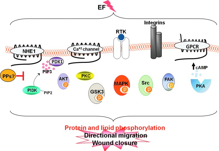
Significance: Skin wounds cause great distress and are a huge economic burden, particularly with an increasingly aging population that heals poorly. There is an urgent need for better therapies that improve repair. Intracellular signaling pathways that regulate wound repair are activated by growth factors, hormones, and cytokines released at the wound. In addition, endogenous electric fields (EFs) are generated by epithelia in response to injury and are an important cue that coordinates cell behavior at wounds. Electrical stimulation (ES), therefore, holds the potential to be effective therapeutically in treating wounds. Recent Advances: ES of wounds is an old idea based on observations of the natural occurrence of EF at wound sites. However, it is now receiving increasing attention, because (1) the underpinning mechanisms are being clarified; (2) devices that measure skin wound currents are in place; and (3) medical devices that apply EF to poorly healing wounds are in clinical use with promising results. Critical Issues: Several signaling proteins transduce the EF influence to cells. However, a bigger picture of the EF-proteome is needed in order to understand this complex process and target it in a controlled manner. Future Directions: Dissecting the signaling pathways driving electrical wound healing will allow further identification of key molecular switches that control the cellular response to EFs. These findings herald the development of a new concept, the use of hydrogel electrodes impregnated with small molecules that target signaling pathways to explore the potential of dual electric-pharmacological therapies to repair wounds.


Figures

None
Cristina Martin-Granados
None
Colin D. McCaig, PhD, FRSE
<b>Figure 1.</b>
Figure 1.
Structure of the mammalian skin. The dermis contains fibroblasts, endothelial cells, and inflammatory cells that are surrounded by an organized extracellular matrix (ECM) of collagen type I, fibronectin, and elastin. The epidermis is a continuously renewing tissue composed of keratinocytes at different stages of differentiation represented by three layers: granular (composed of three cell sheets SG1–SG3), spinous, and basal. The granular layer is overlain by a stratum corneum that represents the endpoint of epidermal differentiation and cell death. The stratum corneum confers the barrier function to the skin and protects internal organs from the environment. As the differentiation process progresses, keratinocytes express proteins involved in both the scaffold function and the eventual formation of the insoluble cornified envelope (CE). The CE replaces the plasma membrane of differentiating keratinocytes and consists of keratins that are enclosed within an insoluble amalgam of proteins, which are crosslinked by transglutaminases and surrounded by a lipid envelope forming a “mortar-brick” structure. Intercellular lipids are primarily generated from exocytosis of lipid-containing granules called lamellar bodies, during the terminal differentiation of keratinocytes. Tight junctions (TJs) are intercellular junctions that are formed by various TJ transmembrane proteins; for example, claudins, occludin, tricellulin, and junctional adhesion molecule, as well as intracellular scaffold proteins, for example, ZO-1, ZO-2, ZO-3, and cingulin. These proteins regulate the passage of ions and molecules through the paracellular pathway in epithelial and endothelial cells. TJs in the skin are located in the granulous SG2 layer. TJ formation is a prerequisite for the formation of the epidermal permeability barrier and the maintenance of barrier function, in addition to the sealing of proteins into CE. To see this illustration in color, the reader is referred to the web version of this article at www.liebertpub.com/wound
<b>Figure 2.</b>
Figure 2.
Ion flows and potentials in intact skin. (A) Diagram of a typical epithelial cell in a monolayer with Na+ and Cl channels localized on the apical plasma membrane and K+ channels localized on the basolateral membranes along with the Na+/K+-ATPase. (B) This asymmetric distribution of ion channels generates a transcellular inflow of positive current that flows back between the cells. This positive current flow coupled to Cl movement in the opposite direction generates a transepithelial potential difference (TEP) of 10–60 mV. TJs between contiguous cells contribute to creating the TEP by providing high electrical resistance and preventing leakage of ions back down their concentration gradients between cells. Consequently, a concentration of positive charge exists behind the junctions at the deeper layers (positive sign in figure). The potential is, thus, relatively negative on the apical side of the junctions. To see this illustration in color, the reader is referred to the web version of this article at www.liebertpub.com/wound
<b>Figure 3.</b>
Figure 3.
Generation of wound-induced electric currents. When the skin is injured, the epithelial layer is disrupted locally, and the ionic gradient across the epithelium collapses at the wound site. A consequence of the disruption of the epithelial integrity is leakage of Na+ ions out of the wound, down their concentration gradient, and an uptake of Cl ions. The respective ion movements give rise to a physiological injury current that flows toward the wound center (defined as the movement of positive charge) and a lateral voltage gradient oriented parallel to the epithelial sheet (EF, electric field; green discontinuous arrow at bottom). The discontinuous red arrows indicate positive ion (current) flux through the cell layers and the return path. The wound is, therefore, more negatively charged and acts as a cathode (negative sign in figure) as compared with healthy tissue (which acts as the anode, positive sign in figure). For simplification, we only show the ionic flux loop and EF on the right side of the figure, although the same applies for the left side. These endogenously generated EFs contribute to the guidance of cells, blood vessels, and nerves toward the wound site. Based on Nuccitelli et al. and Shaw and Martin. To see this illustration in color, the reader is referred to the web version of this article at www.liebertpub.com/wound
<b>Figure 4.</b>
Figure 4.
EFs activate several kinase signaling pathways. Membrane proteins transduce the electric signal by activating several intracellular signaling pathways. Eventually, transmission of the electric signal to downstream kinase effectors induces phosphorylation of proteins at Ser, Thr, and Tyr residues controlling the cell polarization and migration events that are essential for wound healing. The action of protein kinases is reversed by protein phosphatases (PPs). Strikingly, the identity of PPs involved in counteracting the action of kinases during electrotaxis remains largely unstudied. To see this illustration in color, the reader is referred to the web version of this article at www.liebertpub.com/wound

Similar articles

Cited by

References

    1. Sen CK, Gordillo GM, Roy S, Kirsner R, Lambert L, Hunt TK, Gottrup F, Gurtner GC, and Longaker MT: Human skin wounds: a major and snowballing threat to public health and the economy. Wound Repair Regen 2009; 17:763. - PMC - PubMed
    1. Gurtner GC, Werner S, Barrandon Y, and Longaker MT: Wound repair and regeneration. Nature 2008; 453:314. - PubMed
    1. Kloth LC: Electrical stimulation for wound healing: a review of evidence from in vitro studies, animal experiments, and clinical trials. Int J Low Extrem Wounds 2005; 4:23. - PubMed
    1. Balakatounis KC. and Angoules AG: Low-intensity electrical stimulation in wound healing: review of the efficacy of externally applied currents resembling the current of injury. Eplasty 2008; 8:e28. - PMC - PubMed
    1. Ramadan A, Elsaidy M, and Zyada R: Effect of low-intensity direct current on the healing of chronic wounds: a literature review. J Wound Care 2008; 17:292. - PubMed

LinkOut - more resources