Review
. 2014 Feb 1;3(2):184-201.
doi: 10.1089/wound.2013.0442.

The Electrical Response to Injury: Molecular Mechanisms and Wound Healing

Affiliations
Review

The Electrical Response to Injury: Molecular Mechanisms and Wound Healing

Brian Reid et al. Adv Wound Care (New Rochelle). .

Abstract

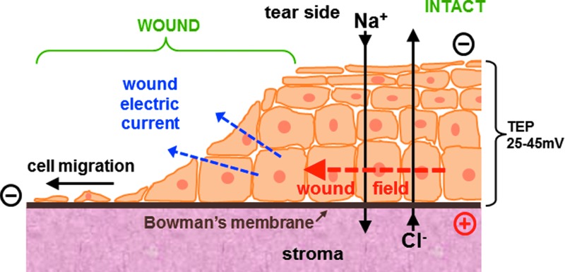
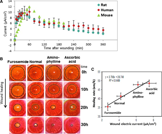
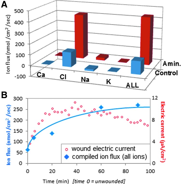
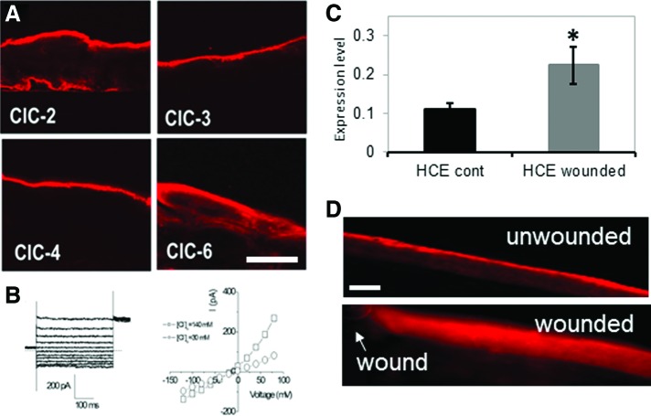
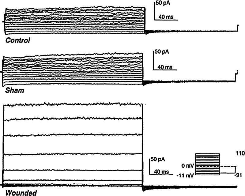
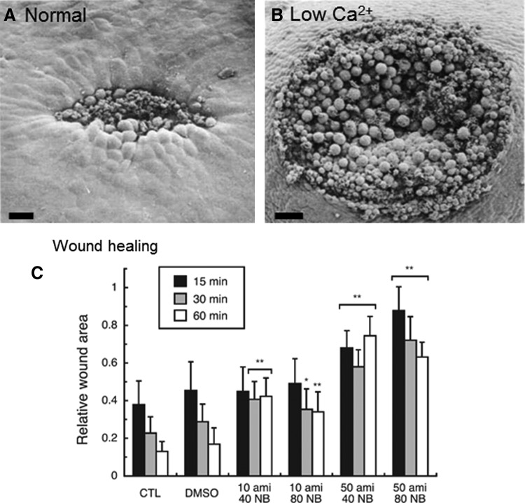
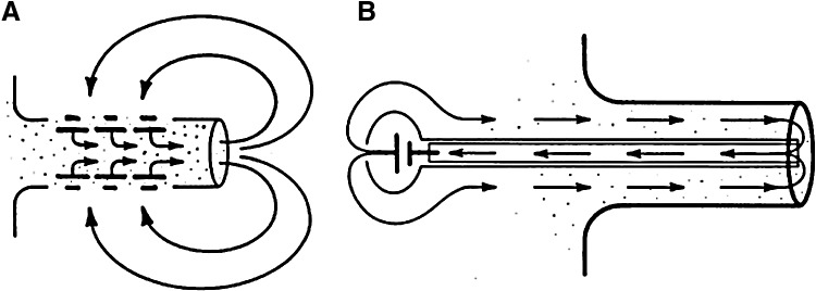
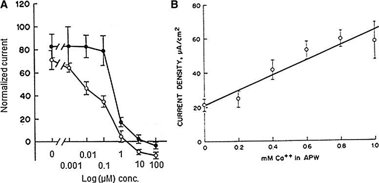
Significance: Natural, endogenous electric fields (EFs) and currents arise spontaneously after wounding of many tissues, especially epithelia, and are necessary for normal healing. This wound electrical activity is a long-lasting and regulated response. Enhancing or inhibiting this electrical activity increases or decreases wound healing, respectively. Cells that are responsible for wound closure such as corneal epithelial cells or skin keratinocytes migrate directionally in EFs of physiological magnitude. However, the mechanisms of how the wound electrical response is initiated and regulated remain unclear. Recent Advances: Wound EFs and currents appear to arise by ion channel up-regulation and redistribution, which are perhaps triggered by an intracellular calcium wave or cell depolarization. We discuss the possibility of stimulation of wound healing via pharmacological enhancement of the wound electric signal by stimulation of ion pumping. Critical Issues: Chronic wounds are a major problem in the elderly and diabetic patient. Any strategy to stimulate wound healing in these patients is desirable. Applying electrical stimulation directly is problematic, but pharmacological enhancement of the wound signal may be a promising strategy. Future Directions: Understanding the molecular regulation of wound electric signals may reveal some fundamental mechanisms in wound healing. Manipulating fluxes of ions and electric currents at wounds might offer new approaches to achieve better wound healing and to heal chronic wounds.


Figures

None
Brian Reid, PhD
<b>Figure 1.</b>
Figure 1.
Ionic basis of cornea wound electrical response. The intact corneal epithelium (right) pumps sodium and chloride in opposite directions to generate and maintain a trans-epithelial electrical potential difference (TEP) of approximately 45 mV, which is necessary to maintain a healthy cornea. Injury to the cornea (left) collapses the TEP locally at the wound to zero and significant electric fields (EFs) build up around the wound (red arrow), which also generate large electric currents flowing out of the wound (blue arrows). The EF is intrinsically directional: It is orientated towards the wound, with the wound the negative cathode. Corneal epithelial cells respond to physiological-strength EFs by migrating to the cathode, suggesting they use the natural, endogenous wound EF as a directional cue to migrate into the wound, thus enhancing healing. Reprinted with permission from Vieira et al. To see this illustration in color, the reader is referred to the web version of this article at www.liebertpub.com/wound
<b>Figure 2.</b>
Figure 2.
Cornea wound electric response and wound healing. (A) The cornea wound electrical activity is consistent and appears to be a regulated response. After corneal injury in mouse, rat, and human, the wound electric current increases rapidly and is maintained for many hours. Note the human and mouse data are smaller than the rat data; these data have been normalized to the maximum rat data point at 40 min to enable a comparison. (B) Drugs that enhance or inhibit ion transport increase or decrease wound current and wound healing, respectively, in rat cornea. (C) There is a strong correlation between wound electric current (X-axis) and wound-healing rate (Y-axis) (linear best fit R2=0.9499). Reprinted with permission from Reid et al. To see this illustration in color, the reader is referred to the web version of this article at www.liebertpub.com/wound
<b>Figure 3.</b>
Figure 3.
Cornea-wound ion fluxes. (A) In normal wounds (blue) the electric current is carried mainly by the flux of chloride ions. Enhancement of wound current with aminophylline is due mainly to increased chloride flux (red). (B) Increasing total ion flux (blue) correlates with increasing electric current (red) after wounding. The ion flux and electric current are plotted on different y-axis: Ion flux (nmol/cm2/s) is plotted on the left axis, and electric current (μA/cm2) is plotted on the right axis, to enable a comparison of the increase of both after wounding. Reprinted with permission from Vieira et al. To see this illustration in color, the reader is referred to the web version of this article at www.liebertpub.com/wound
<b>Figure 4.</b>
Figure 4.
Chloride channels (CLCs) in cornea. (A) CLCs show distinct distributions in human corneal epithelium (HCE). Scale bar 50 μm. (B) Chloride currents recorded from primary cultured HCE cells. Left: whole-cell chloride current at different membrane potentials. Right: i/v plot of current versus voltage showing the outward rectifying chloride current. □: normal Ca (140 mM); ○: low Ca (30 mM). (C) CLC-2 mRNA was up-regulated after wounding in an HCE cell monolayer scratch wound assay (*p<0.05; HCE control vs. HCE wounded). (D) CLC-2 in normal unwounded rat cornea was concentrated in the apical cells (upper panel). After wounding, CLC-2 was re-distributed throughout the corneal epithelium (lower panel). Arrow shows wound edge. Scale bar 50 μm. (A, B) Reprinted with permission from Cao et al.; (C, D) Reprinted with permission from Vieira et al. To see this illustration in color, the reader is referred to the web version of this article at www.liebertpub.com/wound
<b>Figure 5.</b>
Figure 5.
Cation channel activation after wounding. Endothelial cells from normal unwounded (top) and sham wounded (center) rabbit cornea had normal whole-cell cation currents. Injury to the cornea caused these currents to increase significantly (bottom; 70 h [almost 3 days] after wounding). Reprinted with permission from Watsky.
<b>Figure 6.</b>
Figure 6.
Ion channels in Xenopus wound healing. Calcium controls changes in cell shape and contractility that are necessary for Xenopus embryo wound healing. (A) 15 min after wounding in normal solution, cells around the wound edge change shape by elongating into the wound to close it. Scale bar 50 μm (B) Low calcium inhibits wound healing (scale bar 100 μm). (C) Blocking sodium channel ENaC with amiloride (ami) or CLC-3 with NPPB (NB) reduces wound healing. Columns show relative wound area at different time points after wounding, in different concentrations of inhibitor (*p<0.01, **p<0.001). (A, B) Reprinted with permission from Stanisstreet. (C) Reprinted with permission from Fuchigami et al.
<b>Figure 7.</b>
Figure 7.
Circulating currents in limb stumps. (A) Newt limb stump currents are driven by the skin battery. Large currents flow out of the cut stump, flowing inward at intact skin to complete the circuit. (B) In frog limb, an artificial current drawn into the stump by a mercury cell battery via an implanted wick in the center of the limb stump was able to initiate regeneration. Sham implants supplying no current gave no regeneration, just a healing of the cut limb stump. Reprinted with permission from Borgens et al.
<b>Figure 8.</b>
Figure 8.
Newt limp stump currents. (A) Sodium channel blocker amiloride, or one of its analogs, can significantly inhibit, even reverse, the normal limb stump current. Closed circles: amiloride; open circles: amiloride analog B (benzamil). (B) Normal limb stump current requires external calcium. Reprinted with permission from Eltinge et al.
<b>Figure 9.</b>
Figure 9.
Wounding induces cell depolarization. (A, B) After scratch wounding a bovine endothelial cell monolayer (wound at the top), a wave of depolarization passes from cells at the edge and extends to undamaged cells in the intact monolayer (e.g., see arrows in B). (C, D) Blocking sodium channel with phenamil decreases depolarization. (E, F) In a scratch wound assay (wound in the center), the normal monolayer heals almost completely in 6 h (E). In phenamil, healing rate is significantly reduced. Phenamil also appears to reduce actin cable formation (bright labeling at wound edges indicated with arrows). Scale bar 30 μm. Reprinted with permission from Chifflet et al. To see this illustration in color, the reader is referred to the web version of this article at www.liebertpub.com/wound
<b>Figure 10.</b>
Figure 10.
Wound-induced calcium wave. (A) Wounding a corneal epithlial cell monolayer induces an immediate and rapid intracellular calcium wave that is propagated from cell to cell (white asterisk shows site of injury; scale bar 50 μm). (B) Depletion of intracellular calcium stores with thapsigargin (T) or incubation in calcium-free medium significantly reduced wound healing (*p<0.001). Reprinted with permission from Leiper et al. To see this illustration in color, the reader is referred to the web version of this article at www.liebertpub.com/wound
<b>Figure 11.</b>
Figure 11.
Wound calcium wave in Caenorhabditis elegans. (A) Needle wound (arrow) triggers a rapid epidermal calcium response (arrowhead) that eventually spreads to approximately one third of the length of the animal. Scale bars 100 μm. (B) Femptosecond laser wounding (X) induces a calcium wave that traveled at about 25.6 μm/s, a speed consistent with propagation via Ca2+-induced Ca2+ release from internal stores. N=epidermal nuclei. Scale bars 10 μm. Reprinted with permission from Xu and Chisholm. To see this illustration in color, the reader is referred to the web version of this article at www.liebertpub.com/wound
<b>Figure 12.</b>
Figure 12.
Gap junctions in cornea. (A) In normal intact cornea, gap junction connexin 43 (Cx43) (green) was present in the basal cell layer of the corneal epithelium. Basement membrane protein laminin (red) was found in the basement membrane region beneath the epithelium. Scale bar 50 μm. (B) Twelve hours after wounding, Cx43 and laminin were present in low levels at the back of the migrating epithelium (white arrow), but not at the leading edge (left). Black arrowhead shows original site of wound edge. Scale bar 50 μm. Reprinted with permission from Suzuki et al. To see this illustration in color, the reader is referred to the web version of this article at www.liebertpub.com/wound

References

    1. Klyce SD: Electrical profiles in the corneal epithelium. J Physiol 1972; 226:407. - PMC - PubMed
    1. Klyce SD: Transport of Na, Cl, and water by the rabbit corneal epithelium at resting potential. Am J Physiol 1975; 228:1446. - PubMed
    1. Foulds IS. and Barker AT: Human skin battery potentials and their possible role in wound healing. Br J Dermatol 1983; 109:515. - PubMed
    1. Barker AT, Jaffe LF, and Vanable JW, Jr: The glabrous epidermis of cavies contains a powerful battery. Am J Physiol 1982; 242:R358. - PubMed
    1. Mathias RT, Kistler J, and Donaldson P: The lens circulation. J Membr Biol 2007; 216:1. - PubMed